. 2010 Mar;216(3):356-67.
doi: 10.1111/j.1469-7580.2009.01192.x. Epub 2010 Jan 7.

Isolation of a new class of cysteine-glycine-proline-rich beta-proteins (beta-keratins) and their expression in snake epidermis

Affiliations

Isolation of a new class of cysteine-glycine-proline-rich beta-proteins (beta-keratins) and their expression in snake epidermis

Luisa Dalla Valle et al. J Anat. 2010 Mar.

Abstract

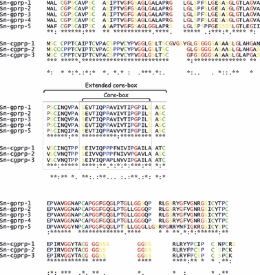
Scales of snakes contain hard proteins (beta-keratins), now referred to as keratin-associated beta-proteins. In the present study we report the isolation, sequencing, and expression of a new group of these proteins from snake epidermis, designated cysteine-glycine-proline-rich proteins. One deduced protein from expressed mRNAs contains 128 amino acids (12.5 kDa) with a theoretical pI at 7.95, containing 10.2% cysteine and 15.6% glycine. The sequences of two more snake cysteine-proline-rich proteins have been identified from genomic DNA. In situ hybridization shows that the messengers for these proteins are present in the suprabasal and early differentiating beta-cells of the renewing scale epidermis. The present study shows that snake scales, as previously seen in scales of lizards, contain cysteine-rich beta-proteins in addition to glycine-rich beta-proteins. These keratin-associated beta-proteins mix with intermediate filament keratins (alpha-keratins) to produce the resistant corneous layer of snake scales. The specific proportion of these two subfamilies of proteins in different scales can determine various degrees of hardness in scales.


Figures

Fig. 1
Fig. 1
Histologic sections of scale epidermis in resting (A) and renewal (B–D) stages. (A) Two overlapped scales are covered by a pale beta-layer (arrows). The epidermis (arrows) has one to two layers. Bar: 20 μm. (B) Scale in renewal phase (stage 4 of the shedding cycle) with the differentiating, inner beta-layer (arrows) within the multilayered epidermis. The arrowhead points to the outer (mature and compact) beta-layer. The double arrowheads indicate the thin oberhautchen layer present in the inner scale surface and hinge region. Bar: 20 μm. (C) Detail of renewing epidermis showing the tapering of the inner beta-layer (arrows) from the outer scale to the tip and down the inner surface. In the latter, only the dark oberhautchen layer is present (arrowheads). Bar: 10 μm. (D) Beta-1 immunolabelled outer (arrowhead) and inner (arrow) beta-layers. The other layers in between the two beta-layers, and the basal layer (above the dashes), are unlabelled. Bar: 10 μm. a, alpha-layer; ba, basal layer of the epidermis; de, dermis; h, hinge region; i, inner scale surface; la, lacunar layer; o, outer scale layer; t; scale tip; The basal layer is underlined with dashes.
Fig. 2
Fig. 2
Nucleotide sequences of cysteine-rich snake beta-keratin transcript (Sn-cgrpr-1), of the genes (Sn-cgrpr-2 and 3), and the deduced amino acid sequences. One-letter symbols of the encoded amino acids are shown below the DNA sequence. The numbers refer to the nucleotide and amino acid positions at the end of each line. Dashed lines indicate missing nucleotides or amino acid loss. Intron sequences are indicated in lowercase. The in-frame translation start codon (ATG) as well as the stop codon (TAA or TAG) and the putative polyadenylation signal (ATTAAA) are boxed. Splice signals (gt and ag) are shown in bold and are boxed. The specific oligonucleotide primers used for the cloning (see Table 1) are underlined and the names are reported in bold over the sequences. The sequences are available at the EMBL/GenBank/DDBJ database under accession number FN297847, FN297848 and FN297849 for Sn-cgprp-1, 2 and 3, respectively.
Fig. 2
Fig. 2
Nucleotide sequences of cysteine-rich snake beta-keratin transcript (Sn-cgrpr-1), of the genes (Sn-cgrpr-2 and 3), and the deduced amino acid sequences. One-letter symbols of the encoded amino acids are shown below the DNA sequence. The numbers refer to the nucleotide and amino acid positions at the end of each line. Dashed lines indicate missing nucleotides or amino acid loss. Intron sequences are indicated in lowercase. The in-frame translation start codon (ATG) as well as the stop codon (TAA or TAG) and the putative polyadenylation signal (ATTAAA) are boxed. Splice signals (gt and ag) are shown in bold and are boxed. The specific oligonucleotide primers used for the cloning (see Table 1) are underlined and the names are reported in bold over the sequences. The sequences are available at the EMBL/GenBank/DDBJ database under accession number FN297847, FN297848 and FN297849 for Sn-cgprp-1, 2 and 3, respectively.
Fig. 3
Fig. 3
Northern blot analysis. (A) Representative Northern blot analysis of total RNA extracted from the skin of Snake-1 (resting), Snake-2 (renewing), and Snake-2 subcutaneous and muscular tissue, detected with a snake DIG-labelled anti-sense cRNA cysteine-rich beta-keratin probe. Exposition time: 5 min. (1) RNA samples from the skin of Snake-1; (2,3) RNA samples from the skin of Snake-2; (4) RNA samples from the subcutaneous and muscular tissue of Snake-2. (B) Methylene blue-stained 28S and 18S ribosomal RNA genes present in each mRNA sample.
Fig. 5
Fig. 5
ClustalW comparison of the amino acid sequences among the cysteine-rich protein sequences (Sn-cgprp-1, 2, 3) and the glycine-rich protein sequences (Sn-gprp-1 to 5, Dalla Valle et al. 2007b). The extended 32 amino acids appear in the centre of the figure, including the core-box region, which shows the highest identity among these proteins. Four key amino acids (glycine in red, proline in blue, serine in yellow, and cysteine in green) are indicated. According to the ClustalW convention, an asterisk denotes the identity in all sequences of the alignment; colons denote conserved substitutions; dots denote semi-conserved substitutions.
Fig. 4
Fig. 4
In situ hybridization of renewing scale epidermis visualized by immunofluorescence (A–D) and colorimetric reaction (red) with the AP-method (E–H). (A) Positive signal (arrows) in beta-keratin cells of the outer (arrows) and in the oberhautchen (arrowheads) of the inner scale surface. Bar: 20 μm. (B) Detail of the labelled beta-cells (arrows) of the outer scale surface and in the oberhautchen of the inner scale surface of the previous scale (arrowhead). Bar: 10 μm. (C) Detail of labelled beta-cells of the stratified inner beta-layer of the outer scale surface (arrow). Bar: 10 μm. (D) Control showing absence of fluorescence in both the beta-layer of the outer (arrow) and the inner (arrowheads) scale surface. Bar: 10 μm. (E) Reactive beta-layer (arrows) and oberhautchen layer of the inner scale surface (arrowhead). Bar: 30 μm. (F) Detail of the reactive cells (arrows) forming a single layer (oberhautchen) in the hinge region (left), which becomes a tetra-stratified beta-layer in the outer scale surface (right). Bar: 15 μm. (G) Reactive oberhautchen (arrowhead) and penta-stratified beta-layer (arrow) located in the outer scale surface. Bar: 15 μm. (H) Control sections showing no reactivity in beta-cells (arrows). The arrowhead indicates the layer of melanocytes located underneath the epidermis. Bar: 15 μm. a, alpha-layer; aR, anti-sense RNA probe detection; c, negative control; de, dermis; the epidermis is underline with dashes.
Fig. 6
Fig. 6
Computer prediction of the secondary structure (using the psipred Protein Structure Prediction Server at http://bioinf.cs.ucl.ac.uk/psipred/) of two Sn-cprp proteins. The central core-box is indicated by squares. The confidence of prediction (%) is indicated.

References

    1. Alibardi L. Presence of acid phosphatase in the epidermis of the regenerating tail of the lizard (Podarcis muralis) and its possible role in the process of shedding and keratinization. J Zool. 1998;246:379–390.
    1. Alibardi L. Keratohyalin-like granules in lizard epidermis: evidence from cytochemical, autoradiographic and microanalytic studies. J Morphol. 2001;248:64–79. - PubMed
    1. Alibardi L. Immunocytochemical analysis of the process of keratinization of the epidermis of snakes. J Zool. 2002;258:541–552.
    1. Alibardi L, Sawyer RH. Immunocytochemical analysis of beta keratins in the epidermis of anapsids, lepidosaurians and archosaurians. J Exp Zool. 2002;293:27–38. - PubMed
    1. Alibardi L, Toni M. Cytochemical, biochemical and molecular aspects of the process of keratinization in the epidermis of reptilian scales. Prog. Histochem Cytochem. 2006;40:73–134. - PubMed

Publication types