

Reprogramming of anaerobic metabolism by the FnrS small RNA
- PMID: 20070527
- PMCID: PMC2941437
- DOI: 10.1111/j.1365-2958.2010.07044.x
Reprogramming of anaerobic metabolism by the FnrS small RNA
Abstract
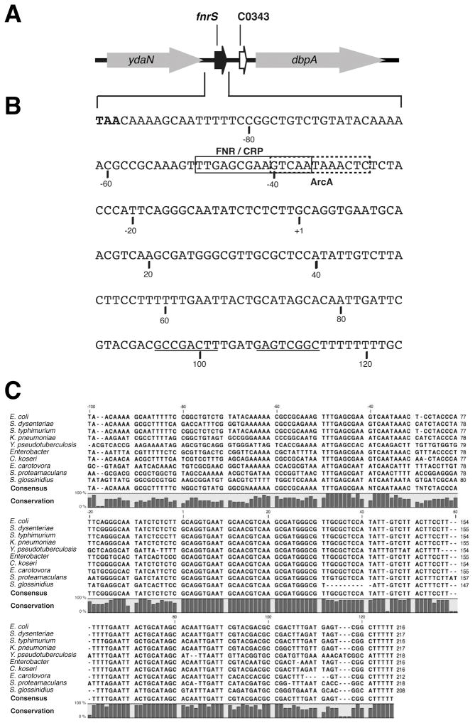
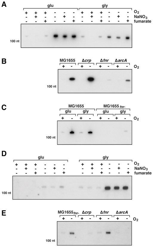
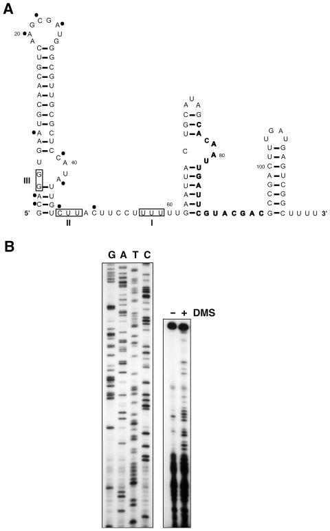
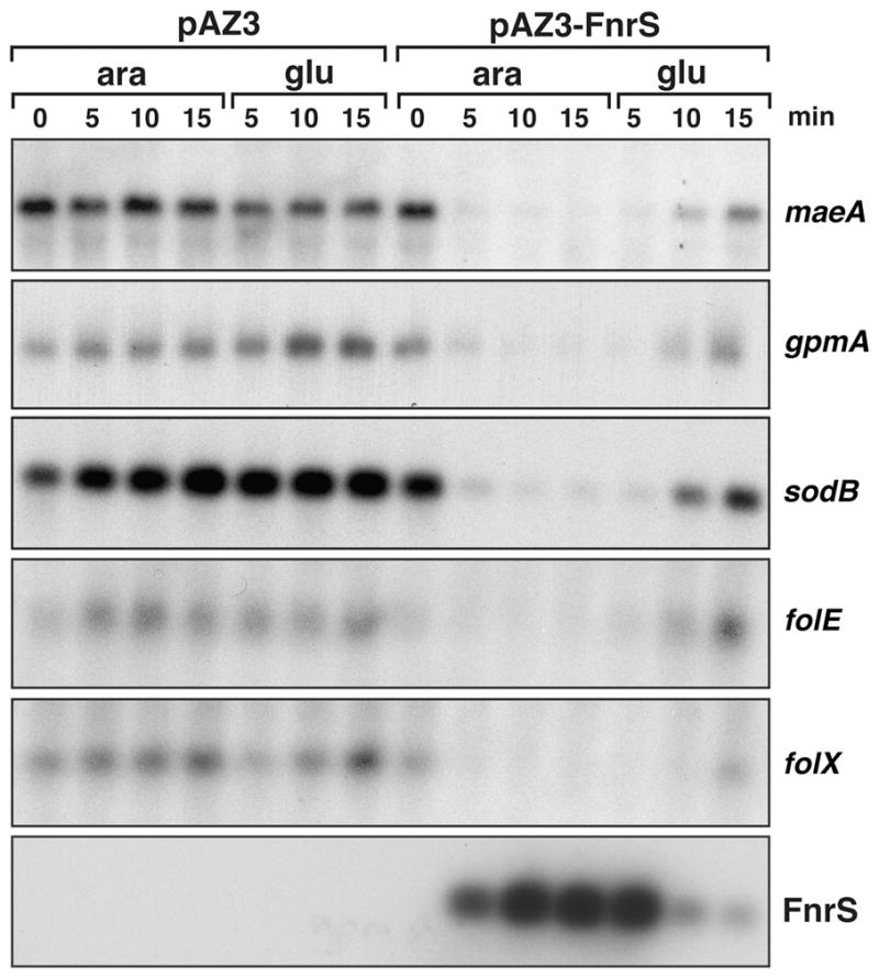
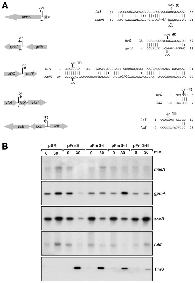
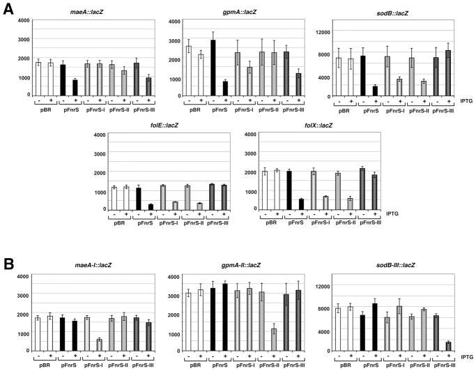
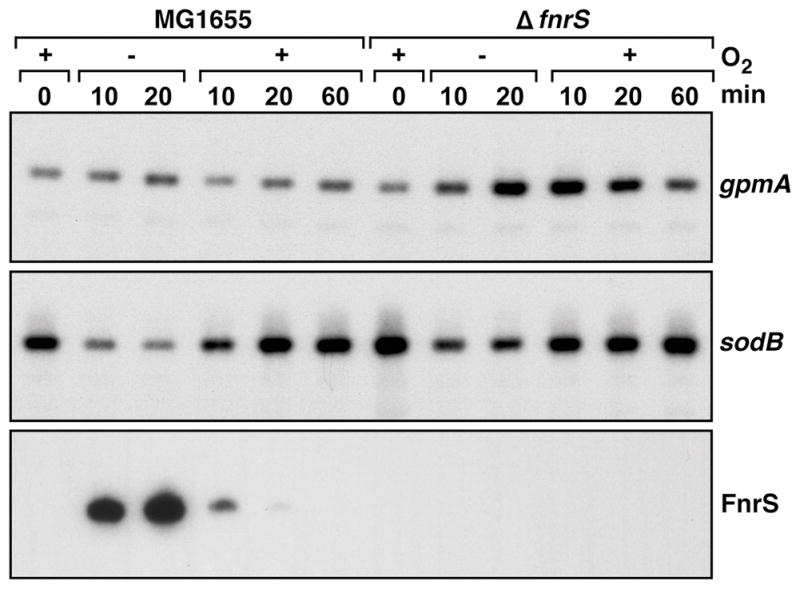
Small RNAs (sRNAs) that act by base pairing with trans-encoded mRNAs modulate metabolism in response to a variety of environmental stimuli. Here, we describe an Hfq-binding sRNA (FnrS) whose expression is induced upon a shift from aerobic to anaerobic conditions and which acts to downregulate the levels of a variety of mRNAs encoding metabolic enzymes. Anaerobic induction in minimal medium depends strongly on FNR but is also affected by the ArcA and CRP transcription regulators. Whole genome expression analysis showed that the levels of at least 32 mRNAs are downregulated upon FnrS overexpression, 15 of which are predicted to base pair with FnrS by TargetRNA. The sRNA is highly conserved across its entire length in numerous Enterobacteria, and mutational analysis revealed that two separate regions of FnrS base pair with different sets of target mRNAs. The majority of the target genes were previously reported to be downregulated in an FNR-dependent manner but lack recognizable FNR binding sites. We thus suggest that FnrS extends the FNR regulon and increases the efficiency of anaerobic metabolism by repressing the synthesis of enzymes that are not needed under these conditions.
Figures
Similar articles
-
Multiple factors dictate target selection by Hfq-binding small RNAs.EMBO J. 2012 Apr 18;31(8):1961-74. doi: 10.1038/emboj.2012.52. Epub 2012 Mar 2. EMBO J. 2012. PMID: 22388518 Free PMC article.
-
Translational regulation of gene expression by an anaerobically induced small non-coding RNA in Escherichia coli.J Biol Chem. 2010 Apr 2;285(14):10690-702. doi: 10.1074/jbc.M109.089755. Epub 2010 Jan 14. J Biol Chem. 2010. PMID: 20075074 Free PMC article.
-
Anaerobic activation of arcA transcription in Escherichia coli: roles of Fnr and ArcA.Mol Microbiol. 1994 Mar;11(5):955-64. doi: 10.1111/j.1365-2958.1994.tb00374.x. Mol Microbiol. 1994. PMID: 8022271
-
Cellular and molecular physiology of Escherichia coli in the adaptation to aerobic environments.J Biochem. 1996 Dec;120(6):1055-63. doi: 10.1093/oxfordjournals.jbchem.a021519. J Biochem. 1996. PMID: 9010748 Review.
-
New aspects of RNA-based regulation by Hfq and its partner sRNAs.Curr Opin Microbiol. 2018 Apr;42:53-61. doi: 10.1016/j.mib.2017.10.014. Epub 2017 Nov 7. Curr Opin Microbiol. 2018. PMID: 29125938 Free PMC article. Review.
Cited by
-
In silico 'fishing' using known small regulatory RNA (sRNA) candidates as the decoy from Escherichia coli, Salmonella typhi and Salmonella typhimurium manifested 14 novel sRNA candidates in the orthologous region of Proteus mirabilis.Mol Biol Rep. 2018 Dec;45(6):2333-2343. doi: 10.1007/s11033-018-4397-z. Epub 2018 Oct 3. Mol Biol Rep. 2018. PMID: 30284142
-
CsrA Shows Selective Regulation of sRNA-mRNA Networks.bioRxiv [Preprint]. 2023 Mar 29:2023.03.29.534774. doi: 10.1101/2023.03.29.534774. bioRxiv. 2023. PMID: 37034808 Free PMC article. Preprint.
-
Differential Regulation of CsrC and CsrB by CRP-cAMP in Salmonella enterica.Front Microbiol. 2020 Oct 14;11:570536. doi: 10.3389/fmicb.2020.570536. eCollection 2020. Front Microbiol. 2020. PMID: 33162952 Free PMC article.
-
Transcriptional and Post-transcriptional Control of the Nitrate Respiration in Bacteria.Front Mol Biosci. 2021 May 7;8:667758. doi: 10.3389/fmolb.2021.667758. eCollection 2021. Front Mol Biosci. 2021. PMID: 34026838 Free PMC article. Review.
-
Multiple factors dictate target selection by Hfq-binding small RNAs.EMBO J. 2012 Apr 18;31(8):1961-74. doi: 10.1038/emboj.2012.52. Epub 2012 Mar 2. EMBO J. 2012. PMID: 22388518 Free PMC article.
References
-
- Argaman L, Altuvia S. fhlA repression by OxyS RNA: kissing complex formation at two sites results in a stable antisense-target RNA complex. J Mol Biol. 2000;300:1101–1112. - PubMed
-
- Argaman L, Hershberg R, Vogel J, Bejerano G, Wagner E, Margalit H, Altuvia S. Novel small RNA-encoding genes in the intergenic regions of Escherichia coli. Curr Biol. 2001;11:941–950. - PubMed
-
- Bouvier M, Sharma CM, Mika F, Nierhaus KH, Vogel J. Small RNA binding to 5′ mRNA coding region inhibits translational initiation. Mol Cell. 2008;32:827–837. - PubMed
-
- Buchet A, Nasser W, Eichler K, Mandrand-Berthelot MA. Positive co-regulation of the Escherichia coli carnitine pathway cai and fix operons by CRP and the CaiF activator. Mol Microbiol. 1999;34:562–575. - PubMed
-
- Busby S, Ebright RH. Transcription activation by catabolite activator protein (CAP) J Mol Biol. 1999;293:199–213. - PubMed
Publication types
MeSH terms
Substances
Grants and funding
LinkOut - more resources
Full Text Sources
Other Literature Sources
Molecular Biology Databases
Research Materials
Miscellaneous
