. 2010 Mar;11(3):332-47.
doi: 10.1111/j.1600-0854.2009.01025.x. Epub 2009 Dec 3.

Mechanisms of function of tapasin, a critical major histocompatibility complex class I assembly factor

Affiliations

Mechanisms of function of tapasin, a critical major histocompatibility complex class I assembly factor

Syed Monem Rizvi et al. Traffic. 2010 Mar.

Abstract

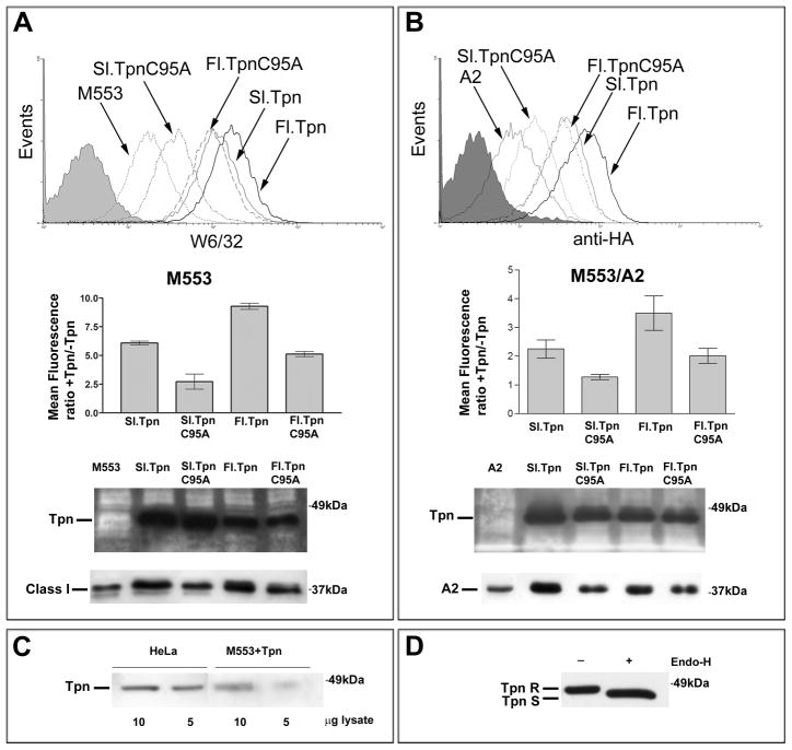
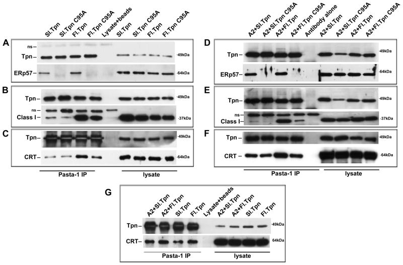
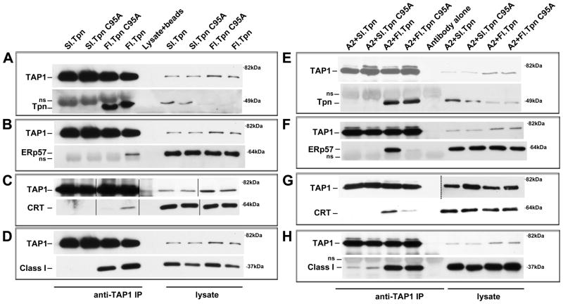
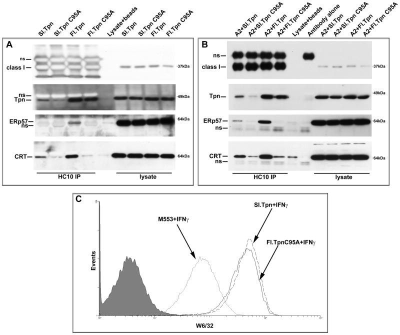
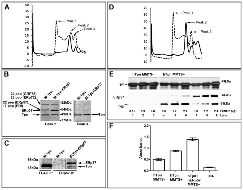
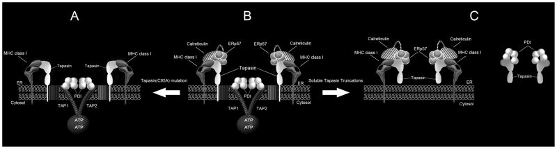
For their efficient assembly in the endoplasmic reticulum (ER), major histocompatibility complex (MHC) class I molecules require the specific assembly factors transporter associated with antigen processing (TAP) and tapasin, as well as generic ER folding factors, including the oxidoreductases ERp57 and protein disulfide isomerase (PDI), and the chaperone calreticulin. TAP transports peptides from the cytosol into the ER. Tapasin promotes the assembly of MHC class I molecules with peptides. The formation of disulfide-linked conjugates of tapasin with ERp57 is suggested to be crucial for tapasin function. Important functional roles are also suggested for the tapasin transmembrane and cytoplasmic domains, sites of tapasin interaction with TAP. We show that interactions of tapasin with both TAP and ERp57 are correlated with strong MHC class I recruitment and assembly enhancement. The presence of the transmembrane/cytosolic regions of tapasin is critical for efficient tapasin-MHC class I binding in interferon-gamma-treated cells, and contributes to an ERp57-independent mode of MHC class I assembly enhancement. A second ERp57-dependent mode of tapasin function correlates with enhanced MHC class I binding to tapasin and calreticulin. We also show that PDI binds to TAP in a tapasin-independent manner, but forms disulfide-linked conjugates with soluble tapasin. Thus, full-length tapasin is important for enhancing recruitment of MHC class I molecules and increasing specificity of tapasin-ERp57 conjugation. Furthermore, tapasin or the TAP/tapasin complex has an intrinsic ability to recruit MHC class I molecules and promote assembly, but also uses generic folding factors to enhance MHC class I recruitment and assembly.


Figures

Figure 1
Figure 1. Tapasin truncations and C95A mutations affect surface expression and steady-state levels of endogenous and exogenous MHC class I molecules
(A and B) Upper panels: Histogram showing cell surface expression of MHC class I in M553 or M553/A2 cells in the presence of the indicated tapasin constructs. Middle panels: Bar graphs shows mean fluorescence ratios of MHC class I expression as assessed by the W6/32 antibody (A) or anti-HA antibody (B) in the presence or absence of the indicated tapasin constructs. FACS data shows average and standard error of mean of two independent analyses for A and four independent analyses for B. Lower panel: Immunoblotting analysis using antibodies against tapasin and MHC class I (HC10 for A and anti-HA for B) of lysates from cells expressing the indicated proteins. (C) Immunoblotting analysis using antibody against tapasin of lysates from HeLa cells and M553 cells expressing full length tapasin. (D) Immunoblotting analysis of lysates from M553 cells expressing soluble tapasin following no treatment or Endo-H digestion. Soluble tapasin and full length tapasin are abbreviated as Sl.Tpn and Fl.Tpn respectively. 2×10⁶ M553 or M553/A2 cells expressing indicated tapasin constructs were used per sample in the FACS analysis.
Figure 2
Figure 2. Tapasin truncations and C95A mutations impact tapasin-ERp57, tapasin-MHC class I, and tapasin-calreticulin interactions
These analyses were conducted following cell treatments with IFN-γ for 48 h (A-G). 1.2–2.4×10⁷ cells were treated with the membrane-permeable cross-linker DTBP immediately prior to lysis. Immunoblotting analysis with anti-tapasin (Pasta-1) immunoprecipitates or lysates from M553 (A–C), M553/A2 (D–F) or both (G) cells expressing indicated tapasin constructs. Antibodies specific for tapasin (Tpn), ERp57, calreticulin (CRT) and MHC class I were used in the immunoblotting analyses. Data are representative of two (A, B, C, D, F, G) or four (E) independent analyses. The lanes labeled lysate+beads corresponds to the background bands obtained when lysates was immunoprecipitated with protein G beads and the lanes labeled antibody alone corresponds to the background bands obtained when antibody was immunoprecipitated with buffer (no cell lysates were used). Ns, indicates non-specific bands that run above the tapasin or MHC class I heavy chain bands in the anti-tapasin and anti-class I blots.
Figure 3
Figure 3. Tapasin truncations and C95A mutations impact the compositions of TAP complexes
The indicated cells were treated with IFN-γ for 48 h. For the analyses shown in panels E-H, 1.2–2.4×10⁷ cells were treated with the membrane-permeable cross-linker DTBP immediately prior to lysis. The analyses in panels A-D did not use a cross-linker. (A–H) Immunoblotting analysis of anti-TAP1 immunoprecipitates or lysates from M553 (A–D) or M553/A2 (E–H) cells expressing indicated tapasin constructs. Antibodies specific for TAP1, tapasin (Tpn), MHC class I heavy chain, ERp57 and calreticulin (CRT) were used. Data are representative of two (A, B, C, D, E, G, H) or four (F) independent analyses. The lanes labeled lysate+beads corresponds to the background bands obtained when lysates was immunoprecipitated with protein G beads and the lanes labeled antibody alone corresponds to the background bands obtained when antibody was immunoprecipitated with buffer (no cell lysates were used). Solid lines in panel C indicate lanes that were cut and pasted from same blot in order to preserve the order of presentation of lanes. Dashed lines in panel G indicate different exposures shown for immunoprecipitates and lysates of TAP1 blots from within the same experiment. Ns indicates antibody-derived non-specific bands that run above the tapasin and class I bands in blots with anti-tapasin, anti-class I, and below the ERp57 band in blots with anti-ERp57 (IP lanes only).
Figure 4
Figure 4. Tapasin truncations and C95A mutations impact MHC class I-tapasin and MHC class I-calreticulin interactions
These analyses were conducted following cell treatments with IFN-γ for 48 h. 1.2–2.4×10⁷ cells were treated with the membrane-permeable cross-linker DTBP immediately prior to lysis. Immunoblotting analysis with anti-MHC class I (HC10) of immunoprecipitates or lysates from M553 (A) or M553/A2 (B) cells expressing indicated tapasin constructs. Samples for all panels of A and B were derived from the same immunoprecipitation experiment respectively, and thus a single MHC class I panel is shown for each. Antibodies specific for MHC class I, tapasin (Tpn), ERp57, and calreticulin (CRT) were used in the immunoblotting analyses. (C) Histograms showing cell surface expression of MHC class I in M553 or M553 cells expressing soluble tapasin or full length tapasin(C95A) following treatment with IFN-γ (500 IU) for 48 h (conditions similar to those under which binding studies were undertaken). Data are representative of one (C) or at least two independent analyses for each panel of A and B. The lanes labeled lysate+beads corresponds to the background bands obtained when lysates was immunoprecipitated with protein G beads and the lanes labeled antibody alone corresponds to the background bands obtained when antibody was immunoprecipitated with buffer (no cell lysates were used). Ns, indicates non-specific bands.
Figure 5
Figure 5. The tapasin(C95A) mutant induces MHC class I assembly independent of an effect on TAP stabilization
(A) Left panel: M553 or full length tapasin(C95A) expressing M553 cells were metabolically labeled for 10 min and chased for the indicated times in fresh media. Lysates were immunoprecipitated with W6/32, then digested with Endo-H or left undigested and analyzed by SDS-PAGE and phosphorimaging analyses. Right panel: The Endo-H resistant MHC class I heavy chain (HC) bands in above gels were quantified using ImageQuant and plotted as percentage of Endo-H resistant HC. Data are representative of two independent analyses. (B) Histograms showing cell surface expression of MHC class I in M553 or M553 cells expressing full length tapasinC95A following no treatment or following incubation with IFN-γ (500 IU) for 20 h. (C) Immunoblotting analysis using HC10 of indicated amounts of total cell lysates from IFN-γ-treated M553 cells or tapasin(C95A)-expressing M553 cells. (D) Immunoblotting analysis of anti-TAP1 immunoprecipitates of IFN-γ-treated or untreated M553 and tapasin(C95A)-expressing cells. Antibodies specific for TAP1 and TAP2 were used.
Figure 6
Figure 6. Tapasin-independent association of PDI with TAP
The indicated cells were treated with IFN-γ for 48h. For panel B, cells were treated with the membrane-permeable cross-linker DTBP immediately prior to lysis, and the analyses of panels A, C and D were undertaken without cross-linker treatment. (A and B) Immunoblotting analysis of anti-TAP1 immunoprecipitates or lysates from M553 (A) or M553/A2 cells (B) expressing indicated tapasin constructs. (C) Immunoblotting analysis of anti-TAP1 immunoprecipitates or lysates from parent M553 cells lacking tapasin expression (No Tpn) or M553 cells expressing full-length tapasin(C95A) as indicated. (D) Immunoblotting analysis of anti-TAP1 immunoprecipitates or lysates from HeLa cells. Antibodies specific for TAP1, PDI were used in the immunoblots. Data are representative of one (C) or two (A, B, D) independent analyses. The lanes labeled lysate+beads corresponds to the background bands obtained when lysates was immunoprecipitated with protein G beads and the lanes labeled antibody alone corresponds to the background bands obtained when antibody was immunoprecipitated with buffer (no cell lysates were used). Ns indicate non-specific antibody-derived band that runs above the tapasin band or a non-specific band that runs below the TAP1 band.
Figure 7
Figure 7. Soluble tapasin truncation induces tapasin-PDI conjugation
M553 cells expressing different tapasin constructs were treated with IFN-γ and lysed in the presence of MMTS (no cross-linker) and immunoprecipitated with Pasta-1 to study tapasin conjugates with ERp57 and PDI. (A–C) Following lysis in the presence of MMTS, Pasta-1 immunoprecipitates or lysates from M553 cells expressing the indicated tapasin constructs were analyzed by immunoblotting, using antibodies specific for tapasin, ERp57 and PDI. Arrows indicate the migration positions of DTT-sensitive bands (conjugates) of approximately 110 kDa. Proteins were separated by reducing (+DTT) and non-reducing (−DTT) SDS-PAGE as indicated, prior to immunoblotting analyses. Non-specific antibody-derived bands that migrate below the tapasin, ERp57 and PDI bands in the corresponding blots are indicated by ns.
Figure 8
Figure 8. PDI forms C95-dependent conjugates with soluble tapasin at reduced levels compared to ERp57
4.8×10⁷ M553/soluble tapasin cells were lysed in the presence of MMTS (no cross-linker) and immunoprecipitated with anti-FLAG beads to study tapasin interaction with ERp57 and PDI by gel filtration analysis. (A) Gel filtration-based separation of proteins derived from M553/soluble tapasin following lysis in MMTS, and anti-FLAG based affinity purification of soluble tapasin. Fractions were analyzed for the presence of tapasin (Tpn), PDI, and ERp57 by immunoblotting analyses, following SDS-PAGE under reducing (left panels) or non-reducing conditions (right panels). (B and C) Following gel filtration and pooling/concentration of fractions containing conjugates, proteins were resolved by SDS-PAGE, coomassie stained, and indicated bands 1 and 2 were subjected to mass spectrometric analyses (B) or titrated against indicated concentrations of purified PDI (C, top panel) and ERp57 (C, Lower panel) in immunoblotting analyses. For B, proteins identified by mass spectroscopic analyses in bands 1 and 2 and the number of peptides identified of each protein are indicated. For C, band intensities were quantified by ImageQuant and plotted as a function of PDI or ERp57 concentration. Linear regression analyses and interpolation from the standard curves allowed estimation of protein amounts in the conjugates. Based on these analyses, 5 μl conjugate is estimated to contain 75.3 ng ERp57 and 9.1 ng PDI.
Figure 9
Figure 9. Purification of different tapasin conjugates from CHO cells and their use in calreticulin binding studies
(A and D) Gel filtration chromatograms of soluble tapasin purified from supernatants (dotted lines) or lysates of MMTS-treated (solid lines) CHO cells expressing soluble tapasin alone (A) or soluble tapasin and ERp57 (D). Peaks corresponding to gel filtration column void volume (peak 1), tapasin-oxidoreductase conjugates (peak 2) and free tapasin (peak 3) are indicated by arrows. (B) Fractions corresponding to the peaks 2 and 3 of the cell lysate-based chromatograms shown in A and D were analyzed by SDS-PAGE and coomassie blue staining. Arrows indicate proteins identified by mass spectroscopic analyses of the peak 2 fractions from CHO cells expressing soluble tapasin, and the number of peptides identified of each protein. C) Metabolic labeling, immunoprecipitation (with indicated antibodies), SDS-PAGE and phosphorimaging analyses of supernatants from CHO cells expressing soluble tapasin and ERp57 or soluble tapasin alone, to indicate that both tapasin and ERp57 or tapasin alone were expressed in the supernatants. Solid line in panel C indicates a lane that was cut and pasted from a different gel within the same experiment. E) Immunoblot analysis of purified tapasin proteins, with antibodies directed against tapasin, ERp57, and PDI as indicated. Lanes 1–3, tapasin purified from CHO-tapasin cells lysed in the absence of MMTS. Lanes 4–6, tapasin purified from CHO-tapasin cells lysed in the presence of MMTS. Lanes 7–9, tapasin purified from CHO-tapasin-human ERp57 cells lysed in the presence of MMTS. Solid lines indicate lanes that were cut and pasted from the same blots. F) 96-well ELISA plates were coated overnight with 1–2 μg of indicated tapasin proteins or BSA as control, and binding to purified calreticulin was analyzed by ELISA. All incubation and wash steps were undertaken in the presence of 0.5 mM CaCl2. Absorbance values obtained from duplicate readings were used to plot the graph. The data is average of two independent analyses.
Figure 10
Figure 10. Two modes of tapasin function Panel A: ERp57-independent mode of tapasin function
Studies with the tapasin C95A mutant indicate that tapasin is able to bind MHC class I molecules, and promote assembly of MHC class I molecules, directly and/or via localization of MHC class I in the vicinity of the TAP transporter. In IFN-γ-treated cells, the C95A mutation reduced the efficiency of tapasin-MHC class I binding compared to wild type tapasin, but to a smaller extent than that induced by the soluble tapasin truncation. Panel B (middle): Combining both modes of tapasin function (A and C) contributes to strong binding interactions and function in the context of wild type tapasin. PDI associates with TAP in a tapasin-independent manner, and may function as an initial acceptor for TAP-translocated peptides and/or promote degradation of unassembled TAP complex components. Panel C (left); ERp57-dependent mode of tapasin function. ERp57 binding to tapasin via C95 promotes assembly of MHC class I molecules directly and/or via the stabilization of calreticulin-MHC class I interaction. This activity of tapasin is observed with a soluble form of tapasin that is unable to associate with TAP, even though removal of the transmembrane and cytosolic domains of tapasin markedly destabilizes tapasin-MHC class I binding. Panel C (right); Removal of the transmembrane and cytosolic domains of tapasin also induces C95-dependent conjugation of soluble tapasin with PDI.

Similar articles

Cited by

References

    1. Raghavan M, Del Cid N, Rizvi SM, Peters LR. MHC class I assembly: out and about. Trends Immunol. 2008;29(9):436–443. - PMC - PubMed
    1. Li S, Sjogren HO, Hellman U, Pettersson RF, Wang P. Cloning and functional characterization of a subunit of the transporter associated with antigen processing. Proc Natl Acad Sci U S A. 1997;94(16):8708–8713. - PMC - PubMed
    1. Ortmann B, Copeman J, Lehner PJ, Sadasivan B, Herberg JA, Grandea AG, Riddell SR, Tampe R, Spies T, Trowsdale J, Cresswell P. A critical role for tapasin in the assembly and function of multimeric MHC class I-TAP complexes. Science. 1997;277(5330):1306–1309. - PubMed
    1. Lehner PJ, Surman MJ, Cresswell P. Soluble tapasin restores MHC class I expression and function in the tapasin-negative cell line. 220. Immunity. 1998;8(2):221–231. - PubMed
    1. Garbi N, Tiwari N, Momburg F, Hammerling GJ. A major role for tapasin as a stabilizer of the TAP peptide transporter and consequences for MHC class I expression. Eur J Immunol. 2003;33(1):264–273. - PubMed

Publication types

Substances