. 2010 Jan 13;12(1):4.
doi: 10.1186/1532-429X-12-4.

Hemodynamic predictors of aortic dilatation in bicuspid aortic valve by velocity-encoded cardiovascular magnetic resonance

Affiliations

Hemodynamic predictors of aortic dilatation in bicuspid aortic valve by velocity-encoded cardiovascular magnetic resonance

P Martijn den Reijer et al. J Cardiovasc Magn Reson. .

Abstract

Background: Congenital bicuspid aortic valve (BAV) is a significant risk factor for serious complications including valve dysfunction, aortic dilatation, dissection, and sudden death. Clinical tools for identification and monitoring of BAV patients at high risk for development of aortic dilatation, an early complication, are not available.

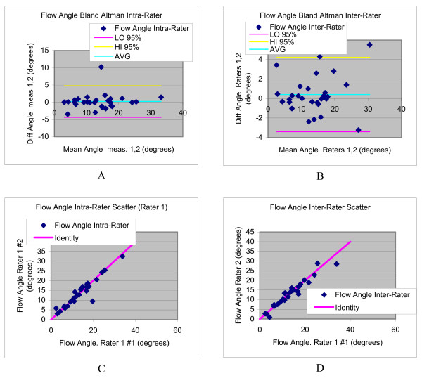
Methods: This paper reports an investigation in 18 pediatric BAV patients and 10 normal controls of links between abnormal blood flow patterns in the ascending aorta and aortic dilatation using velocity-encoded cardiovascular magnetic resonance. Blood flow patterns were quantitatively expressed in the angle between systolic left ventricular outflow and the aortic root channel axis, and also correlated with known biochemical markers of vessel wall disease.

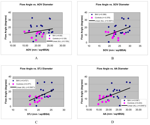
Results: The data confirm larger ascending aortas in BAV patients than in controls, and show more angled LV outflow in BAV (17.54 +/- 0.87 degrees) than controls (10.01 +/- 1.29) (p = 0.01). Significant correlation of systolic LV outflow jet angles with dilatation was found at different levels of the aorta in BAV patients STJ: r = 0.386 (N = 18, p = 0.048), AAO: r = 0.536 (N = 18, p = 0.022), and stronger correlation was found with patients and controls combined into one population: SOV: r = 0.405 (N = 28, p = 0.033), STJ: r = 0.562 (N = 28, p = 0.002), and AAO r = 0.645 (N = 28, p < 0.001). Dilatation and the flow jet angle were also found to correlate with plasma levels of matrix metallo-proteinase 2.

Conclusions: The results of this study provide new insights into the pathophysiological processes underlying aortic dilatation in BAV patients. These results show a possible path towards the development of clinical risk stratification protocols in order to reduce morbidity and mortality for this common congenital heart defect.


Figures

Figure 1
Figure 1
Structural CMR images used in image analysis: (A) an "LVOT1" orthogonal view centrally in the LVOT, (B) a second orthogonal "LVOT2" view, (C) a "candy-cane" view of aortic arch, and (D) a systolic mid-SOV level view of valve leaflets. Figures (A,B) are used to calculate the aortic channel direction, (C) illustrates AAO size measurement, and (D) illustrates how aortic sizes are measured at the levels of AOV, SOV, and STJ.
Figure 2
Figure 2
3-D reconstructions of aortic blood flow during peak systole (A,B) in a normal subject with tricuspid aortic valve, showing a centrally directed systolic flow jet, and (C,D) in a BAV patient with aortic dilatation, showing anteriorly directed off-axis systolic flow. The images in each pair of views mutually cross-reference the location of the perpendicular slice by the color of the intersection lines. The intersection line is ideally located along the LVOT channel axis but this scan planning does not necessarily work out perfectly in all cases, so in the analyses this direction is recalculated (see Figure 3).
Figure 3
Figure 3
Image analysis for calculation of the systolic flow jet angle: (A) 3-D view showing systolic flow velocity vectors in all slices. (B) Systolic flow jet velocities in the central-most slice in the SOV, which are averaged to determine the flow jet direction (orange in E); (C,D) show the aortic channel direction definitions in the LVOT views. Line segments 1-2 and 3-4 in the LVOT1 view (A) and a-b and c-d in the LVOT2 view (B) are manually drawn to connect the proximal and distal boundaries on both sides of the SOV, which are easily detected landmarks. The vector average of each segment pair defines the projection of the outflow channel direction onto each image plane. These two projections are then used to calculate the channel axis (light blue in E) for calculation of the angle between the flow jet direction vector and channel axis, which quantifies the mis-direction of systolic flow.
Figure 4
Figure 4
Comparison of mean flow jet angle and aortic diameters, adjusted for BSA, between BAV patients and control subjects. Errors bars indicate two standard errors of the mean.
Figure 5
Figure 5
Bland Altman plots (a,b) and scatter diagrams (c,d) illustrating the intra- and inter-rater variability in ascending aorta size evaluations.
Figure 6
Figure 6
Scatter diagrams showing distribution systolic blood flow jet angles of aortic outflow as a function of aortic diameters, corrected for BSA, and at the level of the aortic valve (A), sinus of Valsalva (B), sinotubular junction (C) and ascending aorta (D). Regression lines are shown of line fits with all subjects combined. An asterisk with the Pearson correlation coefficient in the figure legend indicates a statistically significant correlation.
Figure 7
Figure 7
Bland Altman plots (a,b) and scatter diagrams (c,d) illustrating the intra- and inter-rater variability in flow angle evaluations.

References

    1. Braverman AC, Guven H, Beardslee MA, Makan M, Kates AM, Moon MR. The bicuspid aortic valve. Curr Probl Cardiol. 2005;30:470–522. doi: 10.1016/j.cpcardiol.2005.06.002. - DOI - PubMed
    1. Ratib O, Perloff JK, Child JS. Images in cardiovascular medicine. Bicuspid aortic valve aneurysm. Circulation. 2004;109:671. doi: 10.1161/01.CIR.0000115643.50179.6E. - DOI - PubMed
    1. Ward C. Clinical significance of the bicuspid aortic valve. Heart. 2000;83:81–85. doi: 10.1136/heart.83.1.81. - DOI - PMC - PubMed
    1. Yener N, Oktar GL, Erer D, Yardimci MM, Yener A. Bicuspid aortic valve. Ann Thorac Cardiovasc Surg. 2002;8:264–267. - PubMed
    1. Bauer M, Bauer U, Siniawski H, Hetzer R. Differences in clinical manifestations in patients with bicuspid and tricuspid aortic valves undergoing surgery of the aortic valve and/or ascending aorta. Thorac Cardiovasc Surg. 2007;55:485–490. doi: 10.1055/s-2007-965387. - DOI - PubMed

MeSH terms