Review
. 2010 Mar;33(3):153-61.
doi: 10.1016/j.tins.2009.12.003. Epub 2010 Jan 12.

Senescent synapses and hippocampal circuit dynamics

Affiliations
Review

Senescent synapses and hippocampal circuit dynamics

Sara N Burke et al. Trends Neurosci. 2010 Mar.

Abstract

Excitatory synaptic transmission is altered during aging in hippocampal granule cells, and in CA3 and CA1 pyramidal cells. These functional changes contribute to age-associated impairments in experimentally-induced plasticity in each of these primary hippocampal subregions. In CA1, plasticity evoked by stimulation shares common mechanisms with the synaptic modification observed following natural behavior. Aging results in deficits in both artificially- and behaviorally-induced plasticity, and this could in part reflect age-related changes in Ca2+ homeostasis. Other observations, however, suggest that increased intracellular Ca2+ levels are beneficial under some circumstances. This review focuses on age-associated changes in synaptic function, how these alterations might contribute to cognitive decline, and the extent to which altered hippocampal circuit properties are detrimental or reflect compensatory processes.


Figures

Figure 1
Figure 1. Long-term potentiation (LTP) and long-term depression (LTD)
A) A schematic of the extracellular excitatory post-synaptic potential (EPSP) recorded in the dendritic region in response to stimulation of presynaptic fibers before (broken lines) and after (solid lines) plasticity is induced. In both A and B the X axis is time and the Y axis EPSP amplitude in mV, the blue component of the trace indicates the 1 ms of stable recording before stimulation, the red is the stimulus artifact, and the black in the evoked response. Long-term potentiation (LTP) reflects an increase in synaptic strength that is measured experimentally as a change in the amplitude of the EPSP following patterned electrical stimulation. The hypothesis is that the brain normally stores information as a pattern of synaptic weights, and that artificially-induced LTP reflects a mechanism that the brain could deploy to modify synapses. If synaptic connections were only strengthened, however, the network would readily approach saturation and the acquisition of new information would be prevented. B) Long-term depression (LTD) and LTP reversal are mechanisms for selectively decreasing synaptic weights, presumably preventing such saturation [for review, see 4]. LTD can be measured as a decrease in EPSP amplitude. In the hippocampus most forms of LTP and LTD require Ca2+ to enter through the NMDA receptor. Depending on intracellular Ca2+ concentrations a cascade of events will occur that will either induce LTP or LTD [73]. C) Changes with age in the maintenance of LTP correlate with spatial memory (green lines – young rats; purple lines – aged rats) [29]. Rats prefer small dark spaces to open bright ones, and thus, over days, learn to find the place where escape is possible below the platform surface (Barnes maze; left panel). When spatial memory accuracy and LTP durability are examined in the same rats, there is a significant correlation, within each age group, such that those rats that show inaccurate spatial behavior for that group, also show faster LTP decay rates (right panel, dentate gyrus) [29].
Figure 2
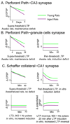
Figure 2. Summary of age-related alterations in LTP and LTD between young and aged animals
The Y-axes reflect the change in the extracellular EPSP (mV) following LTP or LTD induction and the X-axes time following induction of LTP or LTD (green lines - young rats; purple lines - aged rats). A) LTP induction at perforant path–CA3 synapse is intact in aged rats when robust (supra-threshold) stimulation protocols are used, but LTP maintenance over days is impaired [40]. B) LTP induction is also intact at the perforant path–granule cell synapse when robust stimulation parameters are used. Similar to what is observed in CA3, LTP decays more rapidly over days in the dentate gyrus of old rats [left panel; 29]. When weak (peri-threshold) stimulation protocols are used, however, aged rats also show LTP induction deficits at the perforant path dentate gyrus synapse [right panel; 42, 43]. This induction impairment could, in part, result from the higher threshold in LTP induction at this synapse [80]. C) At the Schaffer–CA1 synapse both LTP induction and decay over hours is similar across age groups when robust stimulation protocols are used [top left panel; 41]. As shown above for the dentate gyrus, however, when weak stimulation parameters are used, LTP induction deficits are unmasked in old rats [top right panel; 81, 82]. This LTP induction deficit is not due to a threshold change at Schaffer collateral synapses in old rats [83]. For CA1, aged rats have also been shown to be more susceptible the induction of LTD following low-frequency stimulation (LFS) [bottom left panel; 45]. Moreover, LTP can be reversed with same stimulation protocol that induces LTD. While LTP is not completely reversed by this LFS in young rats, old rats are also more prone to this reversal of LTP [bottom right panel; 45].
Figure 3
Figure 3. Experience-dependent place field expansion plasticity in young and old rats
(A) In young rats CA1 place fields expand asymmetrically during repeated route following, which results in an increase in place field size and a shift in the distribution of place field spiking in the direction opposite to the rat’s trajectory [58, 59]. Specifically, when a rat first traverses a cell’s place field on a fixed path, the firing rate distribution of the place field is Gaussian such that as the rat enters the place field the neuron shows increased spiking that reaches a maxima at the center of the field and then decreases as the rat exits (Lap 1; white). After repeated traversals, the field expands in the direction opposite to that of the rat’s path (Laps >5; grey). This experience-dependent place field expansion plasticity is consistent with neural network models dating back to Hebb's (1949) concept of the 'phase sequence' of cell assemblies, which have suggested that an associative, temporally-asymmetric synaptic plasticity mechanism could serve to encode sequences or episodes of experience [84, 85]. (B) A plot of place field size by traversals through a place field (laps) during track running (green circles – young rats; purple circles - old rats). Young rats show significant place field expansion plasticity while old rats fail to show this form of experience-dependent plasticity as robustly (filled circles; data from [56]). (C) Old rats given memantine (10 mg/kg; red circles) show improved place field expansion plasticity compared to old saline controls (purple circles; data from [64]).
Figure 4
Figure 4. Hypothetical synaptic modification curves for young (green) and aged (purple) CA1 neurons
The X-axis shows different stimulation parameters used to induce LTP or LTD, and the Y-axis is magnitude and sign of synaptic modification. (1) In old CA1 neurons altered Ca2+ homeostasis causes a change in the synaptic modification curve, such that there is a greater probability of LTD following 900 pulses of 1 Hz stimulation (LFS) [45]. (2) This age difference can be eliminated by increasing the ratio of Ca2+ to Mg2+ in the recording medium [44]. (3) Additionally, with weak stimulation parameters (i.e., theta burst stimulation ot 4, 100 Hz pulses), there is a lower probability of LTP induction in aged compared to young CA1 neurons [81, 82]. (4) When more robust stimulation is used (i.e., fifteen 20 ms bursts at 400 Hz) the amount of synaptic modification is equivalent between age groups [41].

Similar articles

Cited by

References

    1. Kinsella K, Velkoff VA. An aging world: 2001. U.S. Census Bureau. 2001 Series P95/01-1.
    1. Hebb D. On watching myself get old. Psychology Today. 1978 November
    1. Moscovitch M, et al. The cognitive neuroscience of remote episodic, semantic and spatial memory. Curr Opin Neurobiol. 2006;16(2):179–190. - PubMed
    1. Rosenzweig ES, Barnes CA. Impact of aging on hippocampal function: plasticity, network dynamics, and cognition. Prog Neurobiol. 2003;69(3):143–179. - PubMed
    1. Wilson IA, et al. Neurocognitive aging: prior memories hinder new hippocampal encoding. Trends Neurosci. 2006;29(12):662–670. - PMC - PubMed

Publication types