. 2010 Mar 12;285(11):8302-15.
doi: 10.1074/jbc.M109.074161. Epub 2010 Jan 13.

Structural and functional characterization of a novel homodimeric three-finger neurotoxin from the venom of Ophiophagus hannah (king cobra)

Affiliations

Structural and functional characterization of a novel homodimeric three-finger neurotoxin from the venom of Ophiophagus hannah (king cobra)

Amrita Roy et al. J Biol Chem. .

Abstract

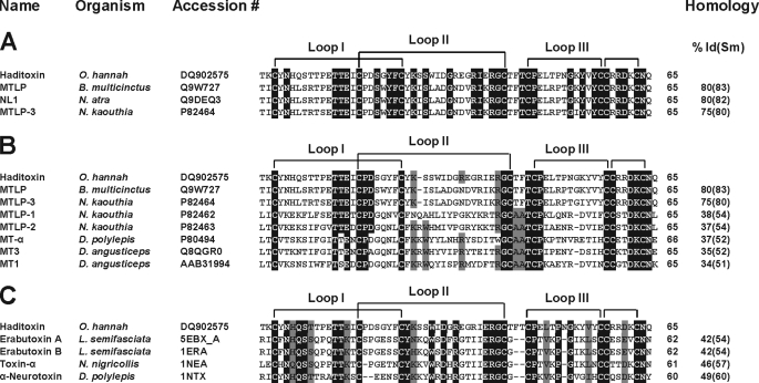
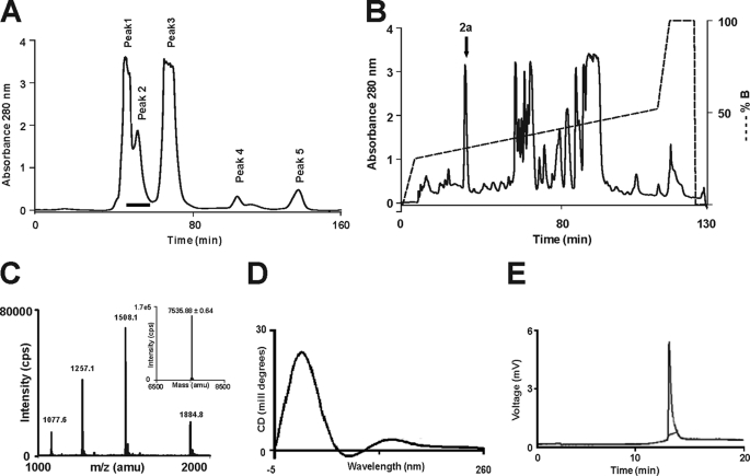
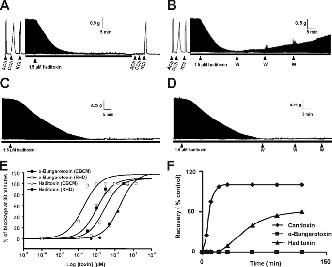
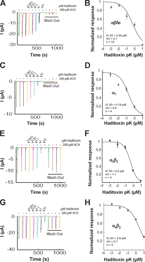
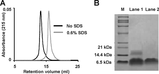
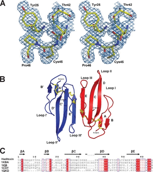
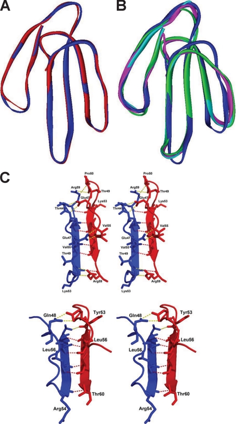
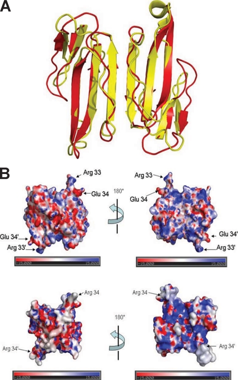
Snake venoms are a mixture of pharmacologically active proteins and polypeptides that have led to the development of molecular probes and therapeutic agents. Here, we describe the structural and functional characterization of a novel neurotoxin, haditoxin, from the venom of Ophiophagus hannah (King cobra). Haditoxin exhibited novel pharmacology with antagonism toward muscle (alphabetagammadelta) and neuronal (alpha(7), alpha(3)beta(2), and alpha(4)beta(2)) nicotinic acetylcholine receptors (nAChRs) with highest affinity for alpha(7)-nAChRs. The high resolution (1.5 A) crystal structure revealed haditoxin to be a homodimer, like kappa-neurotoxins, which target neuronal alpha(3)beta(2)- and alpha(4)beta(2)-nAChRs. Interestingly however, the monomeric subunits of haditoxin were composed of a three-finger protein fold typical of curaremimetic short-chain alpha-neurotoxins. Biochemical studies confirmed that it existed as a non-covalent dimer species in solution. Its structural similarity to short-chain alpha-neurotoxins and kappa-neurotoxins notwithstanding, haditoxin exhibited unique blockade of alpha(7)-nAChRs (IC(50) 180 nm), which is recognized by neither short-chain alpha-neurotoxins nor kappa-neurotoxins. This is the first report of a dimeric short-chain alpha-neurotoxin interacting with neuronal alpha(7)-nAChRs as well as the first homodimeric three-finger toxin to interact with muscle nAChRs.


Figures

FIGURE 1.
FIGURE 1.
Multiple sequence alignment of novel proteins. A–C, sequence alignment of haditoxin with the most homologous sequences (A), muscarinic toxin homologs (B), and short-chain α-neurotoxins (C). Toxin names, species, and accession numbers are shown. Conserved residues in all sequences are highlighted in black. Disulfide bridges and loop regions are also shown. At the end of each sequence, the numbers of amino acids are stated. The homology (sequence identity and similarity (% Id(Sm))) of each protein is compared with haditoxin and shown at the end of each sequence. N. atra, Naja atra; N. kaouthia, Naja kaouthia; D. polylepis, Dendroaspis polylepis; and L. semifasciata, Laticauda semifasciata.
FIGURE 2.
FIGURE 2.
Purification of haditoxin from the venom of O. hannah. A, gel filtration chromatogram of crude venom. Crude venom (100 mg/ml) was fractionated using a Superdex 30 HiLoad (16/60) column. The column was pre-equilibrated with 50 mm Tris-HCl buffer (pH 7.4). Proteins were eluted at a flow rate of 1 ml/min using the same buffer. A black bar at Peak 2 indicates the fractions containing haditoxin. B, RP-HPLC profile of the gel filtration fractions containing haditoxin. Jupiter C18 (5 μ, 300 Å, 4.5 × 250 mm) analytical column was equilibrated with 0.1% (v/v) trifluoroacetic acid. Protein of interest was eluted from the column with a flow rate of 1 ml/min with a gradient of 23–49% buffer B (80% acetonitrile in 0.1% trifluoroacetic acid). The dotted line indicates the gradient of the buffer B. The downward arrow at Peak 2a indicates fractions containing haditoxin. C, ESI-MS profile of the RP-HPLC fraction containing haditoxin. The spectrum shows a series of multiply charged ions, corresponding to a single, homogenous peptide with a molecular mass of 7,535.88 Da. Inset, reconstructed mass spectrum of haditoxin; CPS = counts/s; amu = atomic mass units. D, far-UV CD spectrum of haditoxin. The protein was dissolved in MilliQ water (0.5 mg/ml), and the CD spectra were recorded using a 0.1-cm path length cuvette. E, electropherogram of haditoxin. The sample was injected using pressure mode 5 p.s.i./s, and electrophoresis runs were carried out using a coated capillary (17 cm × 25 μm) at 18 kV, with 0.1 m phosphate buffer (pH 2.5) at 20 °C for 7 min.
FIGURE 3.
FIGURE 3.
Pharmacological profile of haditoxin. A–D, a segment of tracing showing the effect of haditoxin (1.5 μm) on CBCM preparations (A), reversibility of CBCM preparation (B), RHD preparations (C), and reversibility of RHD preparation (D). Contractions were produced by exogenous ACh (300 μm), carbachol (CCh; 10 μm), and KCl (30 mm). The black bar indicates the electrical field stimulation. The point of washing out the toxin with Krebs solution in reversibility studies is indicated by the abbreviation W. E, dose-response curve of haditoxin and α-bungarotoxin on CBCM and RHD. The block is calculated as a percentage of the control twitch responses of the muscle to supramaximal nerve stimulation. Each data point is the mean ± S.E. of at least three experiments. F, comparative reversibility profile of α-bungarotoxin, haditoxin, and candoxin.
FIGURE 4.
FIGURE 4.
Effect of haditoxin on human nACHRs expressed in Xenopus oocytes. A, C, E, and G, inhibition of ACh-induced currents in αβδϵ- (neuromuscular junction) (A), α7- (C), α4β2- (E), and α3β2-nAChRs (G). Neuromuscular junction currents were activated by 10 μm ACh, whereas 200 μm was used to activate α7-, α4β2-, and α3β2-nAChRs. The first three traces are controls, followed by a 2-min exposure to several haditoxin concentrations ranging from 10 nm to 10 μm. Each experiment was terminated by a 8-min wash out. Little or no recovery was observed for αβδϵ- and α7-nAChRs, whereas partial to full recovery was observed for α4β2- and α3β2-nAChRs. Inhibition curves of the fitted data, IC50, and Hill coefficient (nH) for αβδϵ-nAChRs (B) were 0.55 μm and 0.7; for α7-nAChRs (D), they were 0.18 μm and 0.8; for α3β2-nAChRs (F), they were 0.5 μm and 1.1; and for α4β2-nAChRs (H), they were 2.6 μm and 0.7. Error bars indicate S.E.
FIGURE 5.
FIGURE 5.
Dimerization of haditoxin. A, gel filtration profile of haditoxin with (gray) and without (black) SDS. 1 μm haditoxin was loaded onto a Superdex 75 column (1 × 30 cm) equilibrated with 50 mm Tris-HCl buffer (pH 7.4). The protein was eluted out with the 50 mm Tris-HCl buffer (pH 7.4) or 50 mm Tris-HCl buffer (pH 7.4) containing 0.6% SDS at flow rate of 0.6 ml/min. B, Tris-Tricine SDS-PAGE analysis of haditoxin with (lane 1) and without (lane 2) cross linker (BS3). M is the marker lane. The concentration of BS3 is 5 mm.
FIGURE 6.
FIGURE 6.
Overall structure of haditoxin. A, stereo view of a portion of the final 2FoFc map of haditoxin. The map was contoured at a level of 1.0 σ. B, monomers A and B are shown in blue and red, respectively. Disulfide bonds are shown in yellow. N and C termini (N-term and C-term), β-strands, and loops I, II, and III are labeled. C, structure-based alignment of three-finger toxins. Color coding of conserved residues is provided by boxed red text, and color coding of invariant residues is provided by red highlight. Accession numbers are shown on the left, and secondary structural elements of haditoxin are shown on top. Numbering is shown for haditoxin only. Sequence alignment was done by Strap (82) and displayed with ESPript (83).
FIGURE 7.
FIGURE 7.
Structural details of haditoxin. A, superimposition of both subunits of haditoxin. Subunits A and B are shown in blue and red, respectively. B, superimposition subunit A of haditoxin with short-chain α-neurotoxins. Subunit A is shown in blue, erabutoxin-a is shown in magenta, erabutoxin-b is shown in cyan, and toxin-α is shown in green. C, stereo diagram of comparison of dimer interface of haditoxin (top) and κ-bungarotoxin (bottom). The residues to form the hydrogen bonds are labeled. The main chain-main chain hydrogen bonds are shown in red, and the other hydrogen bonds are shown in yellow.
FIGURE 8.
FIGURE 8.
Haditoxin versus κ-bungarotoxin. A, superimposition of haditoxin with κ-bungarotoxin. Haditoxin and κ-bungarotoxin are shown in red and yellow, respectively. B, electrostatic surface of haditoxin (top) and κ-bungarotoxin (bottom). The orientation is the same as in Fig. 6B. The locations of Arg-33 and Glu-34 of haditoxin and Arg-34 of κ-bungarotoxin are indicated.

Similar articles

Cited by

References

    1. Harvey A. L. (1991) Snake Toxins, pp. 1–90, Pergamon Press, New York
    1. Lewis R. J., Garcia M. L. (2003) Nat. Rev. Drug Discov. 2, 790–802 - PubMed
    1. Harvey A. L. (2002) Trends Pharmacol. Sci. 23, 201–203 - PubMed
    1. Langley J. N. (1907) J. Physiol. 36, 347–384 - PMC - PubMed
    1. Katz B., Thesleff S. (1957) J. Physiol. 138, 63–80 - PMC - PubMed

Publication types

MeSH terms

Associated data

LinkOut - more resources