. 2010 Mar 19;285(12):9273-81.
doi: 10.1074/jbc.M109.075218. Epub 2010 Jan 13.

Force-induced myofibroblast differentiation through collagen receptors is dependent on mammalian diaphanous (mDia)

Affiliations

Force-induced myofibroblast differentiation through collagen receptors is dependent on mammalian diaphanous (mDia)

Matthew W C Chan et al. J Biol Chem. .

Abstract

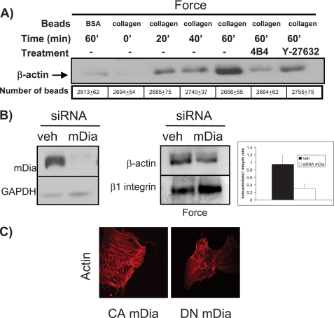
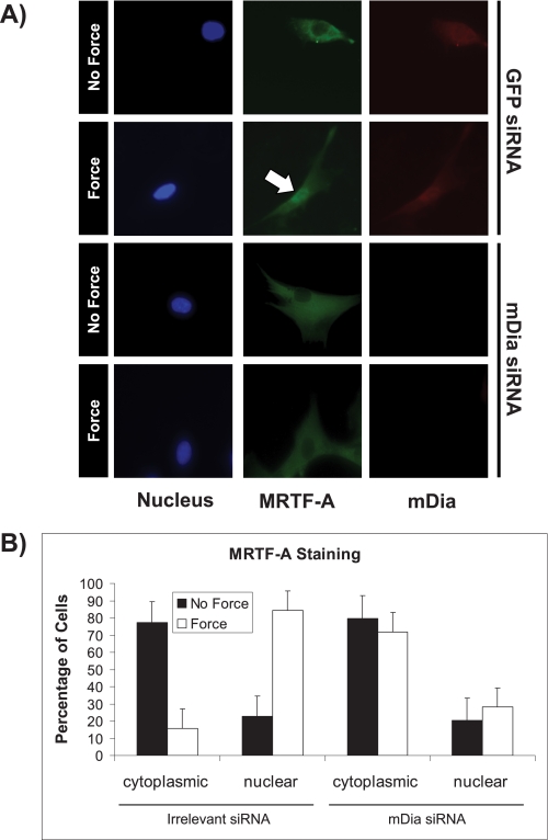
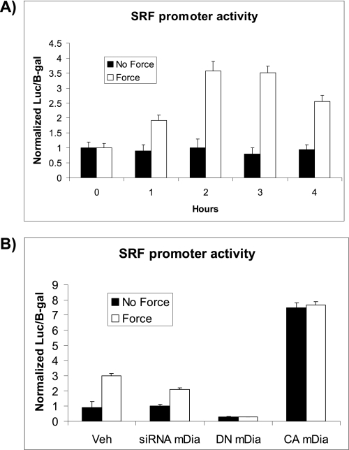
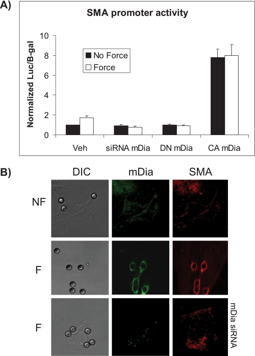
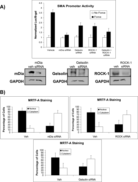
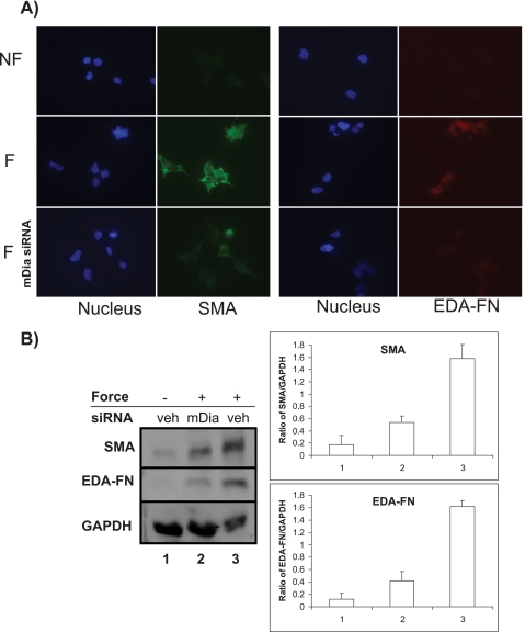
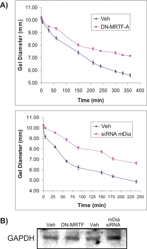
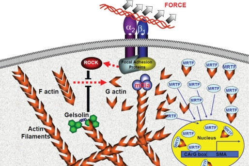
The development of fibrosis promotes the differentiation of myofibroblasts, pro-fibrotic cells, which contribute to tissue dysfunction. Myofibroblast differentiation is dependent on actin assembly, which in response to force, is mediated by various actin-binding proteins including the mammalian Diaphanous-related formins (mDia). We examined the role of mDia in the mechano-sensing pathway that leads to force-induced expression of alpha-smooth muscle actin (SMA), a marker and critical determinant of myofibroblast differentiation. In cells treated with siRNA to knockdown mDia and then subjected to tensile force using collagen-coated magnetite beads attached to beta1 integrins, actin assembly was inhibited at bead contact sites. Force-induced nuclear translocation of MRTF-A, a transcriptional co-activator of SMA, was reduced 50% by mDia knockdown. The expression of the transcriptional co-activator of SMA, serum response factor, was reduced by 50% after siRNA knockdown of mDia or by 100% in cells transfected with catalytically inactive mDia. Force-induced activation of the SMA promoter and SMA expression were blocked by knockdown of siRNA of mDia. In anchored collagen gel assays to measure myofibroblast-mediated contraction, knockdown of mDia reduced contraction by 50%. We conclude that mDia plays an important role in the development of force-induced transcriptional activation of SMA and myofibroblast differentiation.


Figures

FIGURE 1.
FIGURE 1.
Force-induced actin assembly is dependent on mDia. A, immunoblot analysis of collagen-coated bead-associated proteins over a 60-min time course after force application. The numbers of beads isolated in each sample were counted and used to estimate equivalence of protein loading. BSA-coated beads were used as nonspecific adhesion controls. A β1 integrin blocking antibody, 4B4, and the ROCK inhibitor, Y-27632, were used as negative controls. B, cells with mDia knockdown by siRNA were incubated with collagen-coated magnetite beads and subjected to force. Immunoblots of bead-associated proteins show β-actin recruitment. β1-Integrin immunoblots were used as loading controls. Left panel shows efficacy of siRNA knockdown of mDia. The ratios of β-actin to β1-integrin were computed from densitometry measurements of immunoblots (mean ± S.E., right panel). C, confocal images show actin-filament staining with rhodamine-phalloidin in cells transfected with constitutively active (CA)- or dominant negative (DN)-mDia.
FIGURE 2.
FIGURE 2.
Force-induced MRTF-A nuclear translocation is dependent on mDia. A, immunofluorescence images showed the state of nuclear translocation of MRTF-A in cells treated with irrelevant or mDia siRNAs, incubated with collagen-coated magnetite beads and then subjected to force or no force. The white arrow shows the presence of MRTF-A in the nucleus. Cells were stained with DAPI to demonstrate nuclei (blue), and immunostained for MRTF-A (green) and mDia (red). B, immunofluorescence images were quantified after 60 min of force. Data are mean ± S.E. of the percentage of cells exhibiting MRTF-A nuclear translocation. In each experimental group 25 cells were quantified.
FIGURE 3.
FIGURE 3.
mDia is required for force-induced SRF activation. A, force-induced SRF-luciferase promoter activity normalized to β-galactosidase was measured in NIH 3T3 cells. Cells were stimulated with force over a time course (1 to 4 h). Promoter activities are shown as fold-change compared with basal promoter activity. B, SRF promoter activity was measured after 2 h of force application in cells transfected with mDia siRNA, DN-mDia, CA-mDia, or vehicle controls.
FIGURE 4.
FIGURE 4.
Force-induced SMA activation is dependent on mDia. A, force-induced SMA-luciferase promoter activity normalized to β-galactosidase was measured after 4 h of force application in NIH 3T3 cells. Cells were also treated with mDia siRNA, DN mDia, or CA mDia. B, confocal images of cells with (F) or without (NF) 4 h of force application show staining of mDia with FITC-conjugated antibody and SMA with TRITC-conjugated antibody around single collagen-coated beads. DIC images of the same cells show the location of beads. Note that cells treated with siRNA for mDia show no detectable mDia.
FIGURE 5.
FIGURE 5.
Relative contribution of gelsolin, ROCK, and mDia pathways in SMA expression. A, NIH 3T3 cells were pretreated with siRNA to knockdown gelsolin, ROCK, or mDia. Force-induced SMA-luciferase promoter activity normalized to β-galactosidase was measured after 4 h of force application. Immunoblots show efficacy of knockdown of gelsolin, ROCK, and mDia with GAPDH as loading controls. B, measurements of nuclear translocation of MRTF-A in cells. Immunofluorescence images were quantified after 60 min of force in cells with or without siRNA knockdown of gelsolin, ROCK, and mDia. Cells were stained for MRTF-A (green) and with DAPI to identify location of nuclei (blue). Data are mean ± S.E. percentage of cells with MRTF-A nuclear translocation, from 25 cells for each experimental group.
FIGURE 6.
FIGURE 6.
Force-induced myofibroblasts differentiation is dependent on mDia. A, fluorescence images of NIH3T3 cells incubated with collagen-coated magnetite beads, and with (F) or without (NF) exposure to tensile force applied by a ceramic permanent magnet. Cells were immunostained for SMA (green) and ED-A fibronectin (red). Nuclei were stained with DAPI (blue). The presence of SMA and ED-A fibronectin indicates myofibroblastic phenotype. Middle panels show force-loaded cells after treatment with mDia siRNA. B, immunoblots show protein expression of SMA and ED-A fibronectin from experiments conducted in the same conditions as A. GAPDH is shown as loading controls.
FIGURE 7.
FIGURE 7.
Gel contraction by fibroblasts requires MRTF-A and mDia. A, contraction of stress-relaxed collagen gels in cells treated with DN-MRTF-A or siRNA for mDia or vehicle controls. The data are means + S.E. for gel diameter over time after release of the gel from the dish. B, immunoblots of GAPDH from cell lysates isolated from collagen gels indicate equivalent number of cells were analyzed in each collagen gel sample.
FIGURE 8.
FIGURE 8.
Proposed model of mechanotransduction. Application of tensile forces to collagen beads triggers the recruitment and activation of focal adhesion proteins. In turn, focal adhesion proteins promote actin assembly by activating gelsolin and ROCK, which prevent actin filament disassembly. The nucleation of actin barbed ends by mDia enables actin filament growth and elongation. The dissociation of MRTF-A from actin monomers during actin assembly initiates the nuclear translocation of MRTF-A. In the nucleus, MRTF-A acts as transcriptional co-activator for the induction of SMA.

Similar articles

Cited by

References

    1. Tomasek J. J., Gabbiani G., Hinz B., Chaponnier C., Brown R. A. (2002) Nat. Rev. Mol. Cell Biol. 3, 349–363 - PubMed
    1. Leslie K. O., Taatjes D. J., Schwarz J., vonTurkovich M., Low R. B. (1991) Am. J. Pathol. 139, 207–216 - PMC - PubMed
    1. Sun Y., Weber K. T. (1996) Cardiovasc. Res. 31, 518–525 - PubMed
    1. Webber J., Meran S., Steadman R., Phillips A. (2009) J. Biol. Chem. 284, 9083–9092 - PMC - PubMed
    1. MacKenna D. A., Dolfi F., Vuori K., Ruoslahti E. (1998) J. Clin. Invest. 101, 301–310 - PMC - PubMed

Publication types

LinkOut - more resources