. 2010 Mar 15;21(6):885-96.
doi: 10.1091/mbc.e09-09-0811. Epub 2010 Jan 13.

Mutations in Caenorhabditis elegans him-19 show meiotic defects that worsen with age

Affiliations

Mutations in Caenorhabditis elegans him-19 show meiotic defects that worsen with age

Lois Tang et al. Mol Biol Cell. .

Abstract

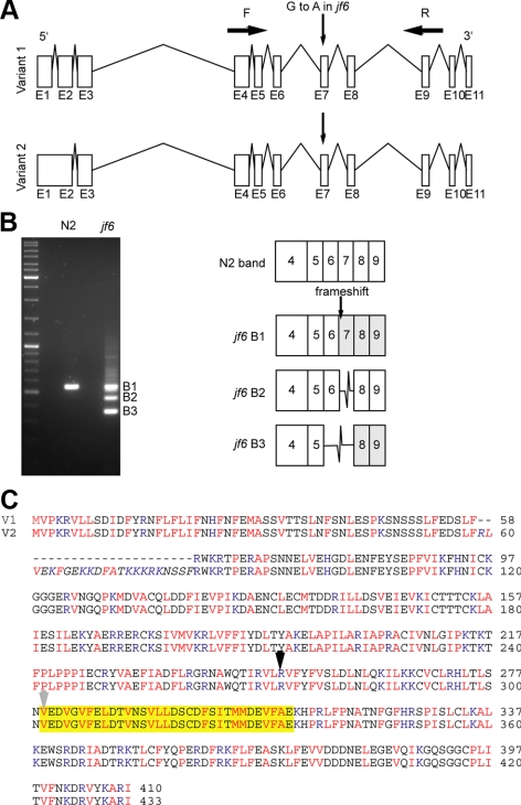
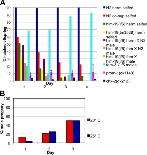
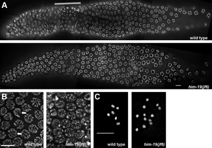
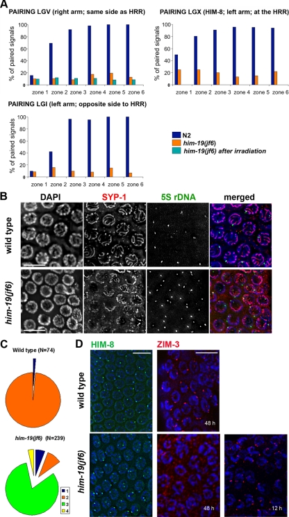
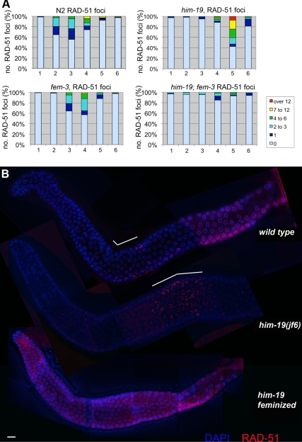
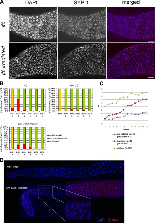
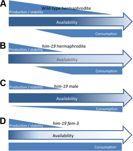
From a screen for meiotic Caenorhabditis elegans mutants based on high incidence of males, we identified a novel gene, him-19, with multiple functions in prophase of meiosis I. Mutant him-19(jf6) animals show a reduction in pairing of homologous chromosomes and subsequent bivalent formation. Consistently, synaptonemal complex formation is spatially restricted and possibly involves nonhomologous chromosomes. Also, foci of the recombination protein RAD-51 occur delayed or cease altogether. Ultimately, mutation of him-19 leads to chromosome missegregation and reduced offspring viability. The observed defects suggest that HIM-19 is important for both homology recognition and formation of meiotic DNA double-strand breaks. It therefore seems to be engaged in an early meiotic event, resembling in this respect the regulator kinase CHK-2. Most astonishingly, him-19(jf6) hermaphrodites display worsening of phenotypes with increasing age, whereas defects are more severe in female than in male meiosis. This finding is consistent with depletion of a him-19-dependent factor during the production of oocytes. Further characterization of him-19 could contribute to our understanding of age-dependent meiotic defects in humans.


Figures

Figure 1.
Figure 1.
Splicing defects induced by the him-19(jf6) mutation. (A) Diagrams of the two naturally spliced variants of Y95B8A.11. Version 1 includes 11 exons. Version 2 has 10 exons. The G-to-A mutation in him-19(jf6) occurs before the exon 7. Primer pairs MJ1976 and MJ1979 were used to amplify the region from jf6 cDNA and to detect the splice variants the mutant. (B) Gel picture of the PCR products amplified with primers F and R (from A). The wild-type product matches the sequence of exon 4–9 consecutively. The three bands in the mutant lane correspond to the various splice versions affected by the disruption of the splice acceptor site. Although band 1 has the same size of the wild-type band, one nucleotide is missing in the exon 7. The next “AG” splice acceptor site is used for exon 7. The product of this band 1 will run into a premature stop codon. Band 2 and band 3 are missing the exon 7 and exon 6 and 7, respectively. (C) Amino acid alignment of the variants 1 and 2. The 23 amino acids unique in version 2 are italicized. Basic amino acids are highlighted in blue: H, K, and R. Hydrophobics in red: V, I, L, M, F, A, and P. The yellow box corresponds to exon 7. B2 in B is missing the 33 amino acids in the highlighted box and continues in frame. The black arrow indicates the start of exon 6 where B3 in Figure 1B and the knockout mutant's disruptions start. The gray arrow indicates where the B1 in Figure 1B starts its frame shift.
Figure 2.
Figure 2.
Aging phenotype of the him-19(jf6) mutant. (A) Graph illustrates the percentages of hatch rate (y-axis) of the assorted strains counted over the reproductive time span (x-axis). Fewer eggs hatched in the later broods in the mutant and in wild type worms injected with DNA encoding Y95B8A.11 leading to transgene-mediated gene silencing (cosuppression). Crosses of wild-type females with him-19(jf6) males are less affected than crosses performed with him-19(jf6) females or self crosses. Viability of the offspring of prom-1(ok1120) or chk-2(gk212) remained steadily low over the time frame investigated. Gray lines in bars for prom-1 and chk-2 indicate the 95% confidence intervals. Detailed numbers of brood size and hatch rates for each time point are shown in Supplemental Table 1. (B) Graph describes the percentage of male progeny (y-axis) in various broods (x-axis). At 20°C, the him-19(jf6) hermaphrodites had 12.87% of male progeny on day 1 (n = 171), 21.67% on day 2 (n = 180), and 50.0% on day 3 (n = 24). At 25°C, the him-19(jf6) hermaphrodites had 4.11% of male progeny on day 1 (n = 243), 25.22% on day 2 (n = 115), and 50.0% on day 3 (n = 6).
Figure 3.
Figure 3.
Gonad organization in wild type and him-19(jf6) mutant. (A) DAPI-stained wild-type gonad and him-19(jf6) mutant gonad. A defined TZ zone is missing in the mutant gonad. Thick gray bar delineates position of the TZ in the wild type. (B) Parallel tracks of synapsed chromosomes are observed in the wild type (left, indicated by arrows) pachytene nuclei. him-19(jf6) (right) pachytene nuclei lack the parallel tracks. (C) Six bivalents are seen in wild-type diakinesis (left). Up to 12 univalents can be detected in the him-19(jf6) mutants (right). Bars, 10 μm.
Figure 4.
Figure 4.
Homologous chromosome pairing is defective in the jf6 mutant. (A) Diagrams representing percentage of nuclei with homologous pairing of chromosome I, V, and X in N2, him-19(jf6), and irradiated him-19(jf6) (FISH performed 6 h after irradiation). Gonads are divided into six equal zones from the distal tip cell to diplotene (x-axes), and the percentage of pairing (y-axes) is determined by FISH probes or α-HIM-8 antibody. Levels of pairing in him-19(jf6) do not rise significantly above those in mitosis. (B) him-19(jf6) shows nonhomologous synapsis. Late pachytene nuclei are first stained with the α-SYP-1 antibody and subsequently tested by FISH with the 5S ribosomal locus probe. In him-19(jf6), a large portion of unpaired FISH signals is associated with SYP-1, indicating nonhomologous synapsis or polymerization on unpaired chromosomes. Bars, 10 μm. (C) Quantitative scoring of the 5S FISH signal in association with SYP-1 stretches in pachytene nuclei. The categories were as follows: 1 (blue), unpaired FISH signals with no association with SYP-1; 2 (orange), paired FISH signals in association with SYP-1; 3 (green), unpaired FISH signals in association with SYP-1; and 4 (yellow), paired FISH with no association with SYP-1. In the him-19(jf6) mutant, the most abundant category is the unpaired FISH signal in association with SYP-1. This either indicates frequent occurrence of nonhomologous synapsis or SYP-1 polymerization along unpaired chromosomes. (D) Immunostaining of HIM-8 (green) and ZIM-3 (red) in wild-type and him-19(jf6) pachytene nuclei. HIM-8 localizes in him-19 gonads in wild-type intensity. However, two signals are often detected in the mutant gonads indicating that the homologous pairing of the X chromosome is impaired. Two foci of ZIM-3, corresponding to the paired chromosome I set and chromosome IV set, are usually detected in wild-type TZ and early pachytene. In the him-19(jf6) background, ZIM-3 foci are detected 12 h post-L4 but not 48 h post-L4. Bars, 10 μm.
Figure 5.
Figure 5.
Lateral HIM-3, and central SYP-1, components of the SC, are loaded in him-19(jf6). Spread pachytene nuclei were stained with an antibody against HIM-3 an axial element component of the SC. Loading of HIM3 occurs at the same time in wild-type and him-19(jf6) individuals. However, more HIM-3 tracks can be observed in the mutant (small arrowhead). Similarly, the central element component SYP-1 is loaded along the entire chromosome axes in wild-type nuclei (indicated by arrows). In him-19 (jf6), the extent of SYP-1 polymerization is reduced as demonstrated by the relatively short SYP-1 stretches (see arrowheads). Also, compare Supplemental Figure 5 for a more detailed view of SYP-1 loading. Bars, 10 μm.
Figure 6.
Figure 6.
Progression of recombination is disrupted in the him-19(jf6) mutant. (A) Bar chart of quantitative time course analysis of RAD-51 foci in various mutant backgrounds. Projected 3D stacks containing the depth of nuclei, whole gonads were sectioned in six parts. Zones 1 and 2 include premeiotic nuclei, zone 3 contains mostly nuclei in transition zone, and zones 4–6 are the pachytene nuclei. RAD-51 foci were counted on deconvolved flattened images. Differently colored sections on the bar represent the percentages of nuclei in a given zone with the number of RAD-51 foci indicated in a color code. In the wild type, RAD-51 foci peak around zone3 and never exceed 12 foci per cell. In him-19(jf6), RAD-51 foci peak around zone 5, and frequently more than 12 foci per cell can be observed. In the feminized fem-3; him19(jf6) double mutant, only very few RAD-51 foci can be detected around zone 4. In the fem-3 single mutant, a normal number of RAD-51 foci was observed that peaked around zone 3 and 4, with a slight delay compared with wild-type. Number of nuclei scored for wild type per zone is as follows: zone 1, 165; zone 2, 185; zone 3, 131; zone 4, 136; zone 5, 133; and zone 6, 79. him-19(jf6): zone 1, 194; zone 2, 239; zone 3, 198; zone 4, 189; zone 5, 165; and zone 6, 144. fem-3(e1996): zone 1, 134; zone 2, 149; zone 3, 143; zone 4, 154; zone 5, 118; and zone 6, 95. him-19(jf6); fem-3(e1996): zone 1, 156; zone 2, 122; zone 3, 133; zone 4, 140; zone 5, 117; and zone 6, 81. (B) Immunostaining of RAD-51 (red) in wild type, him-19(jf6) hermaphrodite, and him-19(jf6) female whole gonads, counterstained with DAPI (blue). White bars on top of the wild-type and the him-19(jf6) hermaphrodite gonads indicate the zones rich in RAD-51 foci. Bar, 10 μm.
Figure 7.
Figure 7.
Artificially induced DSBs restore chromosome clustering and enhance SYP-1 polymerization. (A) High magnification of the meiotic entry area of the him-19(jf6) gonad and him-19(jf6) 6 h post-γ-radiated and stained with α-SYP-1 antibody. SYP-1 loading is restricted in him-19 mutant. Mutant gonads post irradiated show more complete SYP-1 polymerization than nonirradiated gonads. Images are projections of 3D stacks including the whole depth of nuclei shown. Bars, 10 μm. (B) Bar chart showing the presence of nuclei with chromosome clustering. Nuclei with polarized chromosomes (the crescent-shaped nuclei found at the transition zone) peak at zone 2 in wild-type gonads. him-19 mutants have no defined TZ and only a few nuclei in the clustered chromosome configuration. Six hours after irradiation him-19 mutants show more nuclei with polarized chromosomes. Numbers of nuclei scored are as follows: wild type: zone 1, 165; zone 2, 185; zone 3, 131; zone 4, 136; zone 5, 133; and zone 6, 79. him-19(jf6): zone 1, 194; zone 2, 239; zone 3, 198; zone 4, 189; zone 5, 165; and zone 6, 144. him-19(jf6) after irradiation: zone 1, 139; zone 2, 138; zone 3, 190; zone 4, 231; zone 5, 193; and zone 6, 190. (C) Lengths of SYP-1 stretches were measured per cell row upon initiation of SYP-1 loading. Measurements were taken from projected 3D stacks containing the depth of the nuclei. The him-19(jf6) postirradiation gonads have more extensive SYP-1 polymerization than the nonirradiated mutants of the same age. (D) Gamma irradiation restores ZIM-3 loading in him-19(jf6) in parallel to the restoration of clustered chromatin. Worms irradiated 48 h post-L4 were stained for ZIM-3 2 h after irradiation.
Figure 8.
Figure 8.
Model of HIM-19 activity. Production of HIM-19 or a HIM-19-dependent (or HIM-19 stabilized) factor X is initiated in the young gonad. Both the deletion him-19(tm3538) and point mutation him-19(jf6) allele allow production of the aminoterminal 60% of HIM-19, which might still allow residual activity of the protein. The mutated protein might either be less stable or less active leading to reduced activity or production of factor X. Assuming a stabilizing function of HIM-19 on factor X, both truncation or even complete loss of HIM-19 would lead to a more rapid degradation of factor X. (A) In the wild type, HIM-19/Factor X levels are sufficient to support normal meioses (almost) to the end of the reproductive lifespan. (B) In the him-19 hermaphrodite, HIM-19 is less stable, produces less of its dependent factor X, or leads to destabilization of factor X. Consumption starts with the onset of female meioses. The available stock becomes depleted in old animals. (C) Male meiosis is less dependent on HIM-19/factor X; hence, the production to consumption ratio is sufficiently high to ensure mostly normal meioses in him-19 males. (D) In fem-3 worms, HIM-19/factor X consumption by female meioses starts in the young gonad. Depletion of HIM-19/factor X occurs earlier leading to more severely affected meioses.

Similar articles

Cited by

References

    1. Alpi A., Pasierbek P., Gartner A., Loidl J. Genetic and cytological characterization of the recombination protein RAD-51 in Caenorhabditis elegans. Chromosoma. 2003;112:6–16. - PubMed
    1. Andux S., Ellis R. E. Apoptosis maintains oocyte quality in aging Caenorhabditis elegans females. PLoS Genet. 2008;4:e1000295. - PMC - PubMed
    1. Brenner S. The genetics of Caenorhabditis elegans. Genetics. 1974;77:71–94. - PMC - PubMed
    1. Cheng Z., Coller J., Parker R., Song H. Crystal structure and functional analysis of DEAD-box protein Dhh1p. RNA. 2005;11:1258–1270. - PMC - PubMed
    1. Colaiacovo M. P., MacQueen A. J., Martinez-Perez E., McDonald K., Adamo A., La Volpe A., Villeneuve A. M. Synaptonemal complex assembly in C. elegans is dispensable for loading strand-exchange proteins but critical for proper completion of recombination. Dev. Cell. 2003;5:463–474. - PubMed

Publication types

MeSH terms

Substances