. 2010 Jan 13;30(2):449-63.
doi: 10.1523/JNEUROSCI.4992-08.2010.

Postsynaptic clustering and activation of Pyk2 by PSD-95

Affiliations

Postsynaptic clustering and activation of Pyk2 by PSD-95

Jason A Bartos et al. J Neurosci. .

Abstract

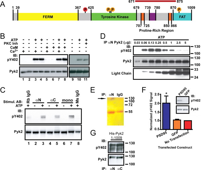
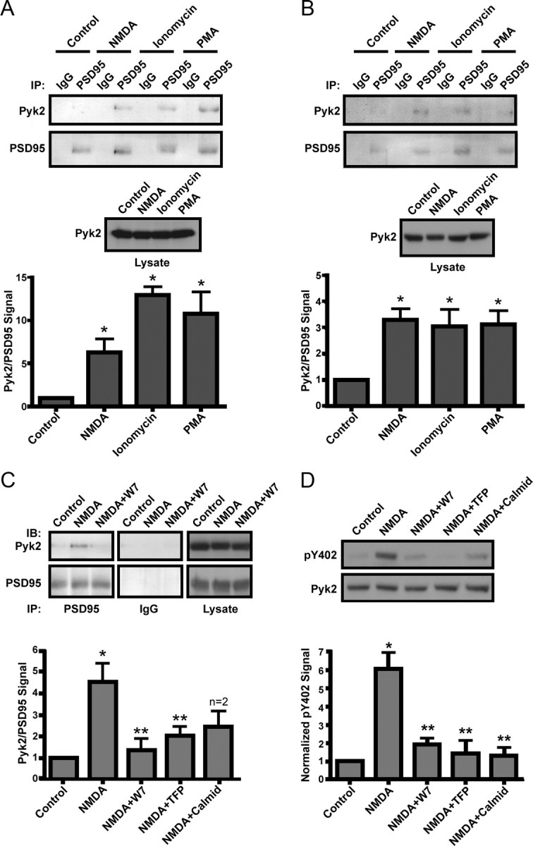
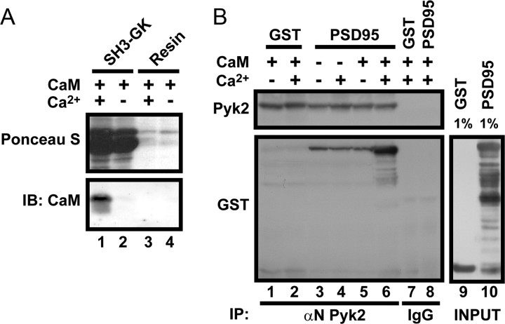
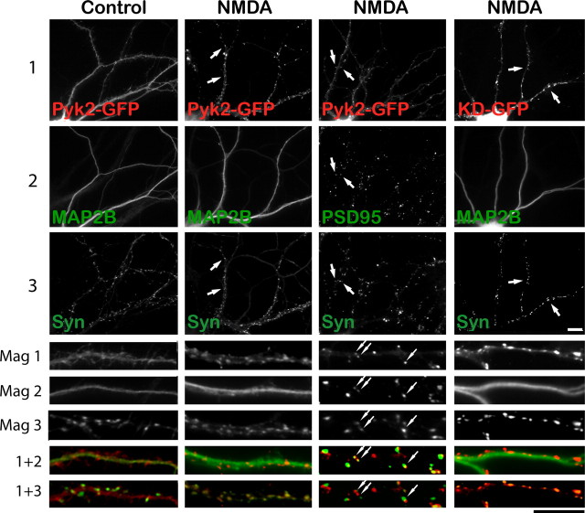
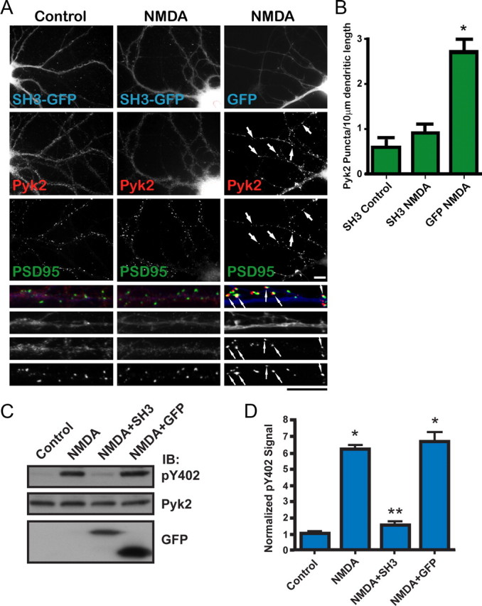
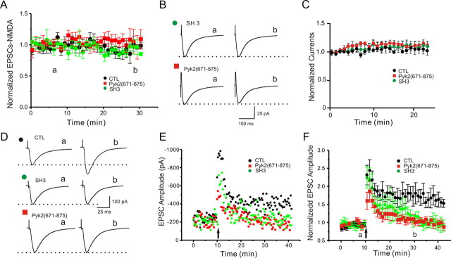
The tyrosine kinase Pyk2 plays a unique role in intracellular signal transduction by linking Ca(2+) influx to tyrosine phosphorylation, but the molecular mechanism of Pyk2 activation is unknown. We report that Pyk2 oligomerization by antibodies in vitro or overexpression of PSD-95 in PC6-3 cells induces trans-autophosphorylation of Tyr402, the first step in Pyk2 activation. In neurons, Ca(2+) influx through NMDA-type glutamate receptors causes postsynaptic clustering and autophosphorylation of endogenous Pyk2 via Ca(2+)- and calmodulin-stimulated binding to PSD-95. Accordingly, Ca(2+) influx promotes oligomerization and thereby autoactivation of Pyk2 by stimulating its interaction with PSD-95. We show that this mechanism of Pyk2 activation is critical for long-term potentiation in the hippocampus CA1 region, which is thought to underlie learning and memory.


Figures

Figure 1.
Figure 1.
Pyk2 dimerization by antibodies or by PSD-95 induces autophosphorylation. A, Schematic of Pyk2 depicting the FERM, tyrosine kinase, and FAT domains. Proline-rich segments are orange. Src phosphorylation sites are denoted as orange circles, and the autophosphorylation site (Tyr402) is red. The magenta bar on top represents the PSD-95 binding region (residues 671-875). B, Pyk2 was immunoprecipitated (αN Pyk2 antibody) from rat brain cytosol and incubated for 1 h at 4°C in the absence or presence of Mg-ATP, Ca2+ (10 mm), calmodulin (10 μm), or a mixture of PKC inhibitors (200 nm bisindolylmaleimide, 5 μm chelerythrine chloride, and 1 μm calphostin C) before immunoblotting with anti-phospho-Tyr402 (pY402), followed by reprobing with the monoclonal Pyk2 antibody. Autophosphorylation occurred independent of Ca2+/calmodulin (compare lanes 3, 4, 7, 8) or PKC activity (lanes 10, 11). C, Rat brain cytosol was incubated with αN, αC, monoclonal Pyk2 antibodies, or nonspecific rabbit or mouse IgG in the absence or presence of Mg-ATP as indicated and directly analyzed by immunoblotting with anti-phospho-Tyr402 and subsequently the monoclonal Pyk2 antibody. D, Increasing amounts of αN Pyk2 antibody were immobilized on protein A Sepharose, washed, incubated with identical amounts of rat brain cytosol, washed, incubated with Mg2+-ATP for 1 h at 4°C, and processed for immunoblotting with anti-phospho-Tyr402 and subsequently monoclonal Pyk2 antibody and an antibody against the light chain of the immunoprecipitating antibody. E, Immunoprecipitation from brain lysate with antibodies against Pyk2 (αN) or control rabbit IgG was followed by SDS-PAGE and silver staining. The thin band near the top of the blot corresponding to the molecular size of Pyk2 is labeled with an arrow and absent in control precipitates. The bands at 55 kDa are the heavy chains of the antibodies used in the immunoprecipitations. F, PC6-3 cells were transfected with PSD-95 24 h before immunoblotting. Immunosignals were quantified by film densitometry and phospho-Tyr402 values (top of inset) per total Pyk2 signal (bottom of inset) normalized to the no-transfection control. Shown are averages ± SEM of n = 3. *p < 0.05, statistically different from GFP-transfected cells by t test. G, E. coli was transformed with pET28a carrying His-tagged Pyk2 and expression induced with IPTG before extraction and immunoblotting with anti-phospho-Tyr402, followed by probing for total Pyk2. The lower band in the phospho-probing is likely a slightly truncated degradation product and was not visible in the reprobing for total Pyk2 because the reprobing was less sensitive than the original phospho-probing. A–G, All blots are representative of three independent experiments. IB, Immunoblots; IP, immunoprecipitation.
Figure 2.
Figure 2.
Stimulation of Pyk2 binding to PSD-95 and Pyk2 autophosphorylation in intact neurons. A, B, Acute cortical slices (A) or primary hippocampal cultures (B; 18 DIV) were incubated with TTX (1 μm) plus vehicle (Control), NMDA (50 μm), ionomycin (1 μm), or PMA (100 nm) for 15 min and extracted (1% deoxycholate) before immunoprecipitation (IP) with control IgG and then anti-PSD-95. Immunoblots (IB) were probed with anti-PSD-95 and reprobed with monoclonal Pyk2 antibody (top). Immunosignals were quantified by film densitometry (bottom). Although the variability in PSD-95 precipitation was low, Pyk2 signals were corrected by dividing each Pyk2 signal by the corresponding PSD-95 signal. The Pyk2/PSD-95 ratio was normalized to control equaling 1. Immunoblotting of lysates illustrates that the same amounts of Pyk2 were present in all samples (middle). Stimulation with NMDA, ionomycin, or PMA induced a several fold increase in Pyk2 coimmunoprecipitation with PSD-95 in slices and cultures. C, Acute cortical slices were preincubated with calmodulin inhibitors (10 μm W7, 20 μm TFP, and 30 μm calmidazolium) for 15 min before incubation with TTX plus vehicle or NMDA, processing, and analysis as in A. Control IgG precipitations (middle column) were from the same blots and exposures as PSD-95 precipitations (left column). All three calmodulin inhibitors prevented the NMDA-induced increase in coprecipitation of Pyk2 with PSD-95 (bottom row) without changing total Pyk2 in lysates (right column). D, Primary hippocampal cultures (18 DIV) were pretreated with calmodulin inhibitors before incubation with TTX plus vehicle or NMDA. Immunoblotting was performed on lysates with the phosphospecific antibody against Pyk2 Tyr402 (pY402) and, after stripping, monoclonal Pyk2 antibody. Signals were quantified by densitometry. The pY402/total Pyk2 ratio was normalized to control equaling 1. NMDA induced a several fold increase in Pyk2 Tyr402 phosphorylation, which was blocked by all three calmodulin antagonists. A–D, Asterisks indicate statistical significance compared with control (*) or NMDA (**) treatment (t test, p < 0.05; n = 4 ± SEM for all experiments).
Figure 3.
Figure 3.
Ca2+/calmodulin stimulates PSD-95 binding to Pyk2. A, Ni2+-agarose was incubated with or without the bacterially expressed His-tagged SH3–GK module of PSD-95 and subsequently with calmodulin (10 μm) in the presence and absence of Ca2+ (10 mm) before immunoblotting (IB). Ponceau S staining indicates immobilization of SH3–GK (lanes 1, 2). Probing with anti-calmodulin antibodies shows specific calmodulin binding to the SH3–GK domain when Ca2+ is present. B, Bacterially expressed Pyk2 was immunoprecipitated (IP) using αN Pyk2 (lanes 1–6) or, as negative control, nonspecific rabbit IgG (lanes 7, 8) before incubation with GST or full-length PSD-95–GST with and without Ca2+ (10 mm) and calmodulin (10 μm) as indicated. Samples were washed with and without Ca2+ corresponding to the binding incubation conditions. Samples underwent immunoblotting with monoclonal Pyk2 and subsequently GST antibodies. GST–PSD-95 (lanes 3–6) but not GST alone (lanes 1, 2) bound to Pyk2. This interaction was increased in the presence of Ca2+ and calmodulin (lane 6). Lanes 9 and 10 show immunoblots with anti-GST of input material. GST–PSD-95 was partially degraded at the C terminus (GST resided at the N terminus). The lack of binding of the truncated polypeptide in the middle region but not the top one-third of the gel (compare lane 10 with lane 6) is consistent with proteolytic removal of SH3 in the more extensively truncated form of GST–PSD-95.
Figure 4.
Figure 4.
Stimulation-induced synaptic clustering of Pyk2 in hippocampal neurons. A, Primary hippocampal cultures (18 DIV) were treated with TTX (100 nm) plus vehicle or glutamate (100 μm) for 15 min or NMDA (50 μm) for 5 min, fixed, and stained with αN Pyk2 (red in overlay at higher magnification at bottom) plus PSD-95, bassoon, or MAP2B antibodies (green). Scale bars at bottom right represent 10 μm for the corresponding magnifications. Arrows indicate Pyk2 puncta that colocalize with PSD-95 or bassoon puncta after stimulation. B, Density of Pyk2 and PSD-95 immunoreactive puncta during treatment with TTX plus vehicle, glutamate (100 μm), PMA (100 nm), ionomycin (1 μm), NMDA (50 μm), glutamate plus EGTA (2 mm), glutamate plus MK801 (10 μm), or glutamate plus W7 (10 μm) for 15 min (5 min for NMDA). C, Colocalization of Pyk2 puncta with PSD-95 puncta indicates that NMDA and glutamate stimulated Pyk2 clustering preferentially at synaptic PSD-95 clusters. A–C, For each experiment, three independent repetitions were performed except for the costaining of Pyk2 with bassoon and with MAP2B after glutamate treatment, which were only performed twice (but see Fig. 5). For quantifications shown in B and C, 5–12 different fields were analyzed for each experiment, and values were averaged before statistical analysis for the means from the three independent experiments (4 experiments for control, glutamate, and NMDA treatments). Asterisks indicate significant differences compared with control (*) or glutamate (**) treatment (t test, p < 0.05; error bars indicate SEM). Both scale bars are 10 μm.
Figure 5.
Figure 5.
NMDA-induced Pyk2–GFP clustering occurs postsynaptically and independently of Pyk2 kinase activity. Primary hippocampal cultures (15 DIV) were transfected with Pyk2–GFP or GFP-tagged kinase-dead Pyk2 (KD-GFP) (72 h; red in all overlays at higher magnification at bottom), treated with TTX plus either vehicle or NMDA (50 μm, 5 min), fixed, and stained with antibodies against synapsin (green in all dual overlays at the very bottom labeled “1 + 3”) and either MAP2B or PSD-95 (green in the respective overlays in the row labeled “1 + 2”). Scale bars, 10 μm for the corresponding magnifications. Pyk2–GFP underwent a dramatic relocation from a fairly smooth distribution under control conditions in dendritic shafts that was primarily colocalized with MAP2B to a pronounced punctate synaptic distribution away from MAP2B staining in dendritic shafts. Arrows illustrate examples of colocalization of Pyk2 and PSD-95 puncta that are juxtaposed to synapsin puncta. Kinase-dead Pyk2 also clusters during NMDA stimulation (right column). All pictures are representatives from three independent experiments.
Figure 6.
Figure 6.
Overexpression of the PSD–95 SH3 domain inhibits NMDA-induced Pyk2 clustering and Pyk2 autophosphorylation. A, Primary hippocampal cultures (15 DIV) were transfected with the GFP-tagged SH3 domain of PSD-95 (SH3-GFP) or GFP alone (72 h; blue in overlay at higher magnification at bottom), treated with TTX plus vehicle or NMDA (50 μm, 5 min), fixed, and stained with antibodies against endogenous Pyk2 (αN; red in overlay) and PSD-95 (green in overlay). Scale bars, 10 μm for the corresponding magnifications. B, Density of Pyk2 immunoreactive puncta. SH3–GFP blocked NMDA-induced Pyk2 clustering seen in the presence of GFP alone. Five to 10 different fields were analyzed for each experiment, and values were averaged before statistical analysis for the means from the three independent experiments. The asterisk indicates statistical significance in Pyk2 clustering in neurons expressing GFP alone versus SH3–GFP (t test, p < 0.05; error bars indicate SEM). C, Primary hippocampal cultures (15 DIV) were infected with packaged FIV pVETL vectors encoding PSD-95 SH3–GFP or GFP alone (72 h) and treated with TTX plus pervanadate (1 mm) plus vehicle or NMDA (50 μm, 15 min) before immunoblotting (IB) with anti-phospho-Tyr402, monoclonal Pyk2, and GFP antibodies. D, Immunosignals were quantified by film densitometry. The pY402/Pyk2 ratios were normalization to control equaling 1. Asterisks denote statistical significance compared with control (*) or NMDA plus GFP (**) (t test, p < 0.05; n = 3 ± SEM).
Figure 7.
Figure 7.
Interfering with the PSD-95–Pyk2 interaction prevents induction of LTP. A, B, Peak currents of NMDAR-mediated EPSCs in CA1 isolated with CNQX are not affected during infusion of GST (n = 5; CTL; black circles), GST–SH3 (n = 3; green circles), or GST–Pyk2(671-875) (n = 5; red squares). C, NMDA-induced whole-cell patch peak currents in acutely isolated CA1 neurons are not affected during infusion of GST (n = 7; CTL; black circles), GST–SH3 (n = 7; green circles), or GST–Pyk2(671-875) (n = 6; red squares). D–F, LTP induced by two tetani of 100 Hz/500 ms, 10 s apart, was blocked by infusion of GST–SH3 (n = 6; green circles) and GST–Pyk2(671-875) (n = 6; red squares) but not GST alone (n = 7; CLT; black circles). Shown are sample traces (D) before (a) and 30 min after (b) tetani, exemplary time courses for each condition (E), and averaged EPSCs that were normalized to baseline for each experiment from all time courses for each condition ±SEM (F). Arrows indicate time of tetani.

References

    1. Ahmed R, Zha XM, Green SH, Dailey ME. Synaptic activity and F-actin coordinately regulate CaMKIIalpha localization to dendritic postsynaptic sites in developing hippocampal slices. Mol Cell Neurosci. 2006;31:37–51. - PubMed
    1. Akyol Z, Bartos JA, Merrill MA, Faga LA, Jaren OR, Shea MA, Hell JW. Apo-calmodulin binds with its C-terminal domain to the N-methyl-D-aspartate receptor NR1 C0 region. J Biol Chem. 2004;279:2166–2175. - PubMed
    1. Altrock WD, tom Dieck S, Sokolov M, Meyer AC, Sigler A, Brakebusch C, Fässler R, Richter K, Boeckers TM, Potschka H, Brandt C, Löscher W, Grimberg D, Dresbach T, Hempelmann A, Hassan H, Balschun D, Frey JU, Brandstätter JH, Garner CC, Rosenmund C, Gundelfinger ED. Functional inactivation of a fraction of excitatory synapses in mice deficient for the active zone protein bassoon. Neuron. 2003;37:787–800. - PubMed
    1. Avraham H, Park SY, Schinkmann K, Avraham S. RAFTK/Pyk2-mediated cellular signalling. Cell Signal. 2000;12:123–133. - PubMed
    1. Avraham S, London R, Fu Y, Ota S, Hiregowdara D, Li J, Jiang S, Pasztor LM, White RA, Groopman JE, Avraham H. Identification and characterization of a novel related adhesion focal tyrosine kinase (RAFTK) from megakaryocytes and brain. J Biol Chem. 1995;270:27742–27751. - PubMed

Publication types

MeSH terms

Substances