. 2010 Jan 13;30(2):491-504.
doi: 10.1523/JNEUROSCI.4961-09.2010.

Loss of Cav1.3 channels reveals the critical role of L-type and BK channel coupling in pacemaking mouse adrenal chromaffin cells

Affiliations

Loss of Cav1.3 channels reveals the critical role of L-type and BK channel coupling in pacemaking mouse adrenal chromaffin cells

Andrea Marcantoni et al. J Neurosci. .

Abstract

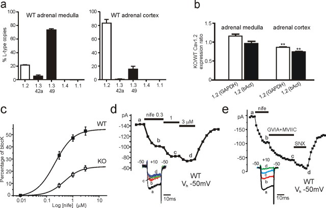
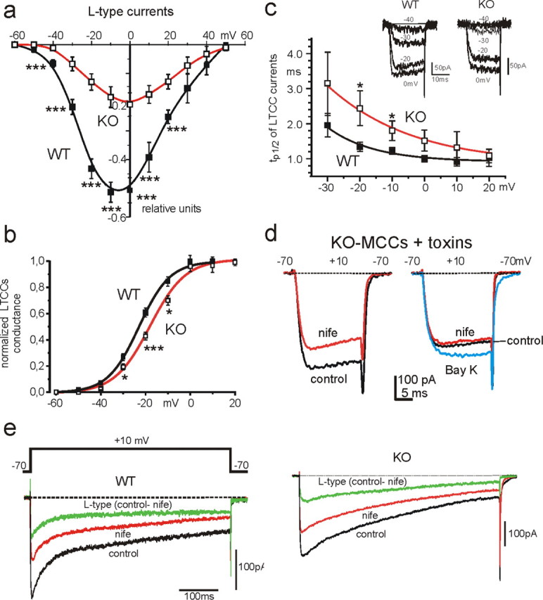
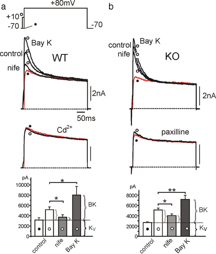
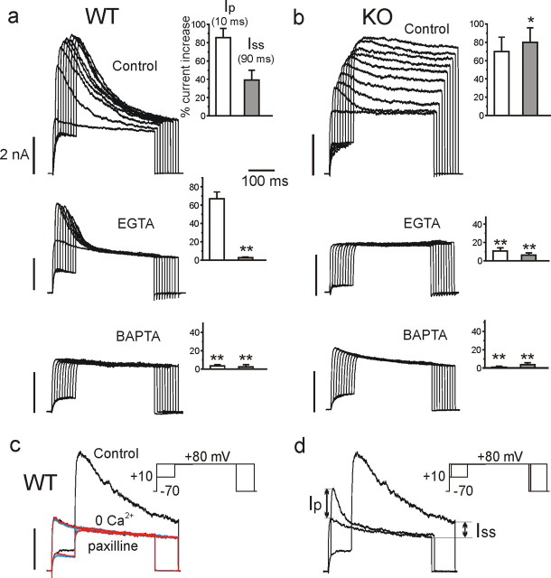
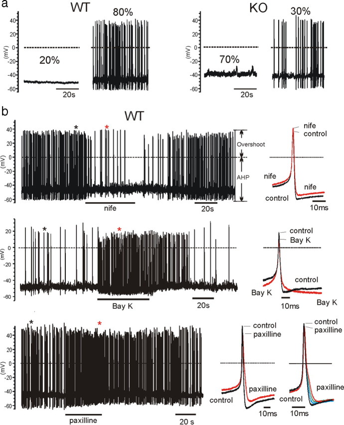
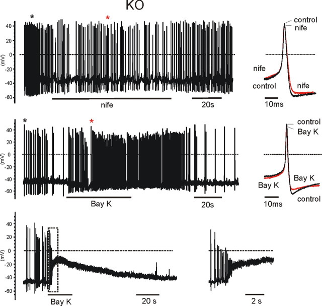
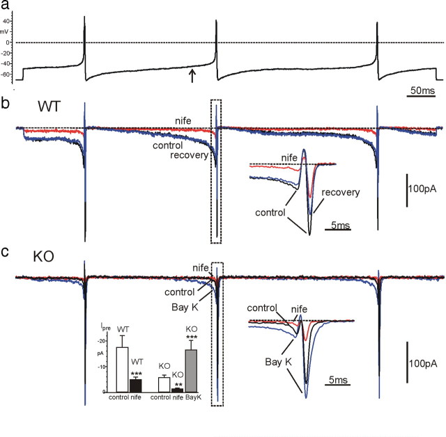
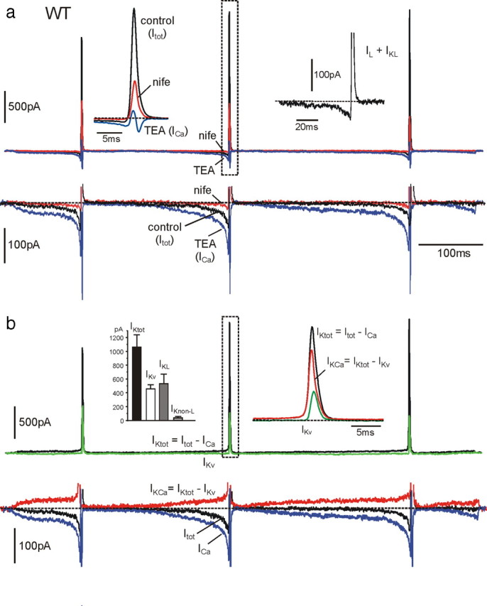
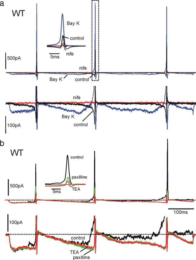
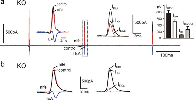
We studied wild-type (WT) and Cav1.3(-/-) mouse chromaffin cells (MCCs) with the aim to determine the isoform of L-type Ca(2+) channel (LTCC) and BK channels that underlie the pacemaker current controlling spontaneous firing. Most WT-MCCs (80%) were spontaneously active (1.5 Hz) and highly sensitive to nifedipine and BayK-8644 (1,4-dihydro-2,6-dimethyl-5-nitro-4-[2-(trifluoromethyl)phenyl]-3-pyridinecarboxylic acid, methyl ester). Nifedipine blocked the firing, whereas BayK-8644 increased threefold the firing rate. The two dihydropyridines and the BK channel blocker paxilline altered the shape of action potentials (APs), suggesting close coupling of LTCCs to BK channels. WT-MCCs expressed equal fractions of functionally active Cav1.2 and Cav1.3 channels. Cav1.3 channel deficiency decreased the number of normally firing MCCs (30%; 2.0 Hz), suggesting a critical role of these channels on firing, which derived from their slow inactivation rate, sizeable activation at subthreshold potentials, and close coupling to fast inactivating BK channels as determined by using EGTA and BAPTA Ca(2+) buffering. By means of the action potential clamp, in TTX-treated WT-MCCs, we found that the interpulse pacemaker current was always net inward and dominated by LTCCs. Fast inactivating and non-inactivating BK currents sustained mainly the afterhyperpolarization of the short APs (2-3 ms) and only partially the pacemaker current during the long interspike (300-500 ms). Deletion of Cav1.3 channels reduced drastically the inward Ca(2+) current and the corresponding Ca(2+)-activated BK current during spikes. Our data highlight the role of Cav1.3, and to a minor degree of Cav1.2, as subthreshold pacemaker channels in MCCs and open new interesting features about their role in the control of firing and catecholamine secretion at rest and during sustained stimulations matching acute stress.


Figures

Figure 1.
Figure 1.
Cav1.2 and Cav1.3 expression in the adrenal medulla and cortex and their DHP sensitivity in WT-MCCs and KO-MCCs. a, Quantitative comparison of different LTCC isoform transcripts in adrenal medulla and cortex of WT mice. Relative abundance of isoforms is given as percentage of the total copy numbers of all LTCC α1-subunit transcripts in each experiment (n = 3). Cav1.1 and Cav1.4 copy numbers did not exceed assay detection limits (n = 2). b, Expression of Cav1.2 α1 subunit in adrenal medulla and cortex in Cav1.3−/− mice relative to WT compared by ΔΔCt analysis using GAPDH and β-actin (bAct) as reference genes (n = 3). c, Dose–response relationship of Ca2+ current block by nifedipine (nife) recorded from WT-MCCs (n = 7) and KO-MCCs (n = 5). Percentage of block was measured using 20 ms pulses to +10 mV from Vh of −50 mV. The smooth curves represent the fit to the data with IC50 of 0.21 ± 0.07 μm (WT) and 0.48 ± 0.12 μm (KO) and Hill slopes of 1.22 ± 0.52 and 1.35 ± 0.48, respectively. d, Time course of peak Ca2+ current recorded from a WT-MCC before, during, and after sequential application of 0.01, 0.3, 1, and 3 μm nifedipine (nife). Step depolarization to +10 mV was applied every 10 s. The inset shows the current traces recorded at the time indicated by the letters. e, Time course of peak Ca2+ current recorded from a WT-MCC during sequential application of 3 μm nifedipine (nife), 3.2 μm ω-CTx-GVIA + 1 μm ω-CTx-MVIIC (GVIA+MVIIC), and 400 nm SNX-482 (SNX). Notice the partial recovery after washing that uncovers the irreversible block of N- and P/Q-type channels by ω-CTx-GVIA and ω-CTx-MVIIC. The inset shows the current traces recorded at the time indicated by the letters.
Figure 2.
Figure 2.
Voltage-dependent characteristics of LTCCs in WT-MCCs and KO-MCCs. a, I–V characteristics of normalized L-type currents in ω-toxin-treated WT-MCCs (filled squares; n = 14) and KO-MCCs (open squares; n = 12). Vh was −70 mV. The L-type current amplitude was determined as the difference between control and nifedipine-insensitive currents, using 3 μm nifedipine to block LTCCs (for the estimated DHP blocking potency at Vh of −70 mV, see text and supplemental Fig. S1, available at www.jneurosci.org as supplemental material). L-type currents are plotted as fractions of the total current. b, Voltage dependence of Cav1.3 and Cav1.2 channel conductance in WT-MCCs and KO-MCCs. The normalized LTCC conductance was calculated as Ipeak/(VVrev) with Vrev = +50 mV from n = 8 WT-MCCs and n = 5 KO-MCCs. The two continuous curves are Boltzmann functions best fitting the data points with V1/2 = −23.0 mV and slope factor k = 6.9 mV for WT-MCCs and V1/2 = −18.1 mV and k = 7.9 mV for KO-MCCs. c, Voltage dependence of tp1/2 taken as an index of LTCC activation in ω-toxin-treated WT-MCCs and KO-MCCs. tp1/2 was smaller in WT-MCCs and significantly prolonged at −20 and −10 mV in KO-MCCs (*p < 0.05). Inset, Time course of two families of L-type currents recorded from a WT-MCC and a KO-MCC during brief step depolarization to −40, −30, −20, and 0 mV from −70 mV Vh. d, KO-MCCs displayed heterogeneous distribution of Cav1.2 channel expression. To the left are shown the blocking effect of nifedipine (nife) on Ca2+ currents in a ω-toxin-treated KO-MCC responding normally to the DHP. To the right are shown the effects of nifedipine and BayK-8644 (Bay K) on a KO-MCC characterized by a weak response to the DHPs. e, Cav1.3 currents inactivate less than Cav1.2 during prolonged membrane depolarization. L-type currents were elicited using pulses of 600 ms to +10 mV and calculated by subtracting nifedipine (nife)-insensitive from control currents. In the WT-MCC (left), the resulting L-type current after an initial fast inactivation of ∼100 ms reached a steady-state value that did not change further for the remaining 500 ms. In the KO-MCC (right), inactivation persists for the entire duration of the pulse reaching a steady-state value after 600 ms. The datasets were compared using one-way ANOVA, followed by Bonferroni's post hoc test (*p < 0.05; ***p < 0.001).
Figure 3.
Figure 3.
LTCC coupling to BK channels in WT-MCCs and KO-MCCs. a, Ca2+-activated and voltage-gated K+ currents recorded from a WT-MCC at control and during application of nifedipine (nife; 3 μm), BayK-8644 (Bay K; 1 μm), or Cd2+ (500 μm). The voltage command consisted of a double-pulse protocol with a test potential of 400 ms to +80 mV preceded (open circle) or not (filled circle) by a 10 ms prepulse to +10 mV to activate maximal inward Ca2+ currents. Ca2+ inward currents were not always visible because of the fast BK and KV channel activation. Below are the same recordings in the presence of 500 μm Cd2+ obtained from the same MCC. To the bottom are the peak amplitude values of K+ currents (n = 20; *p < 0.05). The horizontal dotted line indicates the mean peak amplitude of KV currents estimated at +80 mV without prepulse (filled circle). The amount of currents exceeding the dotted line represents the BK current. b, BK and KV currents recorded from a KO-MCC. The protocol was as in a except that BK currents were blocked by paxilline (1 μm). The recordings are from the same MCC. Addition of Cd2+ (500 μm) or 0 Ca2+ solutions caused similar blocking effects. To the bottom are the mean values of peak KV and BK currents as in a (n = 16; *p < 0.05; **p < 0.01).
Figure 4.
Figure 4.
Effects of varying the duration of Ca2+ loading steps in the absence (control) or in the presence of EGTA-AM or BAPTA-AM on the time course and amplitude of BK currents in WT-MCCs (a) and KO-MCCs (b). a, Recordings from three WT-MCCs in control solution, after cell incubation with 20 μm EGTA for 40 min or 20 μm BAPTA-AM for 40 min, as indicated. Notice the increasing amplitude of fast inactivating BK currents with increasing the duration of preloading steps in control conditions, the persistence of pure fast inactivating BK currents in the presence of EGTA, and the absence of any BK current in the presence of BAPTA. On the insets are reported the values of Ip and Iss measured as indicated in d from n = 12 (control), 11 (EGTA), and 8 (BAPTA) WT-MCCs (**p < 0.01 vs control using Student's paired t test). b, Same as in a but from three different KO-MCCs. Notice how the prolonged Ca2+ preloading steps uncover a large non-inactivating BKs current and how EGTA and BAPTA are both effective in preventing this BK current. Ip and Iss were derived as in a from n = 10 (control), 13 (EGTA), and 6 (BAPTA) KO-MCCs (*p < 0.05, **p < 0.01 vs control using Student's paired t test). c, Overlapped current traces recorded from a WT-MCC in control conditions, 0 Ca2+ solution (red traces), and after adding 1 μm paxilline (blue traces). The double-pulse protocol was as in Figure 3 with a Ca2+ preloading step of 90 ms. Notice the full block of the transient BK current in the two conditions. d, Measure of Ip and Iss from a WT-MCC. The pulse protocol was as indicated in the inset: the test potential was to +80 mV, and the preloading steps of 10 and 90 ms were to +10 mV (Vh of −70 mV).
Figure 5.
Figure 5.
a, Representative recordings and percentages of firing and nonfiring WT-MCCs and KO-MCCs. Only 30% of KO-MCCs showed spontaneous firing, whereas the majority of WT-MCCs (80%) had spontaneous activity. b, Effects of nifedipine (nife), BayK-8644 (Bay K), and paxilline on pacemaking in WT-MCCs and KO-MCCs. The panels show the spontaneous firing of three WT-MCCs before, during, and after application of 3 μm nifedipine (top), 1 μm BayK-8644 (middle), or 1 μm paxilline (bottom). To the right are shown two overlapped action potentials on an expanded timescale corresponding to those indicated by the asterisks. Nifedipine had a full blocking action, which recovered completely after washing, whereas BayK-8644 and paxilline accelerated the firing rate. Paxilline reduced also the AHP and broadened AP width. This is more evident on the overlapped APs to the right recorded during paxilline action in a different WT-MCC. The arrows on the top show how the overshoot and the AHP were determined.
Figure 6.
Figure 6.
Effects of nifedipine and BayK-8644 on three spontaneously firing KO-MCCs. Top and middle, Same protocols and analysis as in Figure 5b. The black and red asterisks indicate the position of the overlapped potentials shown to the right. The two DHPs acted moderately on both the shape and frequency of the AP firing. Bottom, The paradoxical effect of BayK-8644 on a spontaneously firing KO-MCC. Cell firings was rather irregular and application of BayK-8644 first caused an acceleration of the firing, followed by a net depolarization to −10 mV. To the right is the recording inside the dashed rectangle on an expanded timescale. The cell returned spontaneously to the resting potential after washing BayK-8644.
Figure 7.
Figure 7.
Ca2+ currents in WT-MCCs and KO-MCCs during an action potential clamp recorded in current-clamp conditions from a spontaneously firing cell. K+ and Na+ currents were blocked by 135 mm TEA and 0.3 μm TTX in the bath solution. In a is illustrated the voltage-clamp command consisting of a train of three APs separated by different interpulse intervals. The cell was initially held at −70 mV and then clamped at the AP waveform (see Materials and Methods). In b are shown the overlapped Ca2+ currents recorded from a WT-MCC before, during, and after application of 3 μm nifedipine (nife). Right inset, Ca2+ current traces corresponding to the second AP (dashed rectangle) on a more expanded timescale. c, Same as in b except that the recording was from a nonfiring KO-MCC. The cell possessed a small prespike Ca2+ current that was potentiated by BayK-8644 (Bay K; n = 8). Notice the large prespike L-type current increase and how the mean amplitude at −45 mV with BayK-8644 increases to nearly the same size of WT-MCC control current (gray box in b). Left inset, Mean amplitudes of the prespike Ca2+ currents measured from WT-MCCs and KO-MCCs (n = 16) at the time when the interpulse potential reached −45 mV, before the second AP (arrow in a). Notice the strong current reduction induced by nifedipine in both WT-MCCs and KO-MCCs. **p < 0.01, ***p < 0.001 vs control using Student's paired t test.
Figure 8.
Figure 8.
Contribution of BK and voltage-gated K+ and Ca2+ currents during an AP train clamp in a WT-MCC. a, Three overlapped current traces recorded in control conditions, in the presence of nifedipine (nife; 3 μm) and with 135 mm TEA. On the top row are evident the large transient K+ currents of increasing amplitudes during spikes and the small prespike currents preceding AP generation. As shown on a more expanded vertical scale (bottom row), the prespike control current was always net inward and increased markedly after addition of TEA (ICa). Left top inset, K+ and Ca2+ currents just before and during the second spike on a more expanded timescale corresponding to the dashed rectangle. The inward and outward control currents were strongly blocked by nifedipine. Right top inset, Sum of L-type currents (IL) and L-type-activated BK currents (IKL) obtained by subtracting nifedipine-insensitive currents from control currents. The current is net inward before the spike and turns rapidly out during the upstroke. The top part of the outward current is blanked photographically. b, Time courses of the total (IKtot) and the Ca2+-activated K+ current (IKCa) calculated from the recordings of a overlapped to the voltage-gated K+ current (IKv) measured in 0 external Ca2+. IKtot was obtained by subtracting the Ca2+ current recorded in the presence of 135 mm TEA (ICa) from the total control current (Itot), whereas IKCa was obtained by subtracting IKv from IKtot, as indicated. Right top inset, IKtot, IKCa, and IKv during the second spike on an expanded timescale. Left top inset, Mean values of IKtot, IKv, and BK currents activated by L-type (IKL) and non-L-type (IKnon-L) Ca2+ channels (n = 14), calculated as explained in Results. Notice that non-L-type-activated K+ currents are nearly absent in WT-MCCs. In the bottom row is illustrated the time course of the control current (Itot), which is net inward during the interspike intervals and the outward (IKCa) and inward (ICa) components contributing to this current. As shown, the inward Ca2+ current is always larger than IKCa during the interspike intervals.
Figure 9.
Figure 9.
BayK-8644 and paxilline increase the net inward pacemaker current in WT-MCCs. a, In the top row are shown the effects of BayK-8644 (Bay K; 1 μm) and nifedipine (nife; 3 μm) on inward and outward currents during an action potential train clamp. As shown at more expanded vertical (bottom row) and timescales (inset), BayK-8644 increases dramatically the prespike current and generates a dominant BK current component during the three spikes. Both inward and outward currents are primarily blocked by nifedipine. Notice also the brief inward L-type current after the AHP associated with the closing of BayK-8644-modified LTCCs. This current contributed to the quick redepolarization after the increased AHP induced by BayK-8644. b, Effects of paxilline on K+ outward currents in a BayK-8644-treated WT-MCC. The DHP activator induces a large inward L-type current that activates a noisy BK outward current that rises gradually during the interpulse. Paxilline blocks the noisy current, increases the net inward prespike current, and drastically lowers the K+ outward component during the spike (inset). The residual K+ outward current is mainly associated with voltage-gated K+ channels. The block of the slow outward current by paxilline is similar to that induced by TEA, confirming that the current component blocked by paxilline is carried by Ca2+-activated BK channels.
Figure 10.
Figure 10.
Time course and contribution of K+ and Ca2+ currents to the AP firing is markedly different in KO-MCCs. Test solutions and protocols were similar to those used for WT-MCCs (see Fig. 8). a, Overlapped current traces recorded in control conditions, during nifedipine application (3 μm; nife; red trace), and with 135 mm TEA (blue trace) in a KO-MCC. The prespike Ca2+ current was particularly small compared with the postspike current. Left inset, K+ and Ca2+ currents relative to the dashed rectangle on a more expanded timescale. Nifedipine had a marked action on the inward currents but had only minor blocking effects on the outward K+ current, suggesting a small IKL component. Middle inset, Time course of IKtot, IKCa, and IKv, showing the larger contribution of IKv. IKtot and IKCa were obtained as described in Results. Right inset, Mean values of IKtot, IKL, IKnon-L, and IKv (n = 19). Notice the small contribution of IKL compared with WT-MCCs. b, K+ and Ca2+ currents recorded from a KO-MCC in which the BK currents (IKCa) were particularly small. Despite the near full block of the sizeable Cav1.2 current, nifedipine (nife; 3 μm) caused a paradoxical increase of the outward K+ current. As shown to the right, IKv was responsible for most of the outward K+ current.

Similar articles

Cited by

References

    1. Baldelli P, Hernández-Guijo JM, Carabelli V, Novara M, Cesetti T, Andrés-Mateos E, Montiel C, Carbone E. Direct and remote modulation of L-channels in chromaffin cells: distinct actions on alpha1C and alpha1D subunits? Mol Neurobiol. 2004;29:73–96. - PubMed
    1. Bean BP. The action potential in mammalian central neurons. Nat Rev Neurosci. 2007;8:451–465. - PubMed
    1. Brandt BL, Hagiwara S, Kidokoro Y, Miyazaki S. Action potentials in the rat chromaffin cell and effects of acetylcholine. J Physiol. 1976;263:417–439. - PMC - PubMed
    1. Carabelli V, D'Ascenzo M, Carbone E, Grassi C. Nitric oxide inhibits neuroendocrine Ca(V)1 L-channel gating via cGMP-dependent protein kinase in cell-attached patches of bovine chromaffin cells. J Physiol. 2002;541:351–366. - PMC - PubMed
    1. Carabelli V, Marcantoni A, Comunanza V, de Luca A, Díaz J, Borges R, Carbone E. Chronic hypoxia up-regulates alpha1H T-type channels and low-threshold catecholamine secretion in rat chromaffin cells. J Physiol. 2007;584:149–165. - PMC - PubMed

Publication types

MeSH terms

Substances