. 2010 Jan 13;30(2):545-55.
doi: 10.1523/JNEUROSCI.4920-09.2010.

Control of cannabinoid CB1 receptor function on glutamate axon terminals by endogenous adenosine acting at A1 receptors

Affiliations

Control of cannabinoid CB1 receptor function on glutamate axon terminals by endogenous adenosine acting at A1 receptors

Alexander F Hoffman et al. J Neurosci. .

Abstract

Marijuana is a widely used drug that impairs memory through interaction between its psychoactive constituent, Delta-9-tetrahydrocannabinol (Delta(9)-THC), and CB(1) receptors (CB1Rs) in the hippocampus. CB1Rs are located on Schaffer collateral (Sc) axon terminals in the hippocampus, where they inhibit glutamate release onto CA1 pyramidal neurons. This action is shared by adenosine A(1) receptors (A1Rs), which are also located on Sc terminals. Furthermore, A1Rs are tonically activated by endogenous adenosine (eADO), leading to suppressed glutamate release under basal conditions. Colocalization of A1Rs and CB1Rs, and their coupling to shared components of signal transduction, suggest that these receptors may interact. We examined the roles of A1Rs and eADO in regulating CB1R inhibition of glutamatergic synaptic transmission in the rodent hippocampus. We found that A1R activation by basal or experimentally increased levels of eADO reduced or eliminated CB1R inhibition of glutamate release, and that blockade of A1Rs with caffeine or other antagonists reversed this effect. The CB1R-A1R interaction was observed with the agonists WIN55,212-2 and Delta(9)-THC and during endocannabinoid-mediated depolarization-induced suppression of excitation. A1R control of CB1Rs was stronger in the C57BL/6J mouse hippocampus, in which eADO levels were higher than in Sprague Dawley rats, and the eADO modulation of CB1R effects was absent in A1R knock-out mice. Since eADO levels and A1R activation are regulated by homeostatic, metabolic, and pathological factors, these data identify a mechanism in which CB1R function can be controlled by the brain adenosine system. Additionally, our data imply that caffeine may potentiate the effects of marijuana on hippocampal function.


Figures
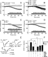
Figure 1.
Figure 1.
Tonic activation of adenosine receptors by eADO controls CB1R modulation of glutamate release in C57BL/6J mice. A, The cannabinoid agonist WIN55,212-2 (500 nm) inhibited glutamatergic fEPSPs in hippocampal slices from SD rats but not in slices from wild-type (CB1+/+) C57BL/6J mice. In this and all subsequent figures, the duration of drug application is indicated by horizontal bars. B, Antagonism of adenosine receptors by caffeine (CAFF, 50 μm), THEO (250 μm), or DPCPX (0.2 μm) reveals robust inhibition of fEPSPs by WIN55,212-2 in the CB1+/+ mouse hippocampus. Antagonists were applied to the brain slices for 15–20 min before WIN55,212-2 application. C, The inhibition of fEPSPs by WIN55,212-2 during THEO application is mediated by CB1Rs. fEPSP inhibition by WIN55,212-2 was compared in hippocampal slices from CB1+/+ and CB1−/− littermates following pretreatment with THEO. Responses were inhibited by WIN55,212-2 only in CB1+/+ slices. D, Catabolism of eADO by ADA (2 U/ml) permits inhibition of fEPSPs by WIN55,212-2 in hippocampal slices from CB1+/+ mice. E, Mean fEPSPs (n = 5 sweeps in all figures) collected from single hippocampal slices demonstrating the effects of WIN55,212-2 during experiments shown in C and D. E1, WIN55,212-2 (WIN, 500 nm) inhibits fEPSPs only in CB1+/+ hippocampal slices following THEO pretreatment. E2, Inhibition of fEPSPs by WIN (500 nm) in hippocampal slices from CB1+/+ mice after a 20 min ADA (2 U/ml) pretreatment. F, Summary showing peak effects of WIN (35–40 min of application) from experiments shown in A–D. **p < 0.01, RM-ANOVA, and Tukey post hoc analysis.
Figure 2.
Figure 2.
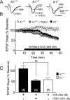
Adenosine A1 receptors control cannabinoid inhibition of glutamatergic synaptic transmission. A, Mean fEPSPs collected during baseline recordings (Control, black traces) and during application of WIN55,212-2 (WIN, 500 nm, gray trace) in hippocampal brain slices from A1+/+ and A1−/− C57BL/6J mice. The center control trace shows the effect of WIN after a 15 min treatment with THEO (250 μm). B, Mean time course of the effects of WIN55,212-2 on the initial slope of fEPSPs in hippocampal slices obtained from A1+/+ and A1−/− mice. Note that WIN55,212-2 inhibited fEPSPs in slices from A1−/− mice and in slices from A1+/+ mice only after theophylline (250 μm) pretreatment. C, Summary of the effects of the treatment conditions in hippocampal slices from A1+/+ and A1−/− mice. **p < 0.01 RM-ANOVA, Tukey post hoc analysis. The mean effects were determined 35–40 min after WIN55,212-2 application and the number of slices in each condition is shown in parentheses.
Figure 3.
Figure 3.
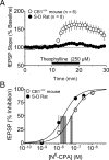
Hippocampal slices from C57BL/6J CB1+/+ mice demonstrate tonic levels of eADO higher than those from SD rats. A, Mean time course of the effects of the adenosine receptor antagonist theophylline on fEPSPs in SD rat or CB1+/+ mouse hippocampal slices. Theophylline caused a significantly larger increase in fEPSPs in mouse brain slices under identical recording conditions (p < 0.001, RM-ANOVA). B, Mean concentration–response curves for the effects of the selective A1R agonist N6-CPA on fEPSPs in hippocampal slices from CB1+/+ mice (n = 3–6 slices per concentration) and SD rats (n = 4 slices per concentration). N6-CPA more potently inhibited fEPSPs in slices from SD rats compared with mice. The EC50 values (dashed vertical lines) were 189 nm (95% CI = 118–301 nm, vertical shaded bar) in rat hippocampal slices and 587 nm (95% CI = 467–737 nm) in mouse slices. The EC50 value CIs obtained in rat and mouse slices did not overlap, indicating a significant difference.
Figure 4.
Figure 4.
The plant-derived cannabinoid receptor agonist Δ9-THC inhibits glutamate release through activation of CB1Rs in SD rat hippocampal brain slices. Top, Traces showing the effect of Δ9-THC on mean fEPSPs in rat hippocampal slices in control aCSF (gray line, left) and following treatment with the CB1R antagonist AM251 (1 μm, gray line at right) in a different rat hippocampal slice. Bottom, Mean time course comparing the effects of Δ9-THC (10 μm) to those of RAMEB alone and to Δ9-THC (10 μm) after a 20 min treatment with AM251 (1 μm). Note that whereas RAMEB alone significantly inhibited glutamate release, the effect of Δ9-THC in the same concentration of vehicle was significantly larger (**p < 0.01, RM-ANOVA). Furthermore, the effect of Δ9-THC on fEPSPs was not significantly different from RAMEB alone, when slices were pretreated with AM251 (p > 0.05, RM-ANOVA).
Figure 5.
Figure 5.
Adenosine A1Rs control CB1R inhibition of hippocampal EPSCs in the C57BL/6J mouse hippocampus. A, Mean evoked EPSCs and currents elicited through photolysis of CNB-caged glutamate in the same CA1 neuron from a CB1+/+ mouse. Both synaptic and photolysis-evoked glutamate responses were blocked by the AMPAR antagonist DNQX (10 μm). B, Mean EPSCs recorded from a pyramidal neuron in CB1+/+ mouse hippocampus showing the effect of the RAMEB vehicle (left) and Δ9-THC (10 μm) after treatment with the selective adenosine A1 receptor antagonist DPCPX (200 nm). C, Mean time course showing the effects of RAMEB on EPSCs and of Δ9-THC (10 μm) dissolved in this vehicle on EPSCs and photolysis-evoked currents in CB1+/+ mouse CA1 pyramidal neurons. The difference between the effects of RAMEB and Δ9-THC plus RAMEB was not statistically significant (p > 0.5, RM-ANOVA), and Δ9-THC plus RAMEB had no effect on the photolysis-evoked glutamate currents. D, Mean time course comparing the effects of RAMEB to Δ9-THC in DPCPX-pretreated pyramidal neurons in hippocampal slices from CB1+/+ and CB1−/− mice. A1R antagonism with DPCPX significantly increased the inhibition of EPSCs by Δ9-THC (p < 0.01, RM-ANOVA) in the CB1+/+ slices, but Δ9-THC effects were not significantly different from vehicle in CB1−/− slices, despite DPCPX pretreatment. In the DPCPX experiments, the data were normalized after the antagonist effects had stabilized, before Δ9-THC application. Therefore, the effect of DPCPX on EPSCs is not shown. E, Summary of the effects of Δ9-THC and RAMEB on EPSCs or photolysis-evoked AMPA currents recorded in mouse hippocampus. The mean effects were obtained 20 min after Δ9-THC or RAMEB application (** = p < 0.01, RM-ANOVA). Picrotoxin was used to block GABAA receptors throughout these experiments.
Figure 6.
Figure 6.
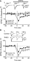
A1R antagonism increases endocannabinoid-dependent DSE in mGluR agonist-treated mouse brain slices. A, DSE was induced using a 3 s voltage step to 0 mV from a −70 mV holding potential (gray vertical bars in A2 and B2). A1, Mean EPSCs collected 6 s before (black line) and 6 s after (gray line) DSE in neurons from CB1+/+ and CB1−/− mice, in the absence or presence of the mGluRI agonist DHPG (10 μm). Note that DSE was observed in neurons from CB1+/+ mice only during DHPG application and was absent in DHPG-treated CB1−/− neurons. A2, Mean time course showing the effect of DHPG on DSE in CB1+/+ and CB1−/− neurons. B, Effects of A1R antagonism by THEO (250 μm) on endocannabinoid-mediated DSE in CA1 pyramidal neurons from CB1+/+ mice pretreated with DHPG (10 μm). B1, Mean EPSCs demonstrating DSE in the absence (Control) and presence of THEO in DHPG-pretreated slices. The EPSCs are averages of three traces collected every 3 s before the indicated time point, before or after the voltage step. B2, Mean time course of the effect of A1R antagonism by THEO on DSE. THEO significantly increased the level of endocannabinoid-mediated DSE (RM-ANOVA; p < 0.01, Tukey post hoc analysis). However, the degree of potentiation of DSE by THEO is likely underestimated due to desensitization of A1Rs by DHPG (de Mendonça and Ribeiro, 1997; Shahraki and Stone, 2003).
Figure 7.
Figure 7.
PCTx (50 μm) increases eADO release and the activation of A1Rs and reduces CB1R signaling in rat hippocampus. A1, Mean fEPSPs recorded before (Control) and during application of the adenosine receptor antagonist theophylline (250 μm, gray lines), in the absence (−PCTx) and presence (+PCTx) of PCTx. A, Mean time course (n = 4) of the effect of theophylline before and during PCTx treatment. The effect of theophylline is significantly larger in during PCTx treatment (p < 0.1, RM-ANOVA). B, The selective A1R antagonist DPCPX (200 nm), but not the GABAB antagonist CGP55845 (2 μm) or the CB1R antagonist AM251 (1 μm), significantly increased fEPSPs during PCTx treatment (p < 0.1, RM-ANOVA). C, Antagonism of A1Rs restores CB1R inhibition of fEPSPs caused by Δ9-THC in PCTx-treated SD rat hippocampal slices. C1, fEPSPs obtained in rat hippocampus during PCTx application. The GABAB antagonist CGP55845 (2 μm) had no effect on the fEPSP, and subsequent application of Δ9-THC (10 μm, dotted line) produced only a small decrease in the response. C2, During PCTx application, DPCPX (200 nm) increased the fEPSP amplitude and permitted a larger inhibition of the fEPSP by Δ9-THC (10 μm, dotted line). C, Mean time course of the effects of Δ9-THC under each condition demonstrating decreased inhibition of fEPSPs by Δ9-THC in PCTx and the restoration of the Δ9-THC inhibition by DPCPX (200 nm; n = 5). Also shown is that GABAB antagonism by CGP55845 (2 μm, n = 6) did not reverse the attenuation of the Δ9-THC effect caused by PCTx (p < 0.01, RM-ANOVA). D, Summary of the data shown in C. Mean effects of 10 μm Δ9-THC were determined by averaging data acquired 35–40 min after its application in slices pretreated with the indicated drugs. **p < 0.01, ANOVA. The number of brain slices under each condition is noted in parentheses.

Similar articles

Cited by

References

    1. Alger BE. Retrograde signaling in the regulation of synaptic transmission: focus on endocannabinoids. Prog Neurobiol. 2002;68:247–286. - PubMed
    1. Berman RF, Fredholm BB, Aden U, O'Connor WT. Evidence for increased dorsal hippocampal adenosine release and metabolism during pharmacologically induced seizures in rats. Brain Res. 2000;872:44–53. - PubMed
    1. Brundege JM, Dunwiddie TV. Modulation of excitatory synaptic transmission by adenosine released from single hippocampal pyramidal neurons. J Neurosci. 1996;16:5603–5612. - PMC - PubMed
    1. Bruns RF, Daly JW, Snyder SH. Adenosine receptors in brain membranes: binding of N6-cyclohexyl[3H]adenosine and 1,3-diethyl-8-[3H]phenylxanthine. Proc Natl Acad Sci U S A. 1980;77:5547–5551. - PMC - PubMed
    1. Burnstock G. Physiology and pathophysiology of purinergic neurotransmission. Physiol Rev. 2007;87:659–797. - PubMed

Publication types

MeSH terms

Substances

LinkOut - more resources