. 2010 Jan 13;30(2):573-82.
doi: 10.1523/JNEUROSCI.3295-09.2010.

P2X7-dependent release of interleukin-1beta and nociception in the spinal cord following lipopolysaccharide

Affiliations

P2X7-dependent release of interleukin-1beta and nociception in the spinal cord following lipopolysaccharide

Anna K Clark et al. J Neurosci. .

Abstract

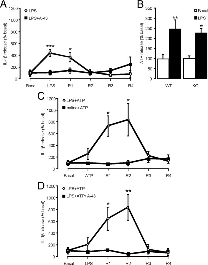
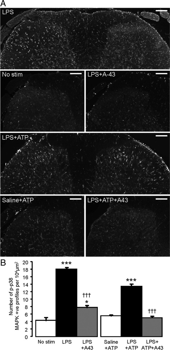
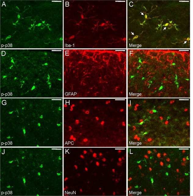
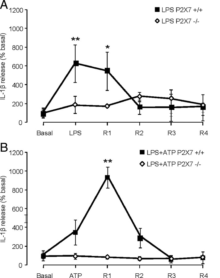
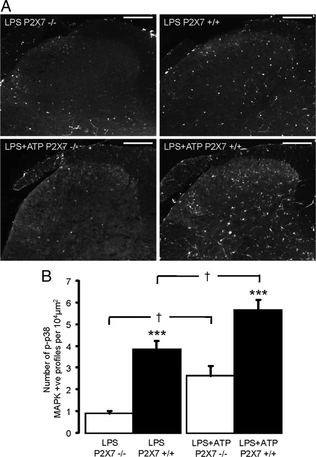
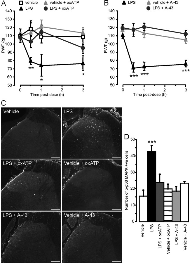
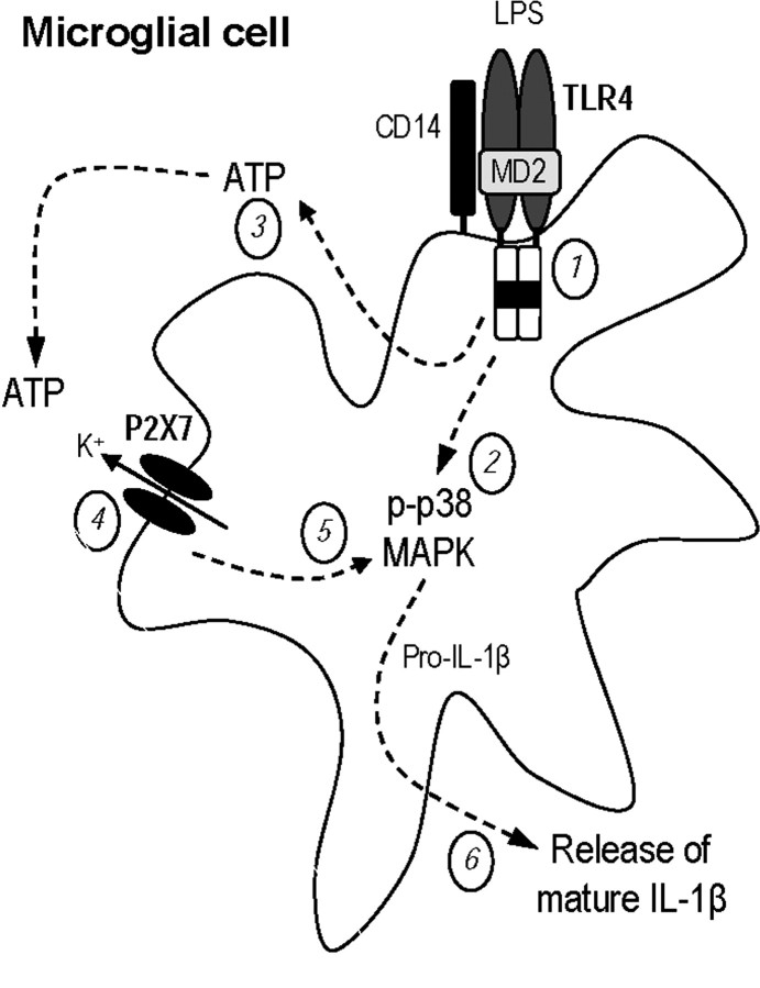
The cytokine interleukin-1beta (IL-1beta) released by spinal microglia in enhanced response states contributes significantly to neuronal mechanisms of chronic pain. Here we examine the involvement of the purinergic P2X7 receptor in the release of IL-1beta following activation of Toll-like receptor-4 (TLR4) in the dorsal horn, which is associated with nociceptive behavior and microglial activation. We observed that lipopolysaccharide (LPS)-induced release of IL-1beta was prevented by pharmacological inhibition of the P2X7 receptor with A-438079, and was absent in spinal cord slices taken from P2X7 knock-out mice. Application of ATP did not evoke release of IL-1beta from the dorsal horn unless preceded by an LPS priming stimulus, and this release was dependent on P2X7 receptor activation. Extensive phosphorylation of p38 MAPK in microglial cells in the dorsal horn was found to correlate with IL-1beta secretion following both LPS and ATP. In behavioral studies, intrathecal injection of LPS in the lumbar spinal cord produced mechanical hyperalgesia in rat hindpaws, which was attenuated by concomitant injections of either a nonspecific (oxidized ATP) or a specific (A-438079) P2X7 antagonist. In addition, LPS-induced hypersensitivity was observed in wild-type but not P2X7 knock-out mice. These data suggest a critical role for the P2X7 receptor in the enhanced nociceptive transmission associated with microglial activation and secretion of IL-1beta in the dorsal horn. We suggest that CNS-penetrant P2X7 receptor antagonists, by targeting microglia in pain-enhanced response states, may be beneficial for the treatment of persistent pain.


Figures

Figure 1.
Figure 1.
Disruption of P2X7 receptor signaling attenuates both LPS- and ATP-evoked IL-1β release in rat dorsal horn slices, but does not alter ATP release. A, LPS superfusion (10 μg/ml for 8 min) induces rapid release of IL-1β from the rat dorsal horn (n = 6 slices). Superfusion of the specific P2X7 receptor antagonist A-438079 (A-43) before and during LPS application (16 min in total) inhibits LPS-evoked IL-1β release (n = 6 slices). Basal release: LPS = 11.7 ± 4.6 pg/8 ml fraction, LPS + A-43 = 13.8 ± 5.6 pg/8 ml fraction. B, LPS incubation (10 μg/ml for 8 min) induces release of ATP from P2X7 wild-type (n = 5) and knock-out (n = 4) dorsal horn slices. Basal release: wild-type = 4.44 ± 0.9 fmol per 100 μl, knock-out = 2.6 ± 0.4 fmol per 100 μl. C, ATP superfusion (1 mm for 8 min) induces significant but delayed release of IL-1β 24 h following an intrathecal LPS priming dose (n = 5 slices). However, in the absence LPS priming, ATP superfusion does not induce significant release of IL-1β (n = 5 slices). Basal release: LPS + ATP = 4.5 ± 1.2 pg/8 ml fraction and saline + ATP = 3.4 ± 1.3 pg/8 ml fraction. D, ATP superfusion (1 mm for 8 min), following LPS priming, induces significant but delayed release of IL-1β from the rat dorsal horn (n = 5 slices). Superfusion of the specific P2X7 receptor antagonist A-43 before and during ATP application (16 min in total) inhibits ATP-evoked IL-1β release (n = 6 slices). Basal release: ATP = 2.6 ± 1.4 pg/8 ml fraction, ATP + A-43 = 6.9 ± 2.3 pg/8 ml fraction. *p < 0.05, **p < 0.01, ***p < 0.001, one-way ANOVA, post hoc Tukey's test.
Figure 2.
Figure 2.
Disruption of P2X7 receptor signaling attenuates both LPS- and ATP-induced p-p38 MAPK phosphorylation in rat dorsal horn slices. A, LPS superfusion results in an increase in p38 MAPK phosphorylation in rat SC slices compared to unstimulated slices, and is prevented by superfusion of the P2X7 antagonist A-438079 (A-43). In addition, following LPS priming ATP superfusion results in an increase in p38 MAPK phosphorylation in rat SC slices compared to ATP alone, and is prevented by A-43 superfusion. Scale bars, 100 μm. B, Quantification of p-p38 immunoreactivity in rat SC slices (n = 4–8 slices per group). *p < 0.05, ***p < 0.001 compared to unstimulated slices, †††p < 0.001 compared to LPS and LPS + ATP slices, one-way ANOVA, post hoc Tukey's test.
Figure 3.
Figure 3.
LPS induces p38 MAPK phosphorylation in spinal microglia. A–C, p-p38 (green, A) colocalizes with Iba-1 (red, B), a marker for microglial cells (merge, C). Arrows indicate cells coexpressing p-p38 and Iba-1. D–F, p-p38 (green, D) does not colocalize with GFAP (red, E; merge, F). G–I, p-p38 (green, G) does not colocalize with APC (red, H; merge, I). J–L, p-p38 (green, J) does not colocalize with NeuN (red, K; merge, L). Scale bars, 25 μm.
Figure 4.
Figure 4.
LPS and ATP-evoked IL-1β release is abolished in P2X7-null mice. A, In SC slices from wild-type (P2X7+/+) mice release of IL-1β from the dorsal horn is significantly increased following LPS superfusion (10 μg/ml for 8 min; n = 6 slices). In contrast, in SC slices from P2X7 knock-out mice (P2X7−/−), LPS superfusion does not induce significant IL-1β release (n = 6 slices). Basal release = 10.9 ± 6.6 pg/8 ml fraction (P2X7+/+) and 6.6 ± 1.8 pg/8 ml fraction (P2X7−/−). B, In wild-type SC slices release of IL-1β is significantly increased by ATP superfusion (1 mm for 8 min, 24 h following intrathecal LPS; n = 5 slices). In contrast, following LPS priming in SC slices from P2X7 knock-out mice, ATP superfusion does not induce significant IL-1β release (n = 5 slices). Basal release = 6.1 ± 2.4 pg/8 ml fraction (P2X7+/+) and 3.0 ± 0.7 pg/8 ml fraction (P2X7−/−). *p < 0.05, **p < 0.01, one-way ANOVA, Tukey post hoc.
Figure 5.
Figure 5.
Disruption of P2X7 receptor signaling attenuates both LPS- and ATP-induced p-p38 MAPK phosphorylation in mouse dorsal horn slices. A, LPS superfusion results in an increase in p38 MAPK phosphorylation in SC slices from wild-type mice, which is absent in SC slices from P2X7 knock-out mice. Similarly, following LPS priming, ATP superfusion results in an increase in p38 MAPK phosphorylation in SC slices from wild-type mice, which is attenuated in P2X7 receptor knock-out mice. Scale bars, 100 μm. B, Quantification of p-p38 immunoreactivity in mouse SC slices (n = 5–6 slices per group). ***p < 0.001 knock-out versus wild-type, p < 0.05 compared to indicated group, one-way ANOVA, post hoc Tukey's test.
Figure 6.
Figure 6.
LPS-induced mechanical hyperalgesia and phosphorylation of p38 MAPK are prevented by P2X7 receptor inhibition. A, B, Intrathecal LPS produces significant mechanical hyperalgesia. Concomitant intrathecal injection of oxATP (A) or A-438079 (A-43) (B) prevented LPS-induced mechanical hyperalgesia (n = 8 rats per group). Each data point represents the mean ± SEM of PWT in grams. C, Intrathecal LPS results in an increase in p38 MAPK phosphorylation which is prevented by both oxATP and A-43. Scale bars, 100 μm. D, Quantification of p-p38-positive cells in the spinal dorsal horn (n = 3–8 per group). *p < 0.05, **p < 0.01, ***p < 0.001 compared to vehicle-treated animals, two-way RM ANOVA (A, B), one-way ANOVA (D), post hoc Tukey's test.
Figure 7.
Figure 7.
LPS-induced mechanical hypersensitivity and p38 MAPK phosphorylation is absent in P2X7 knock-out mice. A, Intrathecal LPS produces significant mechanical allodynia in wild-type (P2X7+/+) mice (black bars, n = 10), but fails to induce changes in mechanical threshold in P2X7 knock-out (P2X7−/−) mice (white bars, n = 12). **p < 0.01 compared to postdose thresholds, two-way RM ANOVA, post hoc Tukey's test. B, Quantification of p-p38-positive cells in the spinal dorsal horn (n = 4 per group). C, Intrathecal LPS results in an increase in p38 MAPK phosphorylation in wild-type, but not P2X7 knock-out mice. Scale bars, 100 μm.
Figure 8.
Figure 8.
Schematic of proposed mechanism of P2X7-mediated IL-1β release from spinal microglia. LPS activates TLR4 on microglia (1), resulting in phosphorylation of p38 MAPK (2) and increased extracellular ATP (3). Other cell types may also contribute to enhanced extracellular ATP. ATP then activates P2X7 receptor signaling (4), leading to further p38 MAPK phosphorylation (5) and release of mature IL-1β (6).

Similar articles

Cited by

References

    1. Abbadie C, Bhangoo S, De Koninck Y, Malcangio M, Melik-Parsadaniantz S, White FA. Chemokines and pain mechanisms. Brain Res Rev. 2009;60:125–134. - PMC - PubMed
    1. Abulafia DP, de Rivero Vaccari JP, Lozano JD, Lotocki G, Keane RW, Dietrich WD. Inhibition of the inflammasome complex reduces the inflammatory response after thromboembolic stroke in mice. J Cereb Blood Flow Metab. 2009;29:534–544. - PubMed
    1. Andrei C, Dazzi C, Lotti L, Torrisi MR, Chimini G, Rubartelli A. The secretory route of the leaderless protein interleukin 1beta involves exocytosis of endolysosome-related vesicles. Mol Biol Cell. 1999;10:1463–1475. - PMC - PubMed
    1. Andrei C, Margiocco P, Poggi A, Lotti LV, Torrisi MR, Rubartelli A. From the cover: phospholipases C and A2 control lysosome-mediated IL-1{beta} secretion: implications for inflammatory processes. Proc Natl Acad Sci U S A. 2004;101:9745–9750. - PMC - PubMed
    1. Baldassare JJ, Bi Y, Bellone CJ. The role of p38 mitogen-activated protein kinase in IL-1{beta} transcription. J Immunol. 1999;162:5367–5373. - PubMed

Publication types

MeSH terms

LinkOut - more resources