. 2010 Jan 13;30(2):600-8.
doi: 10.1523/JNEUROSCI.4264-09.2010.

Conduction block in PMP22 deficiency

Affiliations

Conduction block in PMP22 deficiency

Yunhong Bai et al. J Neurosci. .

Abstract

Patients with PMP22 deficiency present with focal sensory and motor deficits when peripheral nerves are stressed by mechanical force. It has been hypothesized that these focal deficits are due to mechanically induced conduction block (CB). To test this hypothesis, we induced 60-70% CB (defined by electrophysiological criteria) by nerve compression in an authentic mouse model of hereditary neuropathy with liability to pressure palsies (HNPP) with an inactivation of one of the two pmp22 alleles (pmp22(+/-)). Induction time for the CB was significantly shorter in pmp22(+/-) mice than that in pmp22(+/+) mice. This shortened induction was also found in myelin-associated glycoprotein knock-out mice, but not in the mice with deficiency of myelin protein zero, a major structural protein of compact myelin. Pmp22(+/-) nerves showed intact tomacula with no segmental demyelination in both noncompressed and compressed conditions, normal molecular architecture, and normal concentration of voltage-gated sodium channels by [(3)H]-saxitoxin binding assay. However, focal constrictions were observed in the axonal segments enclosed by tomacula, a pathological hallmark of HNPP. The constricted axons increase axial resistance to action potential propagation, which may hasten the induction of CB in Pmp22 deficiency. Together, these results demonstrate that a function of Pmp22 is to protect the nerve from mechanical injury.


Figures
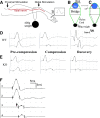
Figure 1.
Figure 1.
Experimental paradigm for nerve compression and mechanically induced CB in mice. A, This diagram shows the placement of stimulation (arrows) and recording (marked by R) electrodes for NCS. B, Compression was delivered by a nylon cord loop attached a 400 g weight. This diagram shows the cross-section at the site of nylon cord. The leg rests on a metal plate (marked as “bridge”) with a width of ∼1 cm. Tibial nerves usually run on the medial side of the ankle (red dot). C, In the second set of experiments, the angle between the nylon cord and the vertical line of leg is reduced by 15 degrees by shortening the length of the bridge (arrow). D, An example of NCS from a compression experiment on a wild-type mouse is shown. On the left, similar amplitudes of CMAP were evoked by stimulations at the distal and proximal sites. After compression was applied, CMAP amplitude from the proximal stimulation was reduced by >60% from the CMAP amplitude by the distal stimulation, called CB (middle). At this point, compression was removed. At day 5 after the compression, CB recovered (right). E, The same experiment was performed on a pmp22+/− mouse. CB did not recover at day 5 following compression (right). Sensitivity = 1 mV; speed = 1 ms. Notice that the onset of CMAP in mice may be obscured by an evolving positive deflection. Thus, peaks of CMAP were often used to calculate the latency and conduction velocity. Nevertheless, this issue does not affect the measurement of CMAP amplitudes or any of our conclusions. F, An original trace recorded at the hypothenar muscle of a patient with HNPP: stimulations on the ulnar nerve inched 1 cm/step across the elbow, a location subject to compression. A focal slowing (a long delay from trace 2 to 3 in contrast to a very short delay from 1 to 2 or from 3 to 4) across the elbow was identified within a 1 cm segment of the nerve, demonstrating the very focal nature of the slowing. CB was conspicuous in this case. The arrow indicates the third response, which had a >50% amplitude drop of motor response and was associated with weakness in muscles innervated by the ulnar nerve.
Figure 2.
Figure 2.
Axonal injuries in compressed PMP22-insufficient nerves. At postcompression days 3 and 5 we obtained semithin sections at transverse 2–3 mm distal to the compression site and examined them under light microscopy. Axons with signs of acute Wallerian degeneration, including extensively collapsed myelin (arrows in A and C), degenerated axons (asterisk in C), and swollen nerve fibers (arrowhead in A), were found in 4 of 11 pmp22+/− mice (n = 11) but not in wild-type mice (n = 12) (B). Two of these four pmp22+/− mice were severe (A). These results suggest that Pmp22 insufficiency may make nerve susceptible to axonal injuries in a small portion of animals.
Figure 3.
Figure 3.
Molecular architecture and septate junctions in naive and compressed nerves. A1–A3, Wild-type mice at 2–3 months old were perfused. Noncompressed sciatic nerves were dissected and teased into individual nerve fibers. Slides were stained with antibodies against MBP (blue) and Caspr (green). The former revealed internodal myelin (blue in A1). Caspr stained paranodes (arrows) that flanked the node of Ranvier. Caspr was also expressed at the Schmidt–Lanterman incisures (arrowhead in A2) and along the inner mesaxon as it spirals around the axon (arrow in A3). Voltage-gated potassium channels (Kv1.2; red in A3) were found in the juxtaparanodes (arrows in A3). Nav were concentrated in the node of Ranvier (arrowhead in A3) and appeared as a narrow band. B–E, From noncompressed pmp22+/− nerves. Nav, Kv1.2, Caspr, and MBP were all localized in their proper regions. In addition, MAG was also correctly localized in the paranodes (D, arrows) and incisures (arrowhead in D) similar to what was observed in the wild-type myelinated nerve fibers. Tomacula were found mainly in the paranodal regions (B, E) and almost always extended beyond the paranodes and into juxtaparanodes and internodes (between arrows in B and E). F, EM was performed on the longitudinal section of a noncompressed pmp22+/− sciatic nerve. Normal paranodal septate junctions (arrowhead array) were observed in the region with tomacula. G, H, Compressed sciatic nerves from the second compression model (surgically exposed and clamped sciatic nerve) (G) and noncompressed sciatic nerves (H) were sectioned into 10 μm thickness and stained with antibodies against Caspr. The localization of Caspr in the paranodes (arrowheads in G and insets and also in supplemental Fig. 1, available at www.jneurosci.org as supplemental material) was normal in compressed nerves. So was that of Kv1.2. However, axons revealed by YFP had smaller diameters and appeared stretched (G). Supporting this notion, we observed the typical undulating “wave” appearance of axons in noncompressed nerve (asterisk array in H). However, these waves disappeared in the compressed nerve (G), suggesting that the compressed nerve was physically stretched during the compression. Notice that the intensity of axonal YFP was much weaker in the compressed nerves. This change has been very helpful for precisely defining the region of compression (please see supplemental Fig. 1, available at www.jneurosci.org as supplemental material, for details). Within the compressed region, there were small islands of axons with strong intensity (arrows in G and in supplemental Fig. 1A, available at www.jneurosci.org as supplemental material), which likely were spared from compression forces. Insets, Caspr localization in paranodes was visualized under high power.
Figure 4.
Figure 4.
Axonal constrictions in tomacula. All data in this figure were derived from noncompressed nerves. pmp22+/− mice were cross-bred with YFPtg+/+ mice, so that all axons were labeled by YFP expressed from the YFP transgene. Sciatic nerves were dissected, fixed, and teased into individual fibers, which were then stained with antibodies against Caspr. A1A3, Three nodes from three myelinated nerve fibers were flanked by the Caspr-stained paranodes. Some Caspr spirals were also visible (arrowheads in A1). In the nodal/paranodal regions, there was a natural decrease of axonal diameter (between arrows in A2). There were no tomacula in these regions under phase-contrast imaging (overlay in A3). B1B3, Another myelinated nerve fiber was examined and showed tomacula in both paranodes that flanked the node of Ranvier (arrowheads in B3). Axonal segments in the tomacula were constricted and the narrowed axonal segments extended far beyond the paranodal regions (between arrows in B3). C1–C3, A pmp22+/− myelinated nerve fiber had an asymmetry in its paranodes. The left paranode was affected by a tomacula with constricted axon (between arrows in C3). In contrast, the right paranode with no tomacula showed a normal axonal diameter. D, In some tomacula, axons may become enlarged (between arrows in D). E, The appearance of the enlarged axon in D is consistent with the EM finding in E. Axonal membranes occasionally became convoluted along the folding of myelin in tomacula (arrowhead array). This membrane would appear enlarged when it is labeled by YFP and viewed under light microscopy. F, A constricted axon on transverse section. G, The lengths of normal paranodal axons and constricted axonal segments were measured in randomly selected fibers (diameters of these selected fibers were comparable between the groups), and showed in this histogram (4.2 ± 0.2 μm for normal paranodal axons vs 19.4 ± 4.9 μm for constricted axonal segments; p < 0.0001).

Similar articles

Cited by

References

    1. Adlkofer K, Martini R, Aguzzi A, Zielasek J, Toyka KV, Suter U. Hypermyelination and demyelinating peripheral neuropathy in Pmp22-deficient mice. Nat Genet. 1995;11:274–280. - PubMed
    1. Adlkofer K, Frei R, Neuberg DH, Zielasek J, Toyka KV, Suter U. Heterozygous peripheral myelin protein 22-deficient mice are affected by a progressive demyelinating tomaculous neuropathy. J Neurosci. 1997;17:4662–4671. - PMC - PubMed
    1. Amici SA, Dunn WA, Jr, Notterpek L. Developmental abnormalities in the nerves of peripheral myelin protein 22-deficient mice. J Neurosci Res. 2007;85:238–249. - PubMed
    1. Bai Y, Ianokova E, Pu Q, Ghandour K, Levinson R, Martin JJ, Ceuterick-de Groote C, Mazanec R, Seeman P, Shy ME, Li J. R69C mutation in P0 gene alters myelination and ion channel subtypes. Arch Neurol. 2006;63:1787–1794. - PubMed
    1. Bishop DL, Misgeld T, Walsh MK, Gan WB, Lichtman JW. Axon branch removal at developing synapses by axosome shedding. Neuron. 2004;44:651–661. - PubMed

Publication types

MeSH terms