. 2010 Jan 13;30(2):714-22.
doi: 10.1523/JNEUROSCI.3852-09.2010.

Sox2 induces neuronal formation in the developing mammalian cochlea

Affiliations

Sox2 induces neuronal formation in the developing mammalian cochlea

Chandrakala Puligilla et al. J Neurosci. .

Abstract

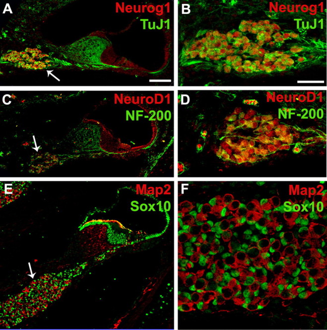
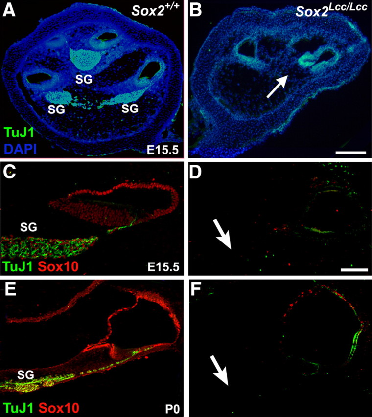
In the cochlea, spiral ganglion neurons play a critical role in hearing as they form the relay between mechanosensory hair cells in the inner ear and cochlear nuclei in the brainstem. The proneural basic helix-loop-helix transcription factors Neurogenin1 (Neurog1) and NeuroD1 have been shown to be essential for the development of otocyst-derived inner ear sensory neurons. Here, we show neural competence of nonsensory epithelial cells in the cochlea, as ectopic expression of either Neurog1 or NeuroD1 results in the formation of neuronal cells. Since the high-mobility-group type transcription factor Sox2, which is also known to play a role in neurogenesis, is expressed in otocyst-derived neural precursor cells and later in the spiral ganglion neurons along with Neurog1 and NeuroD1, we used both gain- and loss-of-function experiments to examine the role of Sox2 in spiral ganglion neuron formation. We demonstrate that overexpression of Sox2 results in the production of neurons, suggesting that Sox2 is sufficient for the induction of neuronal fate in nonsensory epithelial cells. Furthermore, spiral ganglion neurons are absent in cochleae from Sox2(Lcc/Lcc) mice, indicating that Sox2 is also required for neuronal formation in the cochlea. Our results indicate that Sox2, along with Neurog1 and NeuroD1, are sufficient to induce a neuronal fate in nonsensory regions of the cochlea. Finally, we demonstrate that nonsensory cells within the cochlea retain neural competence through at least the early postnatal period.


Figures

Figure 1.
Figure 1.
Neurog1 and NeuroD1 are expressed in spiral ganglion neurons. A, Cross-section through the cochlea at P0 illustrating expression of Neurog1 (red) and ΤuJ1 (green) in the spiral ganglion (arrow). B, High-magnification view of the spiral ganglion labeled as in A. Note that TuJ1 and Neurog1 are coexpressed in neuronal cells. C, Cross-section as in A illustrating expression of NeuroD1 (red) and NF-200 (green) in the spiral ganglion (arrow). D, High-magnification view of the spiral ganglion labeled as in C. NeuroD1 and NF-200 are colocalized in spiral ganglion neurons. E, Cross-section as in A, illustrating expression of Map2 (red) and Sox10 (green). Map2 is expressed in neurons while Sox10 is expressed in nuclei of spiral ganglion glia (arrow) and nonsensory cells in the cochlear duct. F, High-magnification view of the spiral ganglion labeled as in E. Large Map2-positive neurons are surrounded by smaller Sox10-positive glial cells. Scale bars: A (for A, C, E) 50 μm; B (for B, D, F) 20 μm.
Figure 2.
Figure 2.
NeuroD1 and Neurog1 induce neuronal phenotypes in nonsensory cells within the cochlea. A, Low-magnification image of a cochlear explant transfected with NeuroD1.EGFP (green) and labeled with anti-TuJ1 (red). The sensory epithelium (SE, dashed line) and two nonsensory regions, KO and the LER, are indicated. Arrows indicate multiple transfected cells that are positive for TuJ1. B, High-magnification view of three cells located in KO that have been transfected with Neurog1.EGFP (green). Each cell has extended a neurite (arrowheads). C, The same image as in B, except with TuJ1 labeling in red. Note that each cell body and neurite is positive for TuJ1. D, E, Example of a cluster of NeuroD1.EGFP-transfected cells (green) that are TuJ1 positive (red) and have developed neuronal phenotypes including extension of neurites (D, arrowheads) and the formation of a growth cone (arrow and inset in D). F, G, Cells transfected with either Neurog1.EGFP (green in F) or NeuroD1.EGFP (green in G; NeuroD1.EGFP abbreviated as NeuroD1) were also positive for Map2 (red). H, In contrast, cells transfected with Control.EGFP (green) alone were not positive for TuJ1 (red). I, J, In addition, control transfected cells had morphologies that were consistent with epithelial cells including lumenal and basal extensions. K, NeuroD1.EGFP transfected cells (green) were negative for the hair cell marker Myo6 (red) demonstrating that NeuroD1 does not induce a hair cell fate. L, M, In contrast, cells transfected with Atoh1.EGFP (green) were consistently positive for hair cell markers such as Myo7a (red in L) but were negative for TuJ1 (red in M). Scale bars: A, 20 μm; B (for B–G), 10 μm; H, 20 μm; I (for E, J, K–M) 10 μm.
Figure 3.
Figure 3.
Ectopic neurons exhibit electrophysiological characteristics consistent with neurons. Whole-cell recordings from cochlear explant cultures. A, B, Voltage-clamp recording from a Neurog1.EGFP-transfected cell (A) and a non-transfected cell (B). Voltage steps of −70 mV to +10 mV were delivered for duration of 100 or 20 ms (right panel in A). C, D, Current clamp recording from a different Neurog1.EGFP-transfected cell (C) and a non-transfected cell (D) demonstrate the presence of both inward currents and an outward current. Current injections of 50–250 pA were delivered for a duration of 20 ms.
Figure 4.
Figure 4.
SoxB1 gene expression correlates with spiral ganglion development. A–C, Cross-sections through the otocyst from E10.5 mouse embryos demonstrating the expression of Neurog1 (green in A), NeuroD1 (green in B), and Sox2 (green in C) in delaminating neuroblasts of the developing spiral ganglion (arrow in each). Filamentous actin is labeled with Phalloidin (red) in each panel. Note that Neurog1 and Sox2 are also expressed in cells within the otocyst while expression of NeuroD1 is restricted to delaminating neuroblasts. Orientation for sections A–C is indicated to the right of panel C. D–G, Low- and high-magnification cross-sections of the cochlear duct (D, F) or spiral ganglion (E, G). Coimmunolabeling of Sox2 (green in D, E) or Sox1 (green in F, G) with the neuronal marker TuJ1 (red) demonstrates that Sox2 and Sox1 are expressed in spiral ganglion neurons (arrows in D, F) at P0. H, I, Cross-sections as in D,E demonstrating the specificity of TuJ1 as a neuronal marker. There is no overlap between cells labeled with TuJ1 (green) and the glial marker Sox10 (red) in the ganglion. Scale bars: A (for A–C), 50 μm; D (for D, F, H), 50 μm; E (for E, G, I) 20 μm.
Figure 5.
Figure 5.
Sox1 is neither necessary nor sufficient for spiral ganglion neuron formation. A, B, Low-magnification cross-sections through the cochlea of Sox1+/lacZ (control) or Sox1lacZ/lacZ (mutant) mice at P0 double-immunolabeled for ΤuJ1 (green) and Sox10 (red). Spiral ganglion neurons, with processes extending to the sensory epithelium, are present and surrounded by glia in both control and mutant cochleae. Inset, Whole-mount immunolabeling of Sox1+/lacZ (control) (A) or Sox1lacZ/lacZ (mutant) (B) cochlea using anti-NF-200 demonstrates no defects in the pattern of innervation in the absence of Sox1. C, Transfection of nonsensory cells with Sox1.EGFP (green) is not sufficient to induce the expression of TuJ1 (red) or development of a neuronal phenotype. Scale bars: B (for A, B), 50 μm; C, 20 μm.
Figure 6.
Figure 6.
Sox2 is required for formation of the spiral ganglion. A, B, Low-magnification cross-sections through the cochleae of Sox2+/+ (control) and Sox2Lcc/Lcc (mutant) mice at E15.5. Spiral ganglion neurons (SG) are labeled with ΤuJ1 (green) and all cell nuclei are labeled with DAPI (blue). Note the complete absence of spiral ganglion neurons, including cell nuclei (arrow) in the Sox2Lcc/Lcc cochlea. C–F, Double-immunolabeling for TuJ1 (green) and Sox10 (red) on cross-sections of Sox2+/+ (control) and Sox2Lcc/Lcc (mutant) cochlea at E15.5 (C, D) and P0 (E, F) demonstrates the absence of spiral ganglion neurons and glial cells in Sox2 mutants. Arrows in D and F indicate expected position of the spiral ganglion. Scale bars: B (for A, B), 50 μm; D (for C–F), 20 μm.
Figure 7.
Figure 7.
Sox2 induces neuronal phenotypes in cochlear nonsensory cells. A, B, Low- (A) and high-magnification (B) images of Sox2.nucEGFP transfected cells (green) located in KO of cochlear explants labeled with anti-TuJ1 (red in A, B) or anti-Map2 (red in C). A subset of transfected cells are labeled with anti-TuJ1 and have developed a neuronal phenotype. Asterisk in A indicates a Sox2.nucEGFP transfected cell that is TuJ1-positive and has extended a single neurite (enlarged in inset). In contrast with Neurog1.EGFP or NeuroD1.EGFP, the Sox2.nucEGFP construct carries a nuclear localization signal sequence and is therefore restricted to the nucleus. Scale bars: A, 20 μm; B (for B, C), 10 μm.

Similar articles

Cited by

References

    1. Bermingham-McDonogh O, Oesterle EC, Stone JS, Hume CR, Huynh HM, Hayashi T. Expression of Prox1 during mouse cochlear development. J Comp Neurol. 2006;496:172–186. - PMC - PubMed
    1. Bylund M, Andersson E, Novitch BG, Muhr J. Vertebrate neurogenesis is counteracted by Sox1-3 activity. Nat Neurosci. 2003;6:1162–1168. - PubMed
    1. Chu K, Nemoz-Gaillard E, Tsai MJ. BETA2 and pancreatic islet development. Recent Prog Horm Res. 2001;56:23–46. - PubMed
    1. Collignon J, Sockanathan S, Hacker A, Cohen-Tannoudji M, Norris D, Rastan S, Stevanovic M, Goodfellow PN, Lovell-Badge R. A comparison of the properties of Sox-3 with Sry and two related genes, Sox-1 and Sox-2. Development. 1996;122:509–520. - PubMed
    1. Dabdoub A, Puligilla C, Jones JM, Fritzsch B, Cheah KS, Pevny LH, Kelley MW. Sox2 signaling in prosensory domain specification and subsequent hair cell differentiation in the developing cochlea. Proc Natl Acad Sci U S A. 2008;105:18396–18401. - PMC - PubMed

Publication types

MeSH terms

LinkOut - more resources