. 2010 Jan 13;30(2):749-59.
doi: 10.1523/JNEUROSCI.2343-09.2010.

GABAB receptor activation protects neurons from apoptosis via IGF-1 receptor transactivation

Affiliations

GABAB receptor activation protects neurons from apoptosis via IGF-1 receptor transactivation

Haijun Tu et al. J Neurosci. .

Erratum in

Abstract

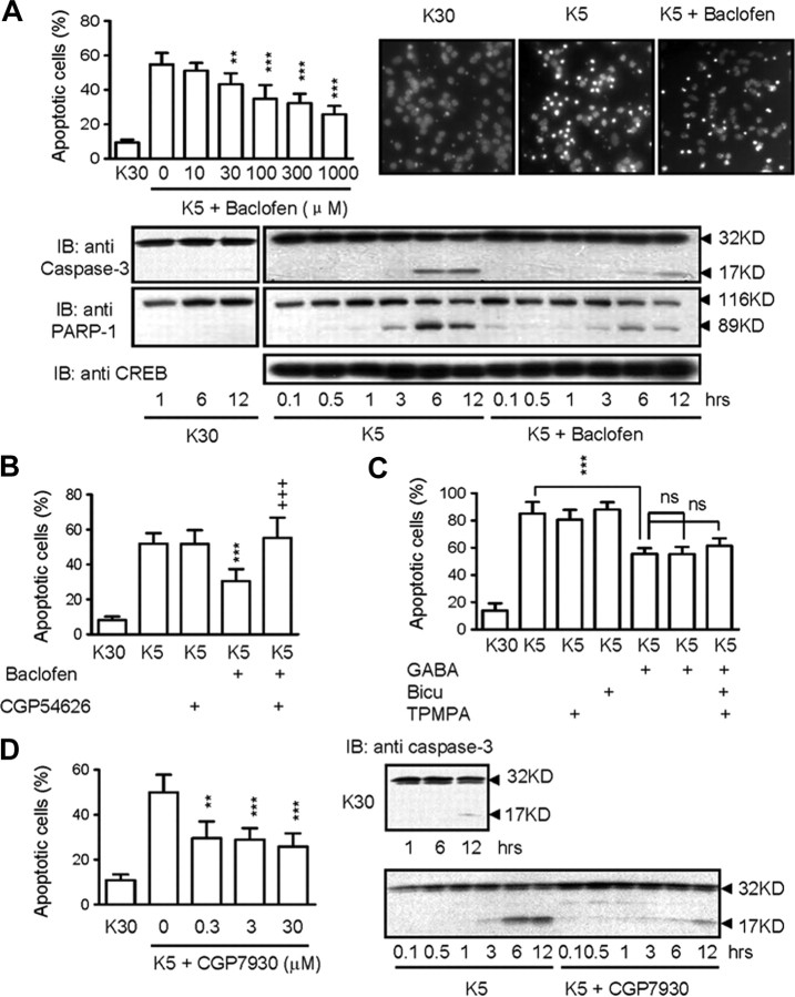
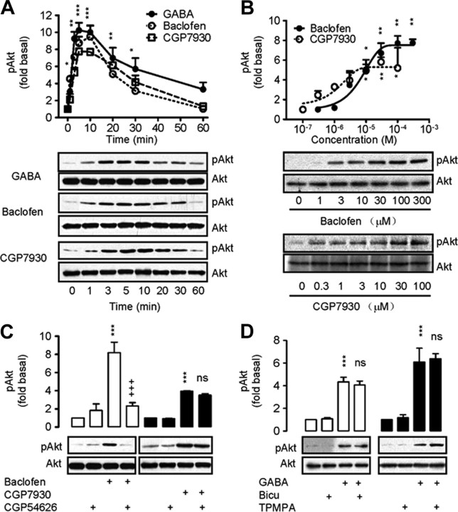
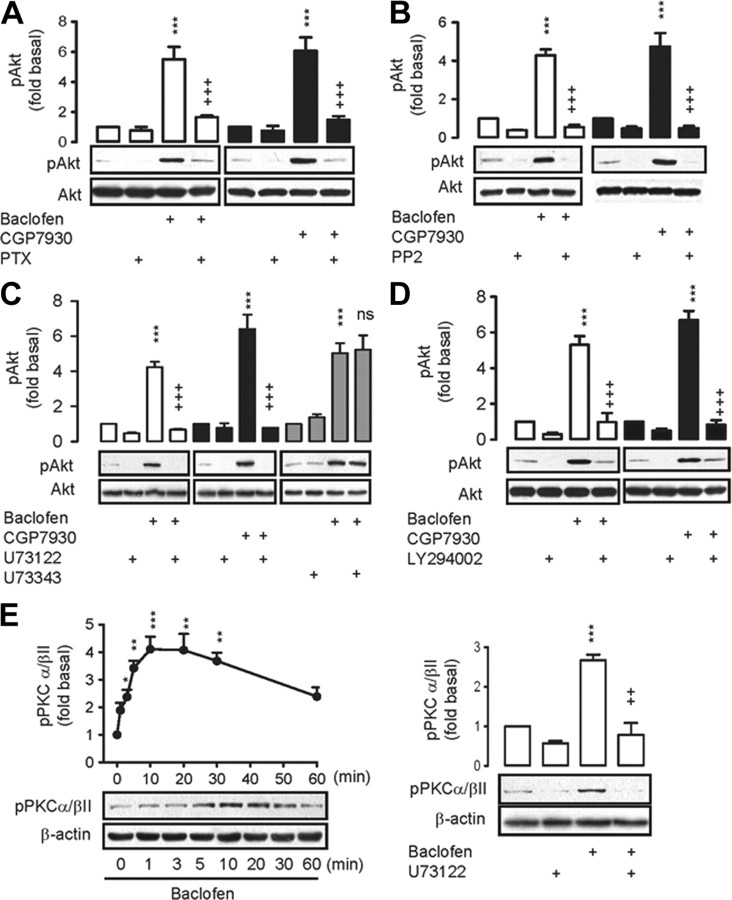
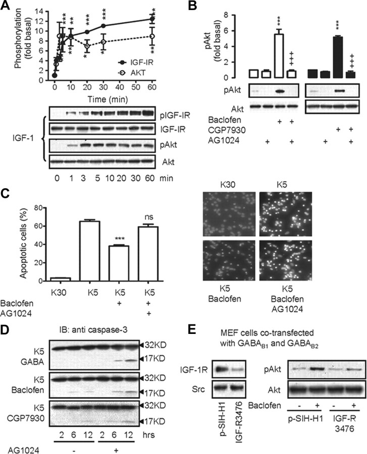
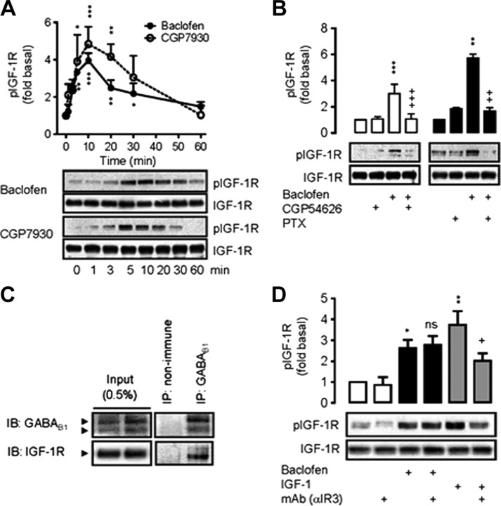
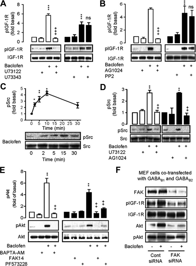
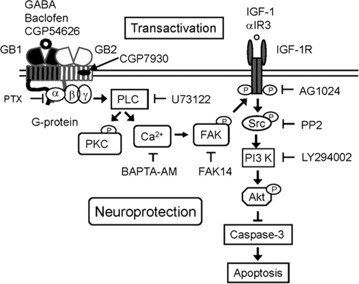
The G-protein-coupled receptors (GPCRs) and receptor tyrosine kinases (RTKs) play key roles in cell-cell communication. Several studies revealed important synergisms between these two types of receptors, with some of the actions of either receptor being mediated through transactivation of the other. Among the large GPCR family, GABA(B) receptor is activated by the neurotransmitter GABA, and is expressed in most neurons where it mediates slow and prolonged inhibition of synaptic transmission. Here we show that this receptor is involved in the regulation of life and death decisions of cerebellar granule neurons (CGNs). We show that specific activation of GABA(B) receptor can protect neurons from apoptosis through a mechanism that involves transactivation of the IGF-1 receptor (IGF-1R). Further work demonstrated that this cross talk was dependent on G(i/o)-protein, PLC, cytosolic Ca(2+), and FAK1 but independent of PKC, while IGF-1R-induced signaling involved Src kinase, PI3 kinase, and Akt activation. These results reveal a new function for this important GPCR and further highlight the importance of functional cross-talk networks between GPCRs and RTKs. Our results reveal GABA(B) receptor as a potential drug target for the treatment of neurodegenerative disorders.


Figures

Figure 1.
Figure 1.
GABAB receptor activation protect neurons from potassium-deprivation-induced apoptosis. A, Dose–response of baclofen on apoptosis when CGNs were transferred from high (30 mm K+, K30) to low (5 mm K+, K5) potassium concentrations for 24 h. Apoptotic cells were measured by TUNEL assay (left) and observed by Hoechst 33258 staining (right). Time course study of the effect of baclofen at 300 μm on caspase-3 activation and PARP activity were shown in the bottom panel and CREB protein expression was used as loading control. B, C, Effect of GABAA [bicuculline (10 μm)] and GABAC [TPMPA (10 μm)] receptor antagonists on apoptotic CGNs after GABA (100 μm) stimulation. For all TUNEL assay or Hoechst 33258 staining, cells were treated with drugs in K5 media for 18–20 h before fixing and staining. For caspase-3 activation, cells were treated with drugs in K5 media for 0.1–12 h. D, Dose–response of the GABAB receptor-positive allosteric modulator CGP7930 on apoptotic CGNs (left) and the effects of CGP7930 at 30 μm on caspase-3 activation (right). **p < 0.01, ***p < 0.001, versus K30 conditions. +++p < 0.001; ns, no significance, versus in the absence of antagonist. The blots shown are representative of three separate experiments.
Figure 2.
Figure 2.
The neuroprotective effect of GABAB receptor is dependent on Gi/o-protein and PI3 kinase but independent of intracellular cAMP levels. A, B, Effect of PTX (200 ng/ml) (A) and LY294002 (20 μm) (B) on the protective effect of baclofen (300 μm) or CGP7930 (50 μm) on apoptotic CGNs (left) and caspase-3 activation (right). For TUNEL assay, cells were treated with drugs in K5 media for 18–20 h. For caspase-3 activation, cells were treated with drugs in K5 media for 1–12 h. ***p < 0.001, versus K30 conditions. +++p < 0.001, versus the absence of PTX or LY294002. The blots shown are representative of three separate experiments. C, GABAB receptor activation does not induce cAMP production in CGNs. Dose–response effect of baclofen or adenylyl cyclase activator forskolin on cAMP production.
Figure 3.
Figure 3.
GABAB receptor activation can increase Akt phosphorylation. A, Time course of Akt activation in CGNs after incubation with GABA (100 μm), baclofen (100 μm), or CGP7930 (50 μm). Representative immunoblots were used to quantify phosphorylated Akt levels (pAkt). *p < 0.05, **p < 0.01, ***p < 0.001, versus basal level for baclofen dose–response. B, Response of baclofen or CGP7930-induced Akt phosphorylation after 10 min incubation. Immunoblots were used to quantify pAkt levels. *p < 0.05, **p < 0.01, versus basal levels. C, D, Cells were treated with CGP54626, bicuculline, or TPMPA at 10 μm for 20 min before the stimulation of baclofen/GABA at 100 μm (10 min). Akt phosphorylation was measured by quantifying pAkt from immunoblots. ***p < 0.001, versus basal levels. +++p < 0.001; ns, nonsignificance, versus treated with baclofen, GABA, or CGP7930 in the absence of antagonist. The blots shown are representative of three separate experiments.
Figure 4.
Figure 4.
Gi/o-protein, Src family kinase, PLC, and PI3 kinase are required for GABAB receptor-mediated Akt phosphorylation. A–D, Cells were pretreated with PTX (200 ng/ml), PP2 (5 μm), U73122 (5 μm), U73343 (5 μm), or LY294002 (10 μm) for 60 min, and Akt phosphorylation was induced by 100 μm baclofen (or 50 μm CGP7930) for 10 min. pAkt levels were quantified by immunoblotting. E, Time course of the PKCα/βII phosphorylation in CGNs after incubation with baclofen at 100 μm. Cells were pretreated with U73122 at 5 μm for 60 min before baclofen stimulation (100 μm). To quantify pAkt or pPKCα/βII, immunoblots from separate experiments (mean ± SEM; n = 3–5) were analyzed, and a representative immunoblot is shown. For all results, *p < 0.05, **p < 0.01, and ***p < 0.001, versus basal levels. ++p < 0.01, +++p < 0.001; ns, nonsignificance, versus treated with baclofen or CGP7930 in the absence of inhibitors.
Figure 5.
Figure 5.
IGF-1R is required for GABAB receptor-mediated neuroprotection. A, Time course of IGF-1 (10 ng/ml)-induced IGF-1R and Akt phosphorylation in CGNs. B, Effect of pretreatment with AG1024 at 0.1 μm for 60 min on Akt phosphorylation, followed by induction by baclofen (100 μm) or CGP7930 (50 μm) for 10 min. To quantify pAkt or pIGF-1R, immunoblots from separate experiments (mean ± SEM; n = 3–5) were analyzed. For all results, *p < 0.05, **p < 0.01, and ***p < 0.001, versus the basal levels, and +++p < 0.001, versus treated with baclofen or CGP7930 in the absence of AG1024. C, Effect of AG1024 (0.1 μm) on apoptotic CGNs after baclofen (100 μm) stimulation. For all TUNEL assay (left) or Hoechst 33258 staining (right), cells were treated with drugs in K5 media for 18–20 h before fixing and staining. ***p < 0.001; ns, no significance, versus K5 conditions. The blots shown are representative of three separate experiments. D, Effect of AG1024 (0.1 μm) on caspase-3 activity in CGNs induced by GABA (100 μm), baclofen (100 μm), or CGP7930 (50 μm). The blots shown are representative of three separate experiments. For caspase-3 activation, cells were treated with drugs in K5 media for 2–12 h. E, Effect of shRNA of IGF-1R (IGF-R3467) on Akt phosphorylation induced by baclofen (100 μm) for 5 min in MEF cells cotransfected GABAB1 and GABAB2. Expression of IGF-R3467 decreased MEF cells endogenous IGF-1R expression but not Src kinase expression (left). The blots shown are representative of three separate experiments.
Figure 6.
Figure 6.
Activation of GABAB receptor transactivated IGF-1R. A, Time course of baclofen- (100 μm) and CGP7930- (50 μm) induced IGF-1R phosphorylation in CGNs. B, Effects of pretreatment with PTX (200 ng/ml) or CGP54626 (10 μm) on IGF-1R phosphorylation induced by baclofen (100 μm) or CGP7930 (50 μm) for 10 min, and representative immunoblots. C, Western blot analysis of IGF-1R coimmunoprecipitated from CGNs with GABAB1 antibody. The membrane was immunoblotted by anti-IGF-1R antibodies. The total lysate (input) was analyzed. D, Effect of the neutralizing antibody anti-IGF-1R (αIR3; 2 μg/ml) on baclofen- (100 μm) or IGF-1 (10 ng/ml) induced IGF-1R phosphorylation for 10 min. For all IGF-IR phosphorylation results, representative immunoblots are shown under the quantified data of pIGF-IR analyzed from separate experiments (mean ± SEM; n = 3–5). For all results, *p < 0.05, **p < 0.01, and ***p < 0.001, versus the basal levels, and +p < 0.05, ++p < 0.01, +++p < 0.001, and ns, nonsignificance, versus treatment with baclofen or CGP7930 in the absence of CGP54626, PTX, IGF-1, or mAb αIR3.
Figure 7.
Figure 7.
PLC downstream effectors FAK1 but not Src kinase is involved in GABAB receptor-mediated IGF-1R transactivation. A, Cells were pretreated with U73122 (5 μm) or U73343 (5 μm) for 60 min and induced by baclofen at 100 μm for 10 min. IGF-1R phosphorylation was measured by immunoblotting. B, PP2 (5 μm) was used to pretreat cells for 60 min or AG1024 (0.1 μm) before baclofen (100 μm) for 10 min. IGF-1R phosphorylation was detected by immunoblotting. C, Time course of baclofen (100 μm)-induced Src kinase phosphorylation in CGNs. D, Effect of pretreatment with U73122 (5 μm) or AG1024 (0.1 μm) on Src kinase phosphorylation induced by baclofen (100 μm) for 10 min. E, Effect of intracellular Ca2+ and FAK1 on baclofen-induced Akt phosphorylation for 10 min. Cells were pretreated with cell membrane-permeable Ca2+ chelator BAPTA-AM (10 μm) for 30 min and FAK1 inhibitors [FAK14 (50 μm) or PF573228 (10 μm)] for 10 min before the CGNs were stimulated with baclofen (100 μm) for 10 min, respectively. Akt phosphorylation was measured by immunoblotting. For all results, representative immunoblots are shown under the quantified data of pIGF-IR, pSrc, or pAkt analyzed from separate experiments (mean ± SEM; n = 3–5). *p < 0.05, **p < 0.01, and ***p < 0.001, versus the basal levels, and ++p < 0.01, +++p < 0.001, ns, no significance, versus treated with baclofen in the absence of inhibitors. F, MEF cells cotransfected with GABAB1 and GABAB2 were pretreated with control RNAi or RNAi of FAK1 for 48 h and then stimulated with baclofen for 5 min, respectively. IGF-1R and Akt phosphorylation were measured by immunoblotting. The blots shown are representative of three separate experiments.
Figure 8.
Figure 8.
Schematic representation of the signaling pathway mediated by GABAB receptor in CGN neuroprotection. Activation of GABAB receptor leads to the release of Gβγ subunits from Gi/o-protein to activate PLC/Ca2+-dependent FAK1 pathway, which in turn transactivates IGF-IR to induce Akt phosphorylation through the Src family kinases.

Similar articles

Cited by

References

    1. Andersson S, D'Arcy P, Larsson O, Sehat B. Focal adhesion kinase (FAK) activates and stabilizes IGF-1 receptor. Biochem Biophys Res Commun. 2009;387:36–41. - PubMed
    1. Belcheva MM, Haas PD, Tan Y, Heaton VM, Coscia CJ. The fibroblast growth factor receptor is at the site of convergence between mu-opioid receptor and growth factor signaling pathways in rat C6 glioma cells. J Pharmacol Exp Ther. 2002;303:909–918. - PubMed
    1. Ben-Ari Y, Gaiarsa JL, Tyzio R, Khazipov R. GABA: a pioneer transmitter that excites immature neurons and generates primitive oscillations. Physiol Rev. 2007;87:1215–1284. - PubMed
    1. Berridge MJ, Lipp P, Bootman MD. The versatility and universality of calcium signaling. Nat Rev Mol Cell Biol. 2000;1:11–21. - PubMed
    1. Bettler B, Kaupmann K, Bowery N. GABAB receptors: drugs meet clones. Curr Opin Neurobiol. 1998;8:345–350. - PubMed

Publication types

MeSH terms

LinkOut - more resources