. 2010 Apr;84(7):3528-41.
doi: 10.1128/JVI.02161-09. Epub 2010 Jan 13.

Human cytomegalovirus infection causes premature and abnormal differentiation of human neural progenitor cells

Affiliations

Human cytomegalovirus infection causes premature and abnormal differentiation of human neural progenitor cells

Min Hua Luo et al. J Virol. 2010 Apr.

Abstract

Congenital human cytomegalovirus (HCMV) infection is a leading cause of birth defects, largely manifested as central nervous system (CNS) disorders. The principal site of manifestations in the mouse model is the fetal brain's neural progenitor cell (NPC)-rich subventricular zone. Our previous human NPC studies found these cells to be fully permissive for HCMV and a useful in vitro model system. In continuing work, we observed that under culture conditions favoring maintenance of multipotency, infection caused NPCs to quickly and abnormally differentiate. This phenotypic change required active viral transcription. Whole-genome expression analysis found rapid downregulation of genes that maintain multipotency and establish NPCs' neural identity. Quantitative PCR, Western blot, and immunofluorescence assays confirmed that the mRNA and protein levels of four hallmark NPC proteins (nestin, doublecortin, sex-determining homeobox 2, and glial fibrillary acidic protein) were decreased by HCMV infection. The decreases required active viral replication and were due, at least in part, to proteasomal degradation. Our results suggest that HCMV infection causes in utero CNS defects by inducing both premature and abnormal differentiation of NPCs.


Figures
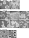
FIG. 1.
FIG. 1.
HCMV infection caused NPC neurospheres to attach and spread on uncoated surfaces, emulating differentiation. Neurospheres were cultured and infected as described in Materials and Methods. (A) Uninfected neurospheres in uncoated dishes. (B) Normal cell migration at 24 h postplating (hpp) on fibronectin-coated dishes. (C) Time course of morphology changes induced by HCMV infection of neurospheres. (D) Infection using UV-irradiated virus did not induce attachment of neurospheres. Magnification for all live-cell images = ×500.
FIG. 2.
FIG. 2.
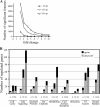
HCMV infection altered mRNA expression levels of multiple cellular genes. Whole-genome expression analyses were performed at 4, 12, and 24 h p.i. as described in Materials and Methods. (A) Numbers of significant genes were plotted versus fold regulation to determine cutoffs for each time point, as described in Materials and Methods. (B) Genes of known function determined to be significantly up- or downregulated at 4, 12, or 24 h p.i. were categorized into functional groupings. The genes in the Neural Related category are included among the eight other functional groups, in addition to being separated for emphasis. TS, transcription factors.
FIG. 3.
FIG. 3.
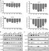
NPC marker proteins were downregulated at the mRNA level and decreased at the protein level by HCMV infection. Monolayer NPCs were infected with HCMV and harvested at the indicated times p.i. for analysis of either the mRNA or protein level. (A) qPCR analysis of NES, DCX, SOX2, and GFAP mRNA levels from 0 to 144 h p.i. Log10 ratios of viral/mock values are given. Calculations were based on absolute starting quantities, using reactions specific for G6PD as normalization controls. The averages of two experiments are shown in the bar charts. Error bars represent the ranges. (B) NPC marker protein steady-state levels at early times p.i. (C) NPC marker protein steady-state levels at late times p.i. Actin was used as a loading control for panels B and C. M, mock infected; V, virus infected.
FIG. 4.
FIG. 4.
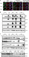
Decreases in NPC markers at the protein level were delayed by GCV treatment. Monolayer NPCs were infected with HCMV, either with or without GCV, as described in Materials and Methods. Cells were analyzed at the indicated times p.i. (A) Cells on poly-d-lysine-coated coverslips were analyzed at the indicated time points, fixed, and stained for IE1 expression and UL44 focus formation and development. Infection without GCV is shown in the left panels, and infection with GCV is shown in the right panels. In the far right panels, brightness levels of UL44 were increased/adjusted (adj.) to show that even after overexposure, no foci were present in GCV-treated cells. At 72 and 96 h p.i., the UL44 exposure time for untreated cells (0.15 s) was half that for GCV-treated (unadjusted) cells (0.3 s) due to increasingly higher levels of UL44 in the untreated cells (as can be observed in panel B). Nuclei were counterstained with Hoechst dye (H). Bar = 5 μm. (B) Viral protein steady-state levels were assessed in the presence and absence of GCV. Viral IE (IE1/IE2), E (UL44), E-L (pp65), and L (gB) antigens were assessed. (C) Steady-state levels of NPC marker proteins were determined for the same time course. Actin was used as a loading control for panels B and C.
FIG. 5.
FIG. 5.
Downregulation of NPC markers at the mRNA level was delayed by GCV treatment. mRNA levels of NPC markers were determined by qPCR at the indicated times p.i. as described in the legend to Fig. 3. Each of the four proteins is represented by three shades of one color. The darkest shade of each color is the untreated viral/mock ratio; the middle shade is the GCV-treated virus/mock ratio; and the lightest shade is the UV-inactivated virus/mock ratio.
FIG. 6.
FIG. 6.
Treatment of cells with MG132 partially blocked NPC marker protein degradation. Monolayer NPCs were mock or virus infected, treated with MG132 (12.5 μM) or left untreated as described in Materials and Methods, and harvested at the indicated times p.i. (A) Viral protein steady-state levels. (B) NPC marker protein steady-state levels. (C) DCX steady-state levels at 48 h p.i. Two exposure times for the same blot are shown. (D) IP using anti-DCX Ab and then Western blotting (WB) using anti-ubiquitin and anti-sumo-1 Abs. (E) Duplicate samples were run in a single gel and transferred to a Protran membrane, and then half the blot was treated with SAP as described in Materials and Methods prior to probing with anti-phospho-DCX Ab. Blots were then probed for actin as loading and staining controls. For panels C, D, and E, MG132 was added to the lysis and IP buffers.

Similar articles

Cited by

References

    1. Aquino, J. B., F. Lallemend, F. Marmigere, I. I. Adameyko, E. A. Golemis, and P. Ernfors. 2009. The retinoic acid inducible Cas-family signaling protein Nedd9 regulates neural crest cell migration by modulating adhesion and actin dynamics. Neuroscience 162:1106-1119. - PMC - PubMed
    1. Bale, J. F., Jr. 1984. Human cytomegalovirus infection and disorders of the nervous system. Arch. Neurol. 41:310-320. - PubMed
    1. Barkovich, A. J., and C. E. Lindan. 1994. Congenital cytomegalovirus infection of the brain: imaging analysis and embryologic considerations. Am. J. Neuroradiol. 15:703-715. - PMC - PubMed
    1. Becroft, D. M. 1981. Prenatal cytomegalovirus infection: epidemiology, pathology and pathogenesis. Perspect. Pediatr. Pathol. 6:203-241. - PubMed
    1. Bergsland, M., M. Werme, M. Malewicz, T. Perlmann, and J. Muhr. 2006. The establishment of neuronal properties is controlled by Sox4 and Sox11. Genes Dev. 20:3475-3486. - PMC - PubMed

Publication types