. 2010 Apr;84(7):3178-88.
doi: 10.1128/JVI.02240-09. Epub 2010 Jan 13.

Viral protein determinants of Lassa virus entry and release from polarized epithelial cells

Affiliations

Viral protein determinants of Lassa virus entry and release from polarized epithelial cells

Katrin Schlie et al. J Virol. 2010 Apr.

Abstract

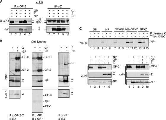
The epithelium plays a key role in the spread of Lassa virus. Transmission from rodents to humans occurs mainly via inhalation or ingestion of droplets, dust, or food contaminated with rodent urine. Here, we investigated Lassa virus infection in cultured epithelial cells and subsequent release of progeny viruses. We show that Lassa virus enters polarized Madin-Darby canine kidney (MDCK) cells mainly via the basolateral route, consistent with the basolateral localization of the cellular Lassa virus receptor alpha-dystroglycan. In contrast, progeny virus was efficiently released from the apical cell surface. Further, we determined the roles of the glycoprotein, matrix protein, and nucleoprotein in directed release of nascent virus. To do this, a virus-like-particle assay was developed in polarized MDCK cells based on the finding that, when expressed individually, both the glycoprotein GP and matrix protein Z form virus-like particles. We show that GP determines the apical release of Lassa virus from epithelial cells, presumably by recruiting the matrix protein Z to the site of virus assembly, which is in turn essential for nucleocapsid incorporation into virions.


Figures

FIG. 1.
FIG. 1.
Entry of Lassa virus into polarized epithelial cells. (A) Polarized MDCK-II cells grown on Transwell filters with a pore size of 3.0 μm were infected with Lassa virus (LASV) at an MOI of 1 via either the apical (black) or basolateral (gray) route. After 48 h, total virus yield from the combined media of the lower and upper chambers was determined by TCID50 (upper panel). Influenza virus and VSV served as controls for apical and basolateral virus infection, respectively, and titers were determined at 8 h p.i. The virus titers of LASV at indicated time points are graphed in the lower panel. Average values were obtained from three independent experiments. The error bars denote standard deviations. ***, P < 0.001; *, P < 0.05 (t test). (B) Localization of the LASV receptor dystroglycan. MDCK-II cells grown to polarity on Transwell filters with a pore size of 0.4 μm were fixed with methanol-acetone and immunostained for expression of the LASV receptor dystroglycan (red) and the tight-junction-specific protein ZO-3 (green) and were subsequently analyzed by confocal microscopy. The optical section in the xy direction was scanned at the height of tight junctions. The yz scan shows distributions of dystroglycan and ZO-3 protein with respect to the apical and basolateral cell surfaces. The scale bar represents 20 μm.
FIG. 2.
FIG. 2.
Directional release of Lassa virus from polarized epithelium. MDCK-II cells grown to polarity on 3.0-μm-pore-size Transwell filters were infected with LASV from the basolateral cell surface at an MOI of 1. Virus released into the apical chambers and into the basolateral chambers were determined by TCID50 titration at 48 h after infection (upper panel). VSV and influenza virus were inoculated from the basolateral or apical side of the cell cultures, respectively, and progeny virus was titrated 8 h p.i., as described in Materials and Methods. Additionally, LASV titers from the cell culture media of the apical or basolateral chamber were measured at the indicated time points and graphed (lower panel). Statistical evaluation was performed as described for Fig. 1.
FIG. 3.
FIG. 3.
Localization of Lassa virus glycoprotein GP in polarized epithelial cells. (A) MDCK-II cells were infected with LASV at an MOI of 1 from the basolateral cell surface. At 72 h p.i. cells were fixed with methanol-acetone and subsequently treated with 4% PFA for 48 h. The LASV glycoprotein subunit GP-1 (red) and the tight-junction resident protein ZO-3 (green) were immunostained. Distributions of GP and ZO-3 were monitored by using confocal laser scanning microscopy in the xy and yz directions. The xy scan is a composite of several images from the apical and middle regions of the image stack because of the irregular height of the cells. The scale bars denote 20 μm. (B) Immunodetection of GP in a stably GP-expressing nonpermeabilized MDCK-II cell line. Cells were fixed with 4% paraformaldehyde and immunostained using an α-GP-2-N antibody (red). The scale bars denote 20 μm. (C) Proteins on the cell surface of stably LASV GP- and VSV G-expressing MDCK-II cells were labeled with sulfo-NHS-biotin on either the apical or basolateral side. After cell lysis, biotinylated proteins were precipitated using streptavidin-coupled Sepharose beads and subsequently subjected to SDS-PAGE and immunoblot analysis using α-GP-2-C or α-VSV G antiserum.
FIG. 4.
FIG. 4.
Release of spike-containing virus-like particles due to solitary expression of Lassa virus glycoprotein GP. (A) Supernatants of stably LASV glycoprotein-expressing MDCK-II cells were collected and pelleted though a 20% sucrose cushion. The pellets were resuspended, fixed with 4% PFA, adsorbed on Formvar-coated copper grids, and negatively stained with 2% phosphotungstate acid before electron microscopic analysis. Scale bars denote 100 nm. (B) Size comparisons of LASV particles and GP-VLPs were performed by electron microscopy using 50 particles each. Particles were measured from the tips of glycoprotein spikes and sorted in size fractions differing by 20 nm. (C) A protease protection assay was performed with three aliquots of a VLP suspension harvested as described for panel A. One aliquot of GP-VLP suspension was left untreated (lane 1), and the second and third aliquots were incubated with proteinase K (lanes 2 and 3). The third aliquot (lane 3) was additionally treated with Triton X-100. The proteins of all three samples were subjected to SDS-PAGE and immunoblotting. LASV glycoprotein was identified by immune detection using the α-GP-2-C antibody.
FIG. 5.
FIG. 5.
Interaction of Lassa virus glycoprotein GP, matrix protein Z, and nucleoprotein NP. (A) VLPs from MDCK-II cells that stably express GP, Z, and NP in various combinations were obtained by pelleting cell culture supernatants through a 20% sucrose cushion. After resuspension and lysis of the VLPs, LASV proteins were coimmunoprecipitated using rabbit α-GP-2-C-specific (lanes 1 to 5) or rabbit α-Z-specific (lanes 6 to 10) antibodies. The precipitated proteins were separated by SDS-PAGE and transferred onto nitrocellulose membranes. LASV GP and Z were identified by immunoblot analyses using a mouse antibody for GP-1 detection and an α-Z rabbit serum. Weak (left panel) and strong (right panel) immunoglobulin (IgG) background bands were also detected. (B) 293 T cells were transfected with different combinations of GP, NP, and Z, as indicated. Cells were lysed, and proteins were immunoprecipitated (IP) using α-GP-2-C, α-NP, or α-Z antibodies. Precipitated proteins (CoIP) as well as cell lysates (Input) were separated by SDS-PAGE. Precipitated proteins were visualized by immunoblotting (IB) using mouse α-GP-1 and α-Z antibodies, whereas immunoblots of cell lysates were stained using rabbit α-GP-2-C, α-NP, and α-Z antibodies. (C) The supernatants of Cos 7 cells which transiently express either GP (lanes 1 to 3), NP (lanes 4 to 6), NP plus GP (lanes 7 to 9), NP plus GP plus Z (lanes 10 to 12), or NP plus Z (lanes 13 to 15) were examined for the presence of nucleoprotein within VLPs by protease protection assay (for the method, see Fig. 4C). Immunoblots of proteinase K-treated and nontreated samples were stained using the α-NP antibody (upper panel). Immunoblots of cell lysates of the various transfected cells (lower panel, lanes 6 to 10) and the corresponding VLPs (lower panel, lanes 1 to 5) are shown. The viral proteins were subjected to SDS-PAGE and subsequently to immunoblotting using α-GP-2-C, α-NP, and/or α-Z antibodies.
FIG. 6.
FIG. 6.
Intracellular colocalization of Lassa virus proteins GP, Z, and NP. Huh 7 cells transiently expressed LASV glycoprotein GPHAtag, NP, and/or Z. Cells were methanol-acetone fixed at 24 h after transfection, and LASV proteins were detected using chicken α-HA tag (green)-, mouse α-m-NP (red)-, and rabbit α-Z (blue)-specific antibodies. The immunofluorescence microscopic analyses were done by sequential scanning using confocal microscopy. Single expression of GP, NP, and Z (A to C); double expression of GP and Z (D to H), NP and Z (I to M), or GP and NP (N to R); and triple expression of GP, NP, and Z (S to W) are shown. Enlarged sections of merged images (H, M, R, and W) that contain fluorescence profile lines are additionally shown. Arrows indicate colocalization.
FIG. 7.
FIG. 7.
Viral determinants for directed virus-like particle release from polarized epithelial cells. MDCK-II cells that stably express GP, Z, and NP in various combinations were grown on Transwell filters (0.4-μm pore size) until polarization was achieved. After cell culture medium replacement with fresh medium containing 2% FCS, cells were further cultivated for 48 h. VLPs from the apical and basolateral chambers were harvested and concentrated by pelleting through a 20% sucrose cushion. The resuspended samples were subjected to SDS-PAGE and immunoblotting using the α-GP-2-C and α-Z antibodies. The viral proteins GP and Z were quantified using the Odyssey infrared quantification system. Average values from four to six independent experiments are expressed as percentage of VLP release. Error bars denote standard deviations. ***, P < 0.001; **, P < 0.01; *, P < 0.05 (t test).

Similar articles

Cited by

References

    1. Agnihothram, S. S., B. Dancho, K. W. Grant, M. L. Grimes, D. S. Lyles, and J. H. Nunberg. 2009. Assembly of arenavirus envelope glycoprotein GPC in detergent-soluble membrane microdomains. J. Virol. 83:9890-9990 - PMC - PubMed
    1. Andrews, B. S., A. N. Theofilopoulos, C. J. Peters, D. J. Loskutoff, W. E. Brandt, and F. J. Dixon. 1978. Replication of dengue and junin viruses in cultured rabbit and human endothelial cells. Infect. Immun. 20:776-781. - PMC - PubMed
    1. Baize, S., J. Kaplon, C. Faure, D. Pannetier, M. C. Georges-Courbot, and V. Deubel. 2004. Lassa virus infection of human dendritic cells and macrophages is productive but fails to activate cells. J. Immunol. 172:2861-2869. - PubMed
    1. Ball, J. M., M. J. Mulligan, and R. W. Compans. 1997. Basolateral sorting of the HIV type 2 and SIV envelope glycoproteins in polarized epithelial cells: role of the cytoplasmic domain. AIDS Res. Hum. Retroviruses 13:665-675. - PubMed
    1. Basak, S., and R. W. Compans. 1989. Polarized entry of canine parvovirus in an epithelial cell line. J. Virol. 63:3164-3167. - PMC - PubMed

Publication types

LinkOut - more resources