. 2010 Apr;84(7):3259-69.
doi: 10.1128/JVI.01715-09. Epub 2010 Jan 13.

Gut mucosal FOXP3+ regulatory CD4+ T cells and Nonregulatory CD4+ T cells are differentially affected by simian immunodeficiency virus infection in rhesus macaques

Affiliations

Gut mucosal FOXP3+ regulatory CD4+ T cells and Nonregulatory CD4+ T cells are differentially affected by simian immunodeficiency virus infection in rhesus macaques

Kristina Allers et al. J Virol. 2010 Apr.

Abstract

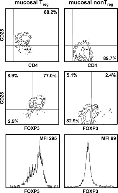
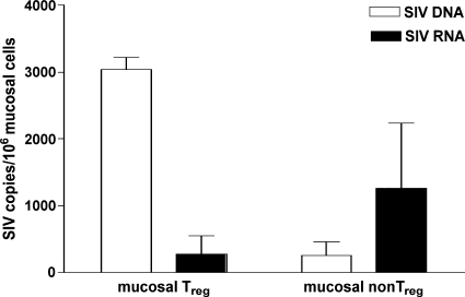
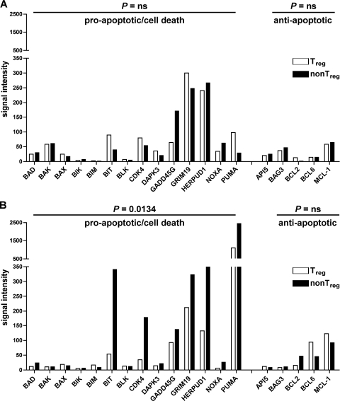
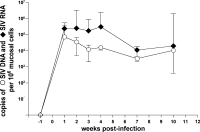
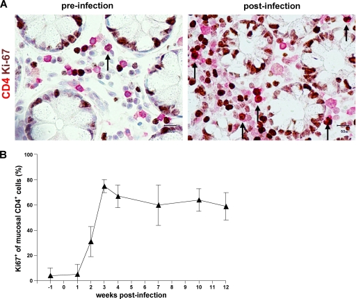
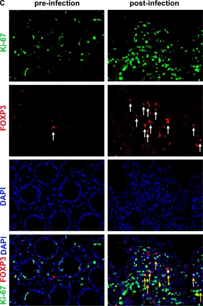
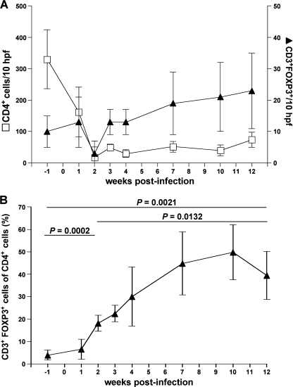
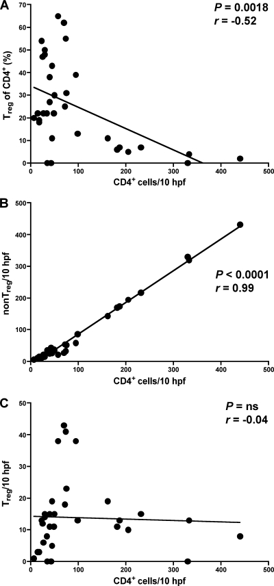
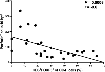
The gastrointestinal tract represents a major site for human and simian immunodeficiency virus (HIV and SIV) replication and CD4(+) T-cell depletion. Despite severe depletion of mucosal CD4(+) T cells, FOXP3(+) regulatory CD4(+) T cells (T(reg)) are highly increased in the gut mucosa of chronically HIV-infected individuals and may contribute to HIV pathogenesis, either by their immunosuppressive function or as a significant target cell population for virus production. Little is known about the susceptibility of mucosal T(reg) to viral infection and the longitudinal effect of HIV/SIV infection on T(reg) dynamics. In this study, we determined the level of SIV infection in T(reg) and nonregulatory CD4(+) T cells (non-T(reg)) isolated from the colon of SIV-infected rhesus macaques. The dynamics of mucosal T(reg) and alterations in the mucosal CD4(+) T-cell pool were examined longitudinally. Our findings indicate that mucosal T(reg) were less susceptible to productive SIV infection than non-T(reg) and thus were selectively spared from SIV-mediated cell death. In addition to improved survival, local expansion of T(reg) by SIV-induced proliferation of the mucosal CD4(+) T-cell pool facilitated the accumulation of mucosal T(reg) during the course of infection. High frequency of mucosal T(reg) in chronic SIV infection was strongly related to a reduction of perforin-expressing cells. In conclusion, this study suggests that mucosal T(reg) are less affected by productive SIV infection than non-T(reg) and therefore spared from depletion. Although SIV production is limited in mucosal T(reg), T(reg) accumulation may indirectly contribute to viral persistence by suppressing antiviral immune responses.


Figures

FIG. 1.
FIG. 1.
Isolation of mucosal Treg and non-Treg subsets. Treg and non-Treg were isolated from colonic lamina propria lymphocytes of chronically SIV-infected rhesus macaques and analyzed by flow cytometry. Cells were gated for lymphocytes by their characteristic forward-sideward-scatter profile and were analyzed for expression of CD4, CD25, and FOXP3. Mean fluorescence intensity (MFI) is given for FOXP3 in the lymphocyte gate.
FIG. 2.
FIG. 2.
SIV RNA production is less efficient in mucosal Treg than in non-Treg. Cell-associated SIV DNA and SIV RNA in mucosal Treg and non-Treg isolated from chronically SIV-infected animals were quantified and are given as number of SIVgag DNA or SIVgag RNA copies per 106 mucosal cells. Data are given in medians, with error bars representing standard deviations.
FIG. 3.
FIG. 3.
In chronic SIV infection, expression levels of cell death-related genes are higher in mucosal non-Treg than in Treg. Microarray analysis of apoptosis- and cell death-related genes in mucosal Treg and non-Treg isolated in parallel from a chronically SIV infected (A) and a healthy SIV-uninfected (B) rhesus macaque. Normalized signal intensities for single genes were analyzed to identify mRNA expression levels of different genes. Genes whose normalized signal intensity values were less than 5 for both cell subsets were excluded from the analysis. Statistical analysis was performed using the Wilcoxon matched-pairs test. ns, not significant.
FIG. 4.
FIG. 4.
Mucosal viral load in the colon of SIV-infected rhesus macaques. Five animals were infected with SIVmac251. SIV DNA and RNA were quantified longitudinally in colonic tissue during the course of SIV infection and are given as number of SIVgag DNA or SIVgag RNA copies per 106 mucosal cells. Data are given in medians, with error bars representing standard deviations.
FIG. 5.
FIG. 5.
Proliferative response in mucosal CD4+ T cells after SIV infection includes Treg. (A) Representative immunohistochemical staining of proliferating CD4+ T cells (arrows) in colonic mucosa identified by double immunoenzymatic labeling for CD4 (red, membranous) and Ki67 (brown, nuclear). Original magnification, ×400. (B) Longitudinal examination of proliferation of colon mucosal CD4+ T cells in five rhesus macaques during the course of SIV infection. Frequency of mucosal CD4+ Ki67+ cells in relation to mucosal CD4+ T cells. Data are given in medians, with error bars representing standard deviations. (C) Representative immunofluorescence staining of proliferating mucosal Treg identified by double staining for FOXP3 (red, white arrows) and Ki67 (green). FOXP3- and Ki67-coexpressing mucosal cells are indicated by yellow arrows. Original magnification, ×400.
FIG. 5.
FIG. 5.
Proliferative response in mucosal CD4+ T cells after SIV infection includes Treg. (A) Representative immunohistochemical staining of proliferating CD4+ T cells (arrows) in colonic mucosa identified by double immunoenzymatic labeling for CD4 (red, membranous) and Ki67 (brown, nuclear). Original magnification, ×400. (B) Longitudinal examination of proliferation of colon mucosal CD4+ T cells in five rhesus macaques during the course of SIV infection. Frequency of mucosal CD4+ Ki67+ cells in relation to mucosal CD4+ T cells. Data are given in medians, with error bars representing standard deviations. (C) Representative immunofluorescence staining of proliferating mucosal Treg identified by double staining for FOXP3 (red, white arrows) and Ki67 (green). FOXP3- and Ki67-coexpressing mucosal cells are indicated by yellow arrows. Original magnification, ×400.
FIG. 6.
FIG. 6.
Mucosal Treg increase during the course of SIV infection in colon despite mucosal CD4+ T-cell depletion. Longitudinal examination of mucosal Treg in five animals following SIV infection. (A) Absolute numbers of mucosal CD4+ T cells and mucosal FOXP3+ Treg per 10 hpf in five rhesus macaques before and after SIV infection. (B) Frequency of mucosal Treg in relation to mucosal CD4+ T cells (relative frequency of mucosal Treg). Data are given in medians, with error bars representing standard deviations, and were analyzed by using the two-tailed Student t test.
FIG. 7.
FIG. 7.
Depletion of mucosal CD4+ T cells correlates with loss of mucosal non-Treg but not of Treg. Correlation between the number of mucosal CD4+ T cells and the relative frequency of mucosal Treg (A), the number of mucosal non-Treg (B), and the number of mucosal Treg (C), respectively. CD4+ T cells and Treg were quantified in colonic tissue, and the absolute number of non-Treg was calculated by subtracting the number of Treg from the number of CD4+ T cells. Pearson's test was used to determine correlation. ns, not significant.
FIG. 8.
FIG. 8.
Increase of Treg in colonic mucosa correlates with a local reduction of perforin-expressing cells. Correlation between the relative frequency of mucosal Treg and the number of perforin-expressing cells in SIV infection. Perforin+ cells were quantified in colonic tissue of SIV-infected rhesus macaques and compared with the corresponding relative frequencies of mucosal Treg (see Fig. 7A). Pearson's test was used to determine the correlation.

Similar articles

Cited by

References

    1. Aandahl, E. M., J. Michaelsson, W. J. Moretto, F. M. Hecht, and D. F. Nixon. 2004. Human CD4+ CD25+ regulatory T cells control T-cell responses to human immunodeficiency virus and cytomegalovirus antigens. J. Virol. 78:2454-2459. - PMC - PubMed
    1. Allers, K., D. Kunkel, V. Moos, M. Eisenblatter, C. Stahl-Hennig, F. J. Kaup, R. Ignatius, and T. Schneider. 2008. Migration patterns of nonspecifically activated versus nonactivated nonhuman primate T lymphocytes: preferential homing of activated autologous CD8+ T cells in the rectal mucosa. J. Immunother. 31:334-344. - PubMed
    1. Andersen, J. L., E. S. Zimmerman, J. L. DeHart, S. Murala, O. Ardon, J. Blackett, J. Chen, and V. Planelles. 2005. ATR and GADD45alpha mediate HIV-1 Vpr-induced apoptosis. Cell Death Differ. 12:326-334. - PubMed
    1. Andersson, J., H. Behbahani, J. Lieberman, E. Connick, A. Landay, B. Patterson, A. Sonnerborg, K. Lore, S. Uccini, and T. E. Fehniger. 1999. Perforin is not co-expressed with granzyme A within cytotoxic granules in CD8 T lymphocytes present in lymphoid tissue during chronic HIV infection. AIDS 13:1295-1303. - PubMed
    1. Andersson, J., S. Kinloch, A. Sonnerborg, J. Nilsson, T. E. Fehniger, A. L. Spetz, H. Behbahani, L. E. Goh, H. McDade, B. Gazzard, H. Stellbrink, D. Cooper, and L. Perrin. 2002. Low levels of perforin expression in CD8+ T lymphocyte granules in lymphoid tissue during acute human immunodeficiency virus type 1 infection. J. Infect. Dis. 185:1355-1358. - PubMed

Publication types

MeSH terms