. 2010 Mar;84(6):2774-86.
doi: 10.1128/JVI.01948-09. Epub 2010 Jan 13.

Predominant clonal accumulation of CD8+ T cells with moderate avidity in the central nervous systems of Theiler's virus-infected C57BL/6 mice

Affiliations

Predominant clonal accumulation of CD8+ T cells with moderate avidity in the central nervous systems of Theiler's virus-infected C57BL/6 mice

Hyun Seok Kang et al. J Virol. 2010 Mar.

Abstract

Induction of antigen-specific CD8(+) T cells bearing a high-avidity T-cell receptor (TCR) is thought to be an important factor in antiviral and antitumor immune responses. However, the relationship between TCR diversity and functional avidity of epitope-specific CD8(+) T cells accumulating in the central nervous system (CNS) during viral infection is unknown. Hence, analysis of T-cell diversity at the clonal level is important to understand the fate and function of virus-specific CD8(+) T cells. In this study, we examined the Vbeta diversity and avidity of CD8(+) T cells specific to the predominant epitope (VP2(121-130)) of Theiler's murine encephalomyelitis virus. We found that Vbeta6(+) CD8(+) T cells, associated with epitope specificity, predominantly expanded in the CNS during viral infection. Further investigations of antigen-specific Vbeta6(+) CD8(+) T cells by CDR3 spectratyping and sequencing indicated that distinct T-cell clonotypes are preferentially increased in the CNS compared to the periphery. Among the epitope-specific Vbeta6(+) CD8(+) T cells, MGX-Jbeta1.1 motif-bearing cells, which could be found at a high precursor frequency in naïve mice, were expanded in the CNS and tightly associated with gamma interferon production. These T cells displayed moderate avidity for the cognate epitope rather than the high avidity normally observed in memory/effector T cells. Therefore, our findings provide new insights into the CD8(+) T-cell repertoire during immune responses to viral infection in the CNS.


Figures
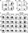
FIG. 1.
FIG. 1.
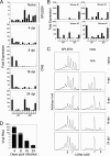
Assessment of Vβ usage by antigen-specific CNS-infiltrating and peripheral T cells. Detection of Db-VP2121-130 versus Db-LCMV-GP33 (negative control) tetramer-positive CD8+ T cells (A) and IFN-γ-producing CD8+ T cells (B) after stimulation with PBS or VP2121-130 peptide in the CNS and spleen. (C) Proportion of CD8+ T cells bearing different Vβs and reactive to VP2 tetramer in the CNS and spleen. Plots represent gated CD8+ T cells. Antibodies to Vβs were selected based on RT-PCR analyses. Three mice were individually analyzed. Data are representative of three independent experiments. All experiments were done at 8 days post TMEV infection.
FIG. 2.
FIG. 2.
Disparity of virus-specific CD8+ T-cell repertoires between the CNS and the periphery. (A) Vβ6-CDR3-Jβ size spectra of isolated tetramer-positive cells from 12 mice. To assess the homogeneity of the identically sized CDR3s, bands (a and b), indicated with arrows, were eluted, reamplified by PCR, and cloned for sequencing. (B) Vβ6-CDR3-Jβ size spectra of isolated IFN-γ producers. Bands (c and d), indicated with arrows, were processed as described for panel A. The spectratypes shown are representative results of two separate experiments. Amino acid sequences were deduced from nucleotide sequences. C, CNS; S, spleen; Freq., frequency. TEVF sequences in the CDR3β loop are part of the Jβ1.1 chain. The values to the left are CDR3 sizes in amino acid numbers.
FIG. 3.
FIG. 3.
Expansion of virus-specific CD8+ T cells with a selective TCR repertoire. (A) Increase in Vβ6-Jβ1.1 mRNAs during the course of viral infection. cDNAs were prepared from splenocytes from naïve mice and CNS mononuclear cells from infected mice (three mice per group) at 4, 8, 16, and 24 dpi. Relative Jβ chain usage was quantitatively measured from primary Vβ6 PCR products using real-time PCR. (B) The dominant use of Jβs other than Jβ1.1 by VP2121-130-specific Vβ6+ cells varies among individual mice. CNS-infiltrating cells were obtained at 8 dpi from four individual mice, and the primary Vβ6-Cβ PCR product was similarly used to measure relative levels of Jβ chains by real-time PCR. (C) Vβ6-Jβ1.1 CDR3 size distribution in the CNS and spleen during the course of viral infection. Spectratyping was carried out using a 32P-labeled Vβ6 primer as described in Materials and Methods. (D) Viral loads during the immune responses. Relative viral loads were determined by real-time PCR using cDNAs prepared from CNS cells at 4, 8, 16, and 24 dpi. N/A, not applicable.
FIG. 4.
FIG. 4.
Tracing clonotypic expansion of the specific CDR3 repertoire in the CNS during viral infection (three mice per group). (A) Diagrammatic presentation of tracing strategy of CDR3 clonotypes. (B) Expression of clonotype-specific CDR3s during viral infection. The relative abundance of CDR3 sequences was measured by real-time PCR using degenerate primers for the MGN, MGF, MGE, RV, and ITPT CDR3 amino acid sequences. The CDR3 clonotypes of naïve mice represent splenic T cells from uninfected mice because there is no significant T-cell infiltration in the CNSs of uninfected mice. Jβ restriction for the CDR3 sequences was further assessed by real-time PCR using the indicated Jβ primers. Arrows indicate sources of the PCR templates. Single and double asterisks indicate P < 0.01 and P < 0.001, respectively, compared with other CDR3 sequences and/or Jβ chains. Additional experimental results obtained by using mice infected with TMEV (three mice per group) at 8 and 16 dpi indicate that the predominant use of Vβ6-MGX-Jβ1.1 is consistent between experiments. In contrast, the use of other CDR3 sequences was somewhat variable. qPCR, quantitative PCR.
FIG. 5.
FIG. 5.
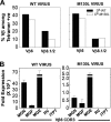
Accumulation of CD8+ T cells with moderate avidity in the CNS during viral infection. (A) Functional avidity of CNS-infiltrating lymphocytes (CNS-ILs) was measured by intracellular IFN-γ staining (ICS); CNS-ILs obtained at 8 and 16 dpi were stimulated by 10-fold serial dilutions of WT VP2121-130 or mutant M130L peptides starting at a 1 μM concentration. Functional avidity was based on the concentration of peptides required to attain 50% of the maximal IFN-γ production by CD8+ T cells. The maximum percentage of IFN-γ-producing cells is set to 100%. Double asterisks represent P < 0.001. (B) Proportions (top) and numbers (bottom) of tetramer-positive (left) and IFN-γ-producing (right) cells against WT and M130L peptides during TMEV infection. Panels A and B are representative of three individual experiments. Pooled CNS cells from 15 mice were used for these analyses. (C) Comparison of WT and M130L tetramer-binding intensities of Vβ6- and Vβ8.1/2-bearing CD8+ T cells. Gated CD8+ and tetramer-positive cells from the CNSs of mice at 8 dpi were analyzed for mean fluorescence intensity (MFI). (D) Assessment of Vβs utilized by WT (VP2121-130)- and M130L-reactive CD8+ T cells by RT-PCR. cDNAs were prepared from CNS cells with each tetramer at 8 and 16 dpi. (E) Sequence analyses of 7-aa CDR3 regions within Vβ6-Jβ1.1. Vβ6-Jβ1.1 PCR products from cells isolated using WT and M130L tetramers at 16 dpi were eluted from a spectratyping gel, reamplified by PCR, cloned, and then sequenced.
FIG. 6.
FIG. 6.
Efficient expansion of Vβ6-MGX bearing moderate-avidity cells in response to WT virus compared with APL-bearing viruses. (A) Percentages of Vβ6- and Vβ8.1/2-bearing cells among WT and M130L tetramer-reactive CD8+ T cells. CNS-CD8+ T cells from three mice infected with WT or M130L virus were stained with WT (VP2121-130) or M130L peptide-loaded tetramers in combinations with anti-CD8 and anti-Vβ6 or -Vβ8.1/2 antibodies. Tetramer- and CD8-positive gates were used to calculate the percentage of Vβ-bearing cells. (B) Measurements of Vβ6-CDR3 mRNAs in the CNS after infection with WT and epitope variant viruses using real-time PCR. After normalization, based on their Cβ mRNA levels, the lowest value was set as onefold expression.
FIG. 7.
FIG. 7.
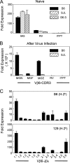
CD8+ T-cell expansion level correlates with naïve T-cell frequency and MHC haplotype. (A) Relative frequency of Vβ6-MGX CDR3 expressed by splenic T cells from naïve mice. The frequency of CDR3 regions was determined by real-time PCR using degenerate primers. (B) Expansion levels of Vβ6-MGX CDR3 in the CNSs of B6 (H-2b) and SJL (H-2s) mice (three mice per group) at 8 days post TMEV infection. (C) Vβ6-Jβ usage by T cells in the CNSs of B6 and 129 (H-2b) mice (three mice per group) at 8 dpi.

Similar articles

Cited by

References

    1. Alexander-Miller, M. A. 2005. High-avidity CD8+ T cells: optimal soldiers in the war against viruses and tumors. Immunol. Res. 31:13-24. - PubMed
    1. Alexander-Miller, M. A., G. R. Leggatt, and J. A. Berzofsky. 1996. Selective expansion of high- or low-avidity cytotoxic T lymphocytes and efficacy for adoptive immunotherapy. Proc. Natl. Acad. Sci. U. S. A. 93:4102-4107. - PMC - PubMed
    1. Altman, J. D., P. A. Moss, P. J. Goulder, D. H. Barouch, M. G. McHeyzer-Williams, J. I. Bell, A. J. McMichael, and M. M. Davis. 1996. Phenotypic analysis of antigen-specific T lymphocytes. Science 274:94-96. - PubMed
    1. Babbe, H., A. Roers, A. Waisman, H. Lassmann, N. Goebels, R. Hohlfeld, M. Friese, R. Schroder, M. Deckert, S. Schmidt, R. Ravid, and K. Rajewsky. 2000. Clonal expansions of CD8(+) T cells dominate the T cell infiltrate in active multiple sclerosis lesions as shown by micromanipulation and single cell polymerase chain reaction. J. Exp. Med. 192:393-404. - PMC - PubMed
    1. Blackman, M. A., H. Gerhard-Burgert, D. L. Woodland, E. Palmer, J. W. Kappler, and P. Marrack. 1990. A role for clonal inactivation in T cell tolerance to Mls-1a. Nature 345:540-542. - PubMed

Publication types

MeSH terms

Substances