. 2010 Mar;84(6):3043-58.
doi: 10.1128/JVI.01655-09. Epub 2010 Jan 13.

Preexisting infection with human T-cell lymphotropic virus type 2 neither exacerbates nor attenuates simian immunodeficiency virus SIVmac251 infection in macaques

Affiliations

Preexisting infection with human T-cell lymphotropic virus type 2 neither exacerbates nor attenuates simian immunodeficiency virus SIVmac251 infection in macaques

Shari N Gordon et al. J Virol. 2010 Mar.

Abstract

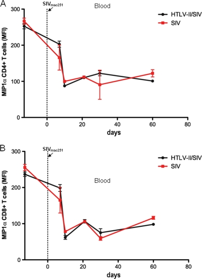
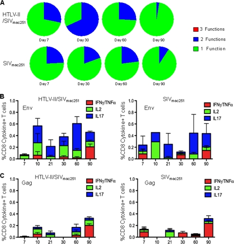
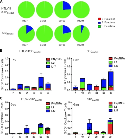
Coinfection with human T-cell lymphotropic virus type 2 (HTLV-2) and human immunodeficiency virus type 1 (HIV-1) has been reported to have either a slowed disease course or to have no effect on progression to AIDS. In this study, we generated a coinfection animal model and investigated whether HTLV-2 could persistently infect macaques, induce a T-cell response, and impact simian immunodeficiency virus SIV(mac251)-induced disease. We found that inoculation of irradiated HTLV-2-infected T cells into Indian rhesus macaques elicited humoral and T-cell responses to HTLV-2 antigens at both systemic and mucosal sites. Low levels of HTLV-2 provirus DNA were detected in the blood, lymphoid tissues, and gastrointestinal tracts of infected animals. Exposure of HTLV-2-infected or naïve macaques to SIV(mac251) demonstrated comparable levels of SIV(mac251) viral replication, similar rates of mucosal and peripheral CD4(+) T-cell loss, and increased T-cell proliferation. Additionally, neither the magnitude nor the functional capacity of the SIV-specific T-cell-mediated immune response was different in HTLV-2/SIV(mac251) coinfected animals versus SIV(mac251) singly infected controls. Thus, HTLV-2 targets mucosal sites, persists, and importantly does not exacerbate SIV(mac251) infection. These data provide the impetus for the development of an attenuated HTLV-2-based vectored vaccine for HIV-1; this approach could elicit persistent mucosal immunity that may prevent HIV-1/SIV(mac251) infection.


Figures

FIG. 1.
FIG. 1.
HTLV-2 establishes an infection in rhesus macaques, replicating in lymphoid and mucosal tissues. (A) Intracellular HTLV-2 Gag p19 expression in the Mo-T and RhM304 cell lines measured by flow cytometry. The isotype control (ctrl) is shown in blue, and p19 Gag expression is shown in black. (B) Number of HTLV-2 genome copies per 106 cells in Mo-T and RhM304 cells. (C) Serum antibodies to p24 Gag measured by ELISA 1 to 3 months after HTLV-2 infection in animals L900, M214, and M304. OD, optical density. (D) HTLV-2 proviral DNA load in the spleen, ileum, and jejunum from animals M214 and M304. (E) Tax protein expression in plasmacytoid dendritic cells infected in vitro with cell-free HTLV-2 (black) or uninfected plasmacytoid dendritic cells (blue).
FIG. 2.
FIG. 2.
HTLV-2 infection stimulates humoral and cell-mediated responses. (A) Serum antibodies specific to several HTLV-2 proteins measured by Western blotting in animals M893, M897, M905, and M906. The months postinfection (p.i.) are shown below the blot. (B) Nested semiquantitative PCR to amplify either HTLV-2 tax or β-actin. DNA was extracted from the lymph nodes of macaques 3 months after HTLV-2 infection, and the mononuclear cells were CD8 enriched (+) or CD8 depleted (−). (C) CD4+ and CD8+ T-cell counts in blood from HTLV-2-infected macaques. Repeated-measure analysis of variance with square root-transformed counts over time demonstrated that CD8+ T cells significantly increase over time with a P value of 0.0017, while the CD4+ cells show a trend toward significance with a P value of 0.049. (D and E) Representative flow cytometric pseudocolor plots showing the frequency of IFN-γ and/or TNF-α production by CD8+ T cells in blood (D) or the gastrointestinal (GI) tract (E) after stimulation with HTLV-2 Tax and Gag overlapping peptides or in unstimulated cells. (F and G) Mean HTLV-2-specific CD4+ and CD8+ T-cell responses, after background subtraction, 10 weeks after HTLV-2 infection in blood (F) and the gastrointestinal tract (G). The frequency of Gag- or Tax-specific cells was measured by intracellular cytokine staining for IFN-γ/TNF-α or IL-2.
FIG. 3.
FIG. 3.
Persistent HTLV-2 infection despite SIV coinfection. (A) Study design. Four macaques were infected with HTLV-2. Ten months postinfection, these 4 macaques along with 4 naïve animals were challenged with SIVmac251. Blood, lymphoid, and mucosal tissues were sampled before infection and for up to 90 days after SIVmac251 infection. (B) Semiquantitative nested PCR for HTLV-2 tax DNA or β-actin amplified from the peripheral blood of HTLV-2-infected animals (M893, M897, M905, and M906) and uninfected control (P182) before and after SIVmac251 infection. The number of weeks after HTLV-2 infection is shown below the blots. (C) Serum antibodies to HTLV-2 measured by Western blotting before and after SIVmac251 infection.
FIG. 4.
FIG. 4.
Similar viral and cellular dynamics in HTLV-2/SIVmac251 coinfected and SIVmac251 monoinfected macaques. (A) SIVmac251 viral load in HTLV-2 coinfected macaques (shown in black) and controls (shown in red) measured by the number of SIVmac251 RNA copies/ml of plasma. (B) Average CD4+ T-cell count in blood following SIVmac251 infection in HTLV-2/SIVmac251-infected macaques (black) and SIVmac251-infected macaques (red). (C) Percentage of baseline CD4+ CCR5+ T-cell count following SIVmac251 infection in HTLV-2/SIVmac251-infected macaques (shown in black) and SIVmac251-infected animals (shown in red). (D) Percentage of baseline CD4+ memory T-cell count in blood following SIVmac251 infection. Memory cells are defined as cells that express both CD28 and CD95. (E) Average CD8+ T-cell count following SIVmac251 infection. (F) Percentage of baseline CD8+ effector/effector memory T-cell count in blood after SIVmac251 infection. CD8+ effector/effector memory T cells are CD8+ CD28 CD95+.
FIG. 5.
FIG. 5.
HTLV-2 preinfection did not significantly affect either the viral load or the rate of CD4+ T-cell loss after SIVmac251 infection. (A) Viral load in the lymph nodes, jejunum, and rectum in HTLV-2/SIVmac251 coinfected macaques (black) and SIVmac251 monoinfected macaques (white) quantified as the number of SIV DNA copies/106 cells 10 and 30 days after SIVmac251 infection. (B) The frequency of CD3+ CD4+ T cells in the bone marrow and lymph nodes of HTLV-2/SIVmac251 coinfected and SIVmac251 singly infected animals before infection (baseline) and 10 and 30 days after SIVmac251 infection. (C) The frequency of CD3+ CD4+ T cells in the jejunum and rectum in HTLV-2/SIVmac251 coinfected and SIVmac251 singly infected animals before infection (baseline) and 10 and 30 days after SIVmac251 infection. (D) Number of CD4+ expressing cells, enumerated by immunohistochemical staining of paraformaldehyde-fixed tissue sections from the rectum. A repeated-measure analysis of variance demonstrated that the difference between the baseline (before infection) values for HTLV-2/SIVmac251 coinfected macaques and SIVmac251 singly infected macaques is significantly different (P < 0.05). (E) Number of CD8+ expressing cells, enumerated by immunohistochemical staining of paraformaldehyde-fixed rectum tissue sections.
FIG. 6.
FIG. 6.
No difference in spontaneous MIPα expression by T cells in blood from HTLV-2/SIVmac251 coinfected and SIVmac251 monoinfected animals. (A) Mean fluorescence intensity (MFI) of MIP1α in CD3+ CD4+ T cells in blood from HTLV-2/SIVmac251 coinfected animals (black) and SIVmac251 singly infected animals (red) before or after SIVmac251 infection. (B) Mean fluorescence intensity of MIP1α in CD3+ CD8+ T cells in blood from HTLV-2/SIVmac251 coinfected animals and SIVmac251 singly infected animals SIVmac251 before or after SIVmac251 infection.
FIG. 7.
FIG. 7.
Proliferation of T cells in blood and tissues from HTLV-2/SIVmac251 coinfected animals and SIVmac251 monoinfected animals. (A and B) Frequency of CD3+ CD4+ T cells (A) and CD3+ CD8+ T cells (B) expressing Ki67 in blood from HTLV-2/SIVmac251 coinfected animals (black) and SIVmac251 singly infected animals (red) after SIVmac251 infection. A Wilcoxon signed-rank test demonstrated that the level of CD8+ Ki67 expression continued to be significantly greater in the chronic phase compared to preinfection levels (B). (C and D) Frequency of CD3+ CD4+ T cells (C) and CD3+ CD8+ T cells (D) expressing Ki67 in the lymphoid tissues (bone marrow and lymph nodes) of HTLV-2/SIVmac251 coinfected animals and SIVmac251 singly infected animals before infection (baseline) and 10 and 30 days after SIVmac251 infection. (E) Number of cells expressing both CD4 and Ki67 in the rectum in HTLV-2/SIVmac251 coinfected macaques and SIVmac251 singly infected macaques before infection (baseline) and 10 and 30 days after SIVmac251 infection. Repeated-measure analysis of variance demonstrated that the difference between HTLV-2/SIVmac251 coinfected macaques and SIVmac251 singly infected macaques at baseline is significantly different (P = 0.0007). (F) Number of cells expressing both CD8 and Ki67 in the rectum in HTLV-2/SIVmac251 coinfected macaques and SIVmac251 singly infected macaques before infection (baseline) and 10 and 30 days after SIVmac251 infection.
FIG. 8.
FIG. 8.
HTLV-2 preinfection does not affect the quality or quantity of the SIVmac251-specific CD8+ T-cell response. Flow cytometric analysis of CD3+ CD8+ T cells expressing IFN-γ and/or TNF-α, IL-2, and IL-17 in blood was performed after stimulation with overlapping pools of SIV Gag and SIV Env peptides or in unstimulated cells. (A) Polyfunctional cytokine response to overlapping SIVmac251 Env peptides produced by CD3+ CD8+ T cells in blood from HTLV-2/SIVmac251 coinfected macaques and SIVmac251 singly infected macaques 7, 30, 60, and 90 days after SIVmac251 infection. The cytokines measured after stimulation were as follows: IL-2, IL-17, and IFN-γ stained along with TNF-α. Within the pie charts, the green slice represents the proportion of cells producing 1 cytokine considered to have 1 function (i.e., either IL-2 or IL-17 or IFN-γ/TNF-α), the blue slice represents the proportion of cells concurrently producing 2 cytokines considered to have 2 functions, and the red slice represents the proportion of cells producing 3 cytokines or 3 functions (i.e., cells concurrently producing IFN-γ and/or TNF-α, IL-2, and IL-17). (B) Average total cytokine (IFN-γ and/or TNF-α, IL-2, IL-17) production, after background subtraction, in CD3+ CD8+ T cells from blood in response to stimulation with SIV Env overlapping peptide pool after SIVmac251 infection. Cytokine production in HTLV-2/SIVmac251 coinfected macaques and SIVmac251 singly infected macaques is shown. (C) Average total cytokine (IFN-γ and/or TNF-α, IL-2, IL-17) production, after background subtraction, in CD3+ CD8+ T cells from blood in response to stimulation with SIV Gag overlapping peptide pool after SIVmac251 infection. Cytokine production in HTLV-2/SIVmac251 coinfected macaques and SIVmac251 singly infected macaques is shown.
FIG. 9.
FIG. 9.
SIVmac251-specific CD4+ T-cell responses are similar in HTLV-2/SIVmac251 coinfected animals and SIVmac251 monoinfected animals. (A) Polyfunctional cytokine response to overlapping SIVmac251 Env peptides produced by CD3+ CD4+ T cells in blood from HTLV-2/SIVmac251 coinfected macaques and SIVmac251 singly infected macaques 7, 30, 60, and 90 days after SIVmac251 infection. The cytokines measured after stimulation were as follows: IL-2, IL-17, and IFN-γ stained along with TNF-α. Within the pie charts, the green slice represents the proportion of cells producing 1 cytokine considered to have 1 function (i.e., either IL-2 or IL-17 or IFN-γ/TNF-α), the blue slice represents the proportion of cells concurrently producing 2 cytokines considered to have 2 functions, and the red slice represents the proportion of cells producing 3 cytokines or 3 functions (i.e., cells concurrently producing IFN-γ and/or TNF-α and IL-2 and IL-17). (C) Average total cytokine (IFN-γ and/or TNF-α, IL-2, IL-17) production, after background subtraction, in CD3+ CD4+ T cells from blood in response to stimulation with SIV Env overlapping peptide pool after SIVmac251 infection. Cytokine production in HTLV-2/SIVmac251 coinfected macaques and SIVmac251 singly infected macaques is shown.(D) Average total cytokine (IFN-γ and/or TNF-α, IL-2, IL-17) production, after background subtraction, in CD3+ CD4+ T cells from blood in response to stimulation with SIV Gag overlapping peptide pool after SIVmac251 infection. Cytokine production in HTLV-2/SIVmac251 coinfected macaques and SIVmac251 singly infected macaques is shown.

Similar articles

Cited by

References

    1. Araujo, A., and W. W. Hall. 2004. Human T-lymphotropic virus type II and neurological disease. Ann. Neurol. 56:10-19. - PubMed
    1. Bartholomew, C., W. Blattner, and F. Cleghorn. 1987. Progression to AIDS in homosexual men co-infected with HIV and HTLV-I in Trinidad. Lancet 2:1469. - PubMed
    1. Bartman, M. T., Z. Kaidarova, D. Hirschkorn, R. A. Sacher, J. Fridey, G. Garratty, J. Gibble, J. W. Smith, B. Newman, A. E. Yeo, and E. L. Murphy. 2008. Long-term increases in lymphocytes and platelets in human T-lymphotropic virus type II infection. Blood 112:3995-4002. - PMC - PubMed
    1. Bassani, S., M. Lopez, C. Toro, V. Jimenez, J. M. Sempere, V. Soriano, and J. M. Benito. 2007. Influence of human T cell lymphotropic virus type 2 coinfection on virological and immunological parameters in HIV type 1-infected patients. Clin. Infect. Dis. 44:105-110. - PubMed
    1. Beilke, M. A., K. P. Theall, M. O'Brien, J. L. Clayton, S. M. Benjamin, E. L. Winsor, and P. J. Kissinger. 2004. Clinical outcomes and disease progression among patients coinfected with HIV and human T lymphotropic virus types 1 and 2. Clin. Infect. Dis. 39:256-263. - PubMed

MeSH terms