A potential role for hypothalamomedullary POMC projections in leptin-induced suppression of food intake
- PMID: 20071607
- PMCID: PMC2838656
- DOI: 10.1152/ajpregu.00619.2009
A potential role for hypothalamomedullary POMC projections in leptin-induced suppression of food intake
Abstract
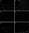
Melanocortin-3/4 receptor ligands administered to the caudal brain stem potently modulate food intake by changing meal size. The origin of the endogenous ligands is unclear, because the arcuate nucleus of the hypothalamus and the nucleus of the solitary tract (NTS) harbor populations of proopiomelanocortin (POMC)-expressing neurons. Here we demonstrate that activation of hypothalamic POMC neurons leads to suppression of food intake and that this suppression is prevented by administration of a melanocortin-3/4 receptor antagonist to the NTS and its vicinity. Bilateral leptin injections into the rat arcuate nucleus produced long-lasting suppression of meal size and total chow intake. These effects were significantly blunted by injection of SHU-9119 into the fourth ventricle, although SHU-9119 increased meal size and food intake during the first, but not the second, 14-h observation period. Leptin effects on meal size and food intake were abolished throughout the 40-h observation period by injection of SHU-9119 into the NTS at a dose that by itself had no effect. Neuron-specific tracing from the arcuate nucleus with a Cre-inducible tract-tracing adenovirus in POMC-Cre mice showed the presence of labeled axons in the NTS. Furthermore, density of alpha-melanocyte-stimulating hormone-immunoreactive axon profiles throughout the NTS was decreased by approximately 70% after complete surgical transection of connections with the forebrain in the chronic decerebrate rat model. The results further support the existence of POMC projections from the hypothalamus to the NTS and suggest that these projections have a functional role in the control of food intake.
Figures


Similar articles
-
Brain stem melanocortinergic modulation of meal size and identification of hypothalamic POMC projections.Am J Physiol Regul Integr Comp Physiol. 2005 Jul;289(1):R247-58. doi: 10.1152/ajpregu.00869.2004. Epub 2005 Mar 3. Am J Physiol Regul Integr Comp Physiol. 2005. PMID: 15746303
-
Activation of the ARCPOMC→MeA Projection Reduces Food Intake.Front Neural Circuits. 2020 Nov 5;14:595783. doi: 10.3389/fncir.2020.595783. eCollection 2020. Front Neural Circuits. 2020. PMID: 33250721 Free PMC article.
-
Pituitary adenylate cyclase-activating polypeptide inhibits food intake in mice through activation of the hypothalamic melanocortin system.Neuropsychopharmacology. 2009 Jan;34(2):424-35. doi: 10.1038/npp.2008.73. Epub 2008 Jun 4. Neuropsychopharmacology. 2009. PMID: 18536705
-
The central melanocortin system and the integration of short- and long-term regulators of energy homeostasis.Recent Prog Horm Res. 2004;59:395-408. doi: 10.1210/rp.59.1.395. Recent Prog Horm Res. 2004. PMID: 14749511 Review.
-
Leptin and the systems neuroscience of meal size control.Front Neuroendocrinol. 2010 Jan;31(1):61-78. doi: 10.1016/j.yfrne.2009.10.005. Epub 2009 Oct 28. Front Neuroendocrinol. 2010. PMID: 19836413 Free PMC article. Review.
Cited by
-
The CNS glucagon-like peptide-2 receptor in the control of energy balance and glucose homeostasis.Am J Physiol Regul Integr Comp Physiol. 2014 Sep 15;307(6):R585-96. doi: 10.1152/ajpregu.00096.2014. Epub 2014 Jul 2. Am J Physiol Regul Integr Comp Physiol. 2014. PMID: 24990862 Free PMC article. Review.
-
Melanocortin signaling in the brainstem influences vagal outflow to the stomach.J Neurosci. 2013 Aug 14;33(33):13286-99. doi: 10.1523/JNEUROSCI.0780-13.2013. J Neurosci. 2013. PMID: 23946387 Free PMC article.
-
Glycemic state regulates melanocortin, but not nesfatin-1, responsiveness of glucose-sensing neurons in the nucleus of the solitary tract.Am J Physiol Regul Integr Comp Physiol. 2015 Apr 15;308(8):R690-9. doi: 10.1152/ajpregu.00477.2014. Epub 2015 Feb 18. Am J Physiol Regul Integr Comp Physiol. 2015. PMID: 25695291 Free PMC article.
-
The physiological control of eating: signals, neurons, and networks.Physiol Rev. 2022 Apr 1;102(2):689-813. doi: 10.1152/physrev.00028.2020. Epub 2021 Sep 6. Physiol Rev. 2022. PMID: 34486393 Free PMC article.
-
Modulation of the central melanocortin system by leptin, insulin, and serotonin: co-ordinated actions in a dispersed neuronal network.Eur J Pharmacol. 2011 Jun 11;660(1):2-12. doi: 10.1016/j.ejphar.2010.11.042. Epub 2011 Jan 3. Eur J Pharmacol. 2011. PMID: 21211525 Free PMC article. Review.
References
-
- Balthasar N, Dalgaard LT, Lee CE, Yu J, Funahashi H, Williams T, Ferreira M, Tang V, McGovern RA, Kenny CD, Christiansen LM, Edelstein E, Choi B, Boss O, Aschkenasi C, Zhang CY, Mountjoy K, Kishi T, Elmquist JK, Lowell BB. Divergence of melanocortin pathways in the control of food intake and energy expenditure. Cell 123: 493–505, 2005 - PubMed
-
- Bereiter D, Berthoud HR, Jeanrenaud B. Hypothalamic input to brain stem neurons responsive to oropharyngeal stimulation. Exp Brain Res 39: 33–39, 1980. - PubMed
-
- Berthoud HR. The caudal brainstem and the control of food intake and energy balance. In: Handbook of Behavioral Neurobiology, edited by Stricker EM, Woods SC. New York: Plenum, 2004, p. 195–240
-
- Berthoud HR, Morrison C. The brain, appetite, obesity. Annu Rev Psychol 59: 55–92, 2008 - PubMed
Publication types
MeSH terms
Substances
Grants and funding
LinkOut - more resources
Full Text Sources
Other Literature Sources
Miscellaneous

