. 2010 Apr;298(4):C817-30.
doi: 10.1152/ajpcell.00460.2009. Epub 2010 Jan 13.

Role of purinergic signaling pathways in V-ATPase recruitment to apical membrane of acidifying epididymal clear cells

Affiliations

Role of purinergic signaling pathways in V-ATPase recruitment to apical membrane of acidifying epididymal clear cells

Clémence Belleannée et al. Am J Physiol Cell Physiol. 2010 Apr.

Abstract

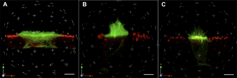
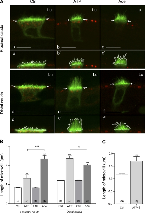
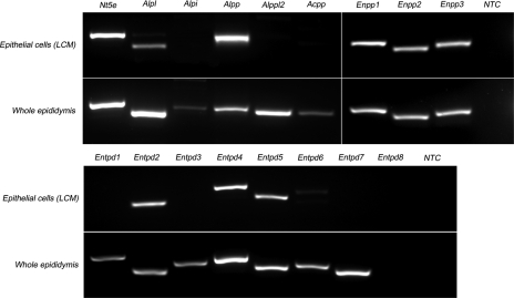
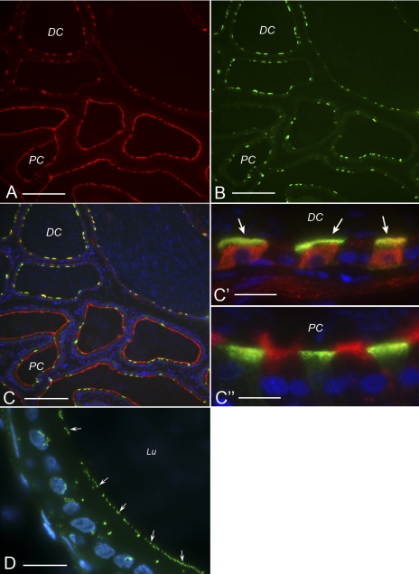
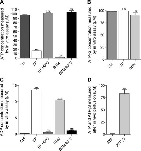
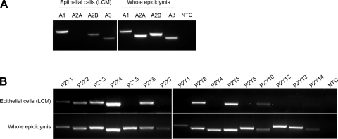
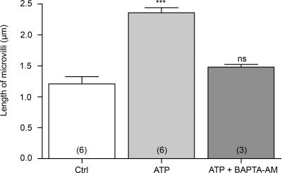
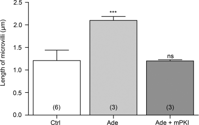
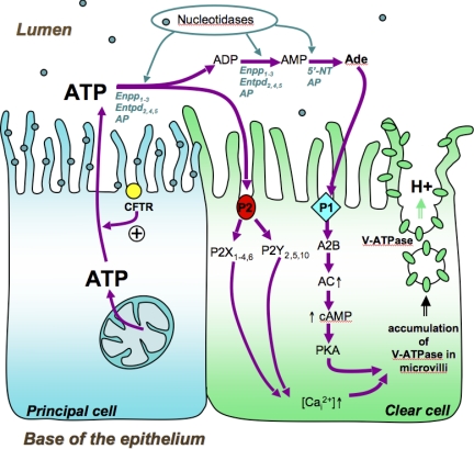
Extracellular purinergic agonists regulate a broad range of physiological functions via P1 and P2 receptors. Using the epididymis as a model system in which luminal acidification is essential for sperm maturation and storage, we show here that extracellular ATP and its hydrolysis product adenosine trigger the apical accumulation of vacuolar H(+)-ATPase (V-ATPase) in acidifying clear cells. We demonstrate that the epididymis can hydrolyze luminal ATP into other purinergic agonists such as ADP via the activity of nucleotidases located in the epididymal fluid and in the apical membrane of epithelial cells. Alkaline phosphatase activity and abundant ecto-5'-nucleotidase protein were detected in the apical pole of principal cells. In addition, we show that nine nucleotidase genes (Nt5e, Alpl, Alpp, Enpp1, 2, and 3, and Entpd 2, 4, and 5), seven ATP P2 receptor genes (P2X1, P2X2, P2X3, P2X4, P2X6, P2Y2, P2Y5), and three adenosine P1 receptor genes (A1, A2B, and A3) are expressed in epithelial cells isolated by laser cut microdissection (LCM). The calcium chelator BAPTA-AM abolished the apical V-ATPase accumulation induced by ATP, supporting the contribution of P2X or P2Y in this response. The PKA inhibitor myristoylated protein kinase inhibitor (mPKI) inhibited adenosine-dependent V-ATPase apical accumulation, indicating the participation of the P1 A2B receptor. Altogether, these results suggest that the activation of P1 and P2 purinergic receptors by ATP and adenosine might play a significant role in luminal acidification in the epididymis, a process that is crucial for the establishment of male fertility.


Figures

Fig. 1.
Fig. 1.
Three-dimensional (3D) reconstruction of clear cells under control conditions or luminally exposed to ATP or adenosine. Ten-micrometer sections of cauda epididymidis were double-stained for the vacuolar H+-ATPase (V-ATPase) B1 subunit (green) and horseradish peroxidase (HRP; red) after luminal perfusion with a control solution (A) or a solution containing 600 μM ATP (B) or 300 μM adenosine (C). 3D reconstructions were performed from a series of 0.1-μm optical sections taken in the z plane. Scale bars, 5 μm. Movies showing 3D rotations of these image stacks are available as supplemental material [Supplemental Videos 1 (A), 2 (B), and 3 (C)].
Fig. 2.
Fig. 2.
A: Double-immunofluorescence staining for V-ATPase B1 subunit (green) and HRP (red) in cryostat sections from rat epididymis perfused in vivo with control solution (Ctrl; a, d), 600 μM ATP (b, e), and 300 μM adenosine (Ade; c, f). Bars, 8 μm. Lu, lumen. V-ATPase apical accumulation was assessed by measuring the mean length of B1-labeled microvilli of clear cells as shown in a′–f′ and described in materials and methods. B: quantification of the length of B1-labeled microvilli in clear cells of the proximal (left) and distal (right) cauda epididymidis under control conditions (Ctrl) and after ATP and adenosine perfusion. C: quantification of the length of B1-labeled microvilli in clear cells of the distal cauda epididymidis under control conditions and after adenosine-5′-(γ-thiotriphosphate) (ATPγS) perfusion. Data are means ± SE; 2-way ANOVA test was used to compare different treatments to the control condition in each cauda region (**P < 0.01, ***P < 0.001) and to compare the different treatments to each other (###P < 0.001; ns, nonsignificant) (B). A Student's t-test was used to compare paired experiments (C). Numbers of rats per group are indicated in parentheses.
Fig. 3.
Fig. 3.
RT-PCR analysis of ectonucleotidase mRNA expression in epididymal epithelial cells isolated by laser cut microdissection (LCM) and in whole epididymis. Top: primers were designed for rat ecto-5′-nucleotidase (Nt5e), 4 isoforms of alkaline phosphatase (Alpl, Alpi, Alpp, Alpp2), prostatic acid phosphatase (Acpp), and 3 isoforms of ectonucleotide pyrophosphatase/phosphodiesterase (Enpp1, Enpp2, Enpp3). Bottom: 8 isoforms of ectonucleotide triphosphate diphosphohydrolase (Entpd1, Entpd2, Entpd3, Entpd4, Entpd5, Entpd6, Entpd7, Entpd8) were analyzed. All enzymes except Entpd8 were detected in whole epididymis mRNA extracts. In epithelial cells, 9 enzymes were detected: Alpl, Alpp, Enpp1, Enpp2, Enpp3, Entpd2, Entpd4, and Entpd5. NTC, no-template control.
Fig. 4.
Fig. 4.
Cellular localization of ecto-5′-nucleotidase and endogenous alkaline phosphatase activity in rat epididymis. Double-staining for ecto-5′-nucleotidase (A) and the V-ATPase B1 subunit (B) revealed a region-specific localization of ecto-5′-nucleotidase. C: merge picture; C′ and C″ show higher magnification of some tubules shown in C. In the proximal cauda (PC) ecto-5′-nucleotidase is highly expressed in the apical membrane of principal cells (negative for V-ATPase). In contrast, in the distal cauda (DC) it is localized in the cytoplasm of clear cells (positive for V-ATPase, arrows) and is absent from principal cells. D: alkaline phosphatase activity was detected in the apical membrane of epididymal epithelial cells (arrows) with the ELF 97 substrate. Lu, Lumen. Bars: 120 μm for A, B, and C; 14 μm for C′ and C″; 10 μm for D.
Fig. 5.
Fig. 5.
In vitro and in vivo detection of ATP hydrolysis in the epididymis. Nucleotidase activity was assessed in vitro in the epididymal fluid (EF) and in brush-border membranes (BBM) by measuring the hydrolysis of 100 μM ATP (A) or 100 μM ATPγS (B) after in vitro incubation with a control solution (Ctrl), EF, BBM, or EF and BBM that had been preheated to 90°C before the incubation (EF 90°C and BBM 90°C, respectively). C: production of ADP was measured after incubation of 50 μM ATP with a control solution (Ctrl), EF, BBM, or EF and BBM preheated to 90°. D: nucleotidase activity was also assessed in vivo by perfusing the epididymal lumen with 100 μM ATP or 100 μM ATPγS and measuring the concentration of ATP and ATPγS in the collected luminal fluid. Data are means ± SE. One-way ANOVA test was used to compare different conditions (A–C) to control. ***P < 0.001; ns, nonsignificant. A Student's t-test was used to compare paired experiments in D. (***P < 0.001).
Fig. 6.
Fig. 6.
RT-PCR analysis of P1 and P2 receptors in epididymal epithelial cells isolated by LCM and in whole epididymis. A: whereas all P1 receptor subtypes were detected in the whole epididymis, only A1, A2B, and A3 transcripts were detected in epithelial cells. B: P2 receptor primers were designed to detect 7 members of the P2X family (P2x1, P2x2, P2x3, P2x4, P2x5, P2x6, P2x7) and 9 members of the P2Y family (P2y1, P2y2, P2y4, P2y5, P2y6, P2y10, P2y12, P2y13, P2y14). All P2 subtypes were detected in the whole epididymis, but only 8 subtypes (P2x1–4, P2x6, P2y2, 5, 10) were detected in epididymal epithelial cells.
Fig. 7.
Fig. 7.
Role of calcium in luminal ATP-dependent V-ATPase apical accumulation in clear cells. Rat epididymides were luminally perfused in vivo with a control solution (Ctrl), 600 μM ATP, or 600 μM ATP in the presence of the calcium chelator BAPTA-AM (10 μM) (ATP + BAPTA-AM). Quantification of the length of V-ATPase-labeled microvilli in clear cells from the distal cauda epididymidis showed a significant elongation by ATP compared with control and inhibition of the ATP-elicited response by BAPTA-AM. Data are means ± SE. One-way ANOVA test was used to compare different treatments to control. ***P < 0.001. Numbers of rats per group are indicated in parentheses.
Fig. 8.
Fig. 8.
Role of protein kinase A (PKA) in luminal adenosine-dependent V-ATPase apical accumulation in clear cells. Rat epididymides were luminally perfused in vivo with a control solution (Ctrl), 300 μM adenosine (Ade), or 300 μM adenosine in the presence of 10 μM of the PKA inhibitor myristoylated protein kinase inhibitor (Ade + mPKI). Quantification of the length of V-ATPase-labeled microvilli in clear cells from the distal cauda epididymidis showed a significant elongation by adenosine compared with control and a complete inhibition of the adenosine response by mPKI. Data are means ± SE. One-way ANOVA test was used to compare different treatments to control. ***P < 0.001. Numbers of rats per group are indicated in parentheses.
Fig. 9.
Fig. 9.
Proposed model of purinergic-dependent V-ATPase apical accumulation in clear cells. The P2 purinergic receptor subtypes P2X1,4,6 and P2Y2,5,10 are expressed in clear cells. Extracellular luminal ATP induces a calcium-dependent V-ATPase accumulation in well-developed microvilli in clear cells. Extracellular ATP is also rapidly degraded into adenosine by ecto-nucleotidases that are expressed in the apical membrane of principal cells or by nucleotidases located in the epididymal fluid. AP, alkaline phosphatase; 5′-NT, 5′-nucleotidase. Extracellular adenosine activates the P1 receptor subtype A2B, which is also expressed in clear cells. Luminal adenosine triggers a PKA-dependent V-ATPase microvillus accumulation in clear cells. Cystic fibrosis transmembrane conductance regulator (CFTR) is expressed in the apical membrane of principal cells and has been proposed to regulate ATP secretion in several cell types. We propose that principal cells release ATP into the epididymal lumen via a mechanism that requires the involvement of CFTR. Future studies will be required to examine this hypothesis.

Similar articles

Cited by

References

    1. Acott TS, Carr DW. Inhibition of bovine spermatozoa by caudal epididymal fluid. II. Interaction of pH and a quiescence factor. Biol Reprod 30: 926–935, 1984 - PubMed
    1. Beaulieu V, Da Silva N, Pastor-Soler N, Brown CR, Smith PJ, Brown D, Breton S. Modulation of the actin cytoskeleton via gelsolin regulates vacuolar H+-ATPase recycling. J Biol Chem 280: 8452–8463, 2005 - PubMed
    1. Biber J, Stieger B, Haase W, Murer H. A high yield preparation for rat kidney brush border membranes. Different behaviour of lysosomal markers. Biochim Biophys Acta 647: 169–176, 1981 - PubMed
    1. Blomqvist SR, Vidarsson H, Soder O, Enerback S. Epididymal expression of the forkhead transcription factor Foxi1 is required for male fertility. EMBO J 25: 4131–4141, 2006 - PMC - PubMed
    1. Breton S, Brown D. New insights into the regulation of V-ATPase-dependent proton secretion. Am J Physiol Renal Physiol 292: F1–F10, 2007 - PubMed

Publication types

MeSH terms