. 2010 Feb;120(2):433-45.
doi: 10.1172/JCI39733. Epub 2010 Jan 11.

Cerebrovascular dysfunction and microcirculation rarefaction precede white matter lesions in a mouse genetic model of cerebral ischemic small vessel disease

Affiliations

Cerebrovascular dysfunction and microcirculation rarefaction precede white matter lesions in a mouse genetic model of cerebral ischemic small vessel disease

Anne Joutel et al. J Clin Invest. 2010 Feb.

Abstract

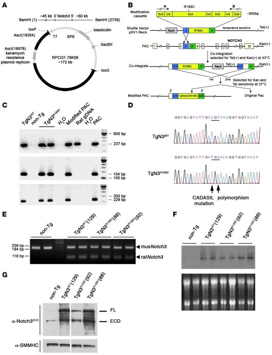
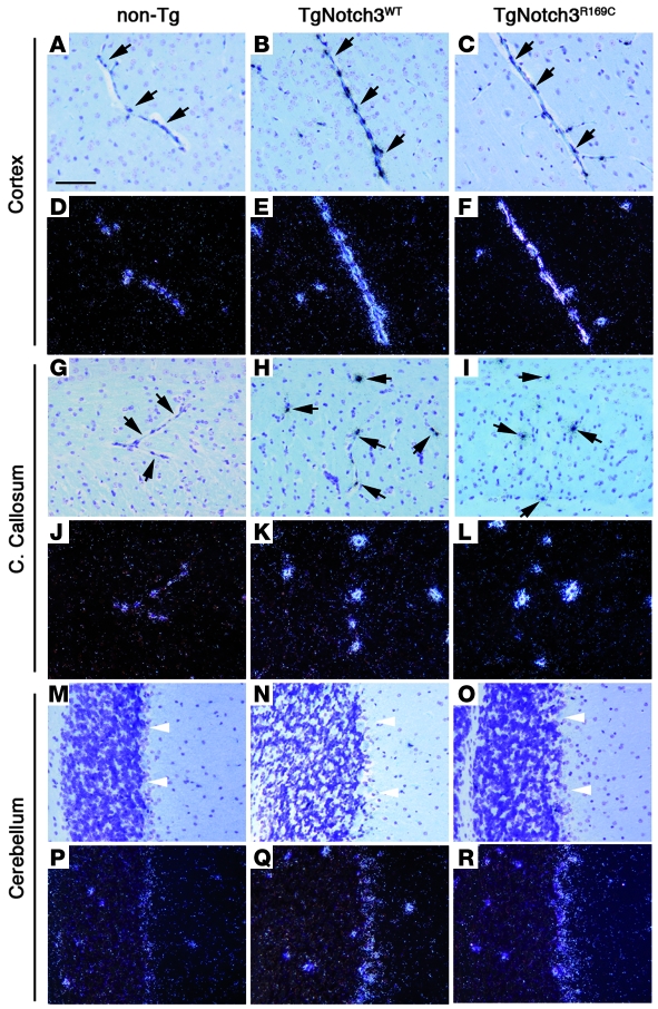
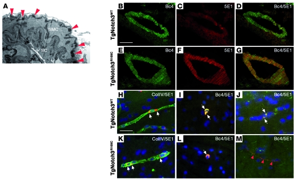
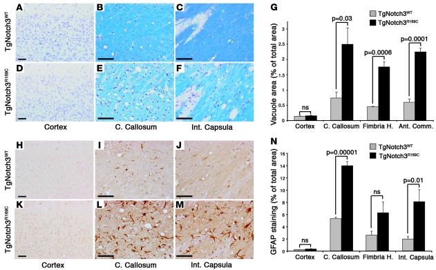
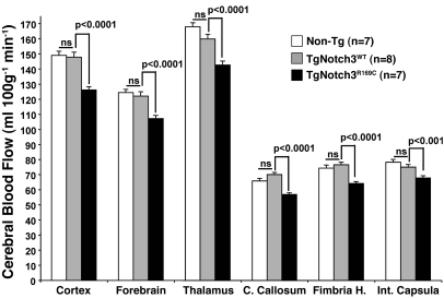
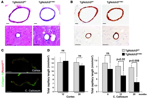
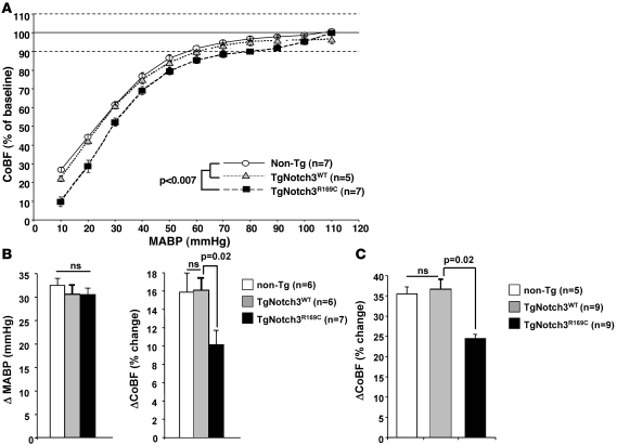
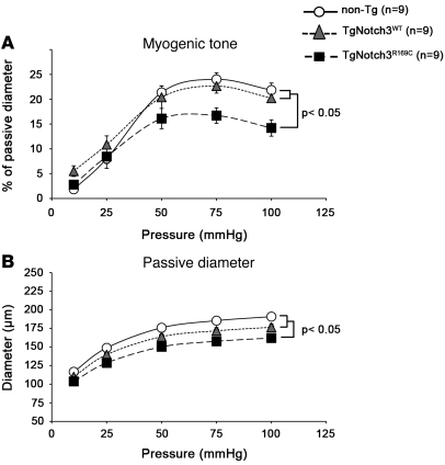
Cerebral ischemic small vessel disease (SVD) is the leading cause of vascular dementia and a major contributor to stroke in humans. Dominant mutations in NOTCH3 cause cerebral autosomal dominant arteriopathy with subcortical infarcts and leukoencephalopathy (CADASIL), a genetic archetype of cerebral ischemic SVD. Progress toward understanding the pathogenesis of this disease and developing effective therapies has been hampered by the lack of a good animal model. Here, we report the development of a mouse model for CADASIL via the introduction of a CADASIL-causing Notch3 point mutation into a large P1-derived artificial chromosome (PAC). In vivo expression of the mutated PAC transgene in the mouse reproduced the endogenous Notch3 expression pattern and main pathological features of CADASIL, including Notch3 extracellular domain aggregates and granular osmiophilic material (GOM) deposits in brain vessels, progressive white matter damage, and reduced cerebral blood flow. Mutant mice displayed attenuated myogenic responses and reduced caliber of brain arteries as well as impaired cerebrovascular autoregulation and functional hyperemia. Further, we identified a substantial reduction of white matter capillary density. These neuropathological changes occurred in the absence of either histologically detectable alterations in cerebral artery structure or blood-brain barrier breakdown. These studies provide in vivo evidence for cerebrovascular dysfunction and microcirculatory failure as key contributors to hypoperfusion and white matter damage in this genetic model of ischemic SVD.


Figures

Figure 1
Figure 1. Generation and characterization of PAC transgenic mice expressing wild-type or CADASIL-linked R169C rat Notch3.
(A) Rat PAC clone and vector map (27, 52). (B) Strategy for targeted PAC modification according to ref. . This panel was adapted from ref. with permission of Nature biotechnology. The modification cassette, inserted in the shuttle vector, contains flanking rat Notch3 genomic sequence of exons 3 and 6 on both sides of the engineered CADASIL mutation in exon 4. Two PAC modification steps are illustrated: co-integration of the shuttle vector into the PAC (1st and 2nd) and the resolution of the co-integrant by a second homologous recombination event to eliminate the shuttle vector and other exogenous sequences leaving the modified PAC carrying a point mutation. (C) Full-length integration of wild-type and modified PACs in transgenic mice. Shown are PCR products corresponding to rat Notch3 exon 1 (top), SP6 site (middle), and T7 site (bottom) of the PAC clone. (D) Sequence analysis of the PCR product from the rat Notch3 transgene in TgNotch3WT (TgN3WT) and TgNotch3R169C (TgN3R169C) mice. (E) RT-PCR assay of brain from nontransgenic, TgNotch3WT, and TgNotch3R169C (lines 88 and 92) mice. PCR products were cleaved with RsaI and fractionated on agarose gel, yielding an uncleaved 206-bp fragment from endogenous mouse Notch3 mRNA and 108- and 98-bp cleaved fragments from rat Notch3 mRNA. (F) Northern blot analysis of brain from nontransgenic, TgNotch3WT, and TgNotch3R169C mice. (G) Representative immunoblots of brain lysates prepared from 1-month-old TgNotch3R169C (lines 88 and 92), TgNotch3WT, and nontransgenic mice probed with the 5E1 anti-Notch3ECD antibody, which recognizes endogenous mouse and exogenous rat Notch3 proteins, and the anti-SMMHC antibody. White lines indicate that the lanes were run on the same gel but were noncontiguous.
Figure 2
Figure 2. Transgenic mice overexpress Notch3 in an endogenous-like expression pattern.
In situ hybridization with a Notch3 antisense riboprobe on sections of the cortex (AF), corpus callosum (GL), and cerebellum (MR) from 1-month-old nontransgenic, TgNotch3WT (line 129), and TgNotchR169C (line 88) mice. Bright- and dark-field images are shown on the upper and lower panel of each structure, respectively. In the cortex and corpus callosum, Notch3 expression was essentially detected in the capillaries (black arrows), with a robust signal in transgenic mice and lesser signal in non-transgenic mice. Weaker expression was also detected in the Bergmann glial cells of the cerebellum (white arrowheads) in both nontransgenic and transgenic mice, with higher expression in the latter. Note that brains were formalin fixed by immersion without prior transcardiac perfusion of the mice. Scale bar: 60 μm.
Figure 3
Figure 3. Notch3ECD aggregates and GOM deposits in brain arteries and capillaries of TgNotch3R169C mice.
(A) Electron micrograph of a pial artery from a 5-month-old TgNotch3R169C mouse demonstrating abundant GOM deposits (red arrowheads) within the basement membrane of smooth muscle cells. (BG) Brain arteries from 2-month-old TgNotch3R169C and TgNotch3WT mice were stained with Notch3 antibodies specific to the intracellular (Bc4, green) or extracellular (5E1, red) domain. Microscopic aggregates of Notch3ECD are shown in mutant artery. (H, I, K, and L) Brain sections of 2-month-old TgNotch3WT and TgNotch3R169C were double labeled with antibodies to Notch3 extracellular domain (5E1, red) and collagen IV (ColIV, green) (H and K) or double labeled with Bc4 (green) and 5E1 (red) Notch3 antibodies (I and L). Nuclei were stained by DAPI (blue). Perinuclear inclusions, strongly labeled by Notch3 intracellular and extracellular antibodies, were seen in wild-type and mutant capillaries (white arrows). (J and M) Brain sections of 12-month-old TgNotch3WT (J) and TgNotch3R169C (M) were double labeled with Bc4 (green) and 5E1 (red) Notch3 antibodies. Shown are dot-like Notch3ECD aggregates unstained by the Bc4 antibody in the mutant capillary (red arrowheads), while wild-type capillary exhibited discrete perinuclear inclusions labeled by Notch3 intracellular and extracellular antibodies (white arrows). Scale bars: 1 μm (A), 50 μm (BG) and 30 μm (HM).
Figure 4
Figure 4. White matter lesions and astrogliosis in aged TgNotch3R169C mice.
(AF) Klüver-Barrera Luxol fast blue staining in the cortex, corpus callosum, and internal capsula of 20-month-old TgNotch3WT and TgNotch3R169C mice revealing vacuolization in white matter bundles as well as pallor and disorganization of the myelin in mutant mice. (G) Quantification of the vacuole area in the cortex, corpus callosum, fimbria of hippocampus, and anterior commissural (Ant. Comm.) of 20-month-old TgNotch3R169C compared with TgNotch3WT mice (n = 3–4 mice per group). (HM) GFAP staining in the cortex, corpus callosum, and internal capsula of 20-month-old TgNotch3WT and TgNotch3R169C mice showing astrogliosis in the white matter bundles of TgNotch3R169C mice. (N) Quantification of astrogliosis in the indicated structures of 20-month-old TgNotch3R169C mice compared with TgNotch3WT mice (n = 4 mice per group). Scale bars: 70 μm.
Figure 5
Figure 5. Decreased resting CBF in aged TgNotch3R169C mice.
Quantitative measurement of CBF through the neocortex, forebrain (including the striatum, pallidum, and amygdala), thalamus, corpus callosum, fimbria of the hippocampus, and internal capsula in 18-month-old TgNotch3R169C compared with TgNotch3WT and nontransgenic mice showed diffuse cerebral hypoperfusion in mutant mice. Each of these large areas included 8–10 gray matter regions or 4–5 white matter regions.
Figure 6
Figure 6. Arterial structure is preserved, but capillary density is progressively reduced in the white matter of TgNotch3R169C mice.
(A) Masson’s trichrome staining showing intact pial artery (upper panels) and small artery (lower panels) of 20-month-old TgNotch3R169C mice. (B) Labeling for smooth muscle myosin heavy chain revealing a continuous rim of smooth muscle cells in pial and small artery of 20-month-old TgNotch3R169C mice. (C) Double labeling for Notch3ECD (5E1, red) and collagen IV (green) of capillaries in the cortex and the corpus callosum of 12-month-old TgNotch3R169C mice showed robust aggregation of Notch3ECD in both capillaries. (D) Mean total capillary length (mm of CD31+ structures per mm2) in the cortex of 12- and 20-month-old TgNotch3R169C mice and the corpus callosum of 6-, 12-, and 20-month-old TgNotch3R169C mice compared with age-matched TgNotch3WT mice showed progressive age-related reduction in capillary density in the white matter of mutant mice (n = 4–5 mice per group). Scale bars: 25 μm.
Figure 7
Figure 7. Impaired cerebrovascular autoregulation and attenuated functional hyperemia in young TgNotch3R169C mice.
(A) Autoregulation curves of CoBF in 5-month-old nontransgenic, TgNotch3WT, and TgNotch3R169C mice in response to provoked hypotension showed that CoBF remained above 90% (lower dotted line, considered as the lower limit of autoregulation) up to 60 mmHg in TgNotch3WT and nontransgenic mice, but only up to 80 mmHg in TgNotch3R169C mice. (B) CoBF (in percent change) changes in response to arterial blood pressure increase (ΔMABP, in mmHg) induced by phenylephrine infusion (40 μg/kg), in 5-month-old nontransgenic, TgNotch3WT, and TgNotch3R169C mice showing a smaller CoBF increase in mutant mice. (C) CoBF increase (%) in the somatosensory cortex in response to whisker stimulation in 5- to 6-month-old nontransgenic, TgNotch3WT, and TgNotch3R169C mice showing attenuation of functional hyperemia in mutant mice.
Figure 8
Figure 8. Altered mechanical properties and myogenic responses in young TgNotch3R169C mice.
(A) Response of cerebral artery to stepwise increase in pressure (myogenic tone) in 6-month-old nontransgenic, TgNotch3WT, and TgNotch3R169C mice showed that myogenic tone was significantly attenuated in TgNotch3R169C mice. (B) Internal diameter/pressure relationships in cerebral arteries during maximal dilation with EGTA and sodium nitroprusside (passive diameter) in 6-month-old nontransgenic, TgNotch3WT, and TgNotch3R169C mice. Passive diameters were significantly less in TgNotch3R169C mice compared with both nontransgenic and TgNotch3WT mice.

References

    1. Ringelstein EB, Nabavi DG. Cerebral small vessel diseases: cerebral microangiopathies. Curr Opin Neurol. 2005;18(2):179–188. doi: 10.1097/01.wco.0000162861.26971.03. - DOI - PubMed
    1. Hainsworth AH, Markus HS. Do in vivo experimental models reflect human cerebral small vessel disease? A systematic review. J Cereb Blood Flow Metab. 2008;28(12):1877–1891. doi: 10.1038/jcbfm.2008.91. - DOI - PubMed
    1. Joutel A, et al. Notch3 mutations in CADASIL, a hereditary adult-onset condition causing stroke and dementia. Nature. 1996;383(6602):707–710. doi: 10.1038/383707a0. - DOI - PubMed
    1. Dichgans M. Genetics of ischaemic stroke. Lancet Neurol. 2007;6(2):149–161. doi: 10.1016/S1474-4422(07)70028-5. - DOI - PubMed
    1. Chabriat H, Joutel A, Dichgans M, Tournier-Lasserve E, Bousser MG. Cadasil. Lancet Neurol. 2009;8(7):643–653. doi: 10.1016/S1474-4422(09)70127-9. - DOI - PubMed

Publication types