. 2010 Feb;120(2):508-20.
doi: 10.1172/JCI40045. Epub 2010 Jan 11.

Beta-catenin blocks Kras-dependent reprogramming of acini into pancreatic cancer precursor lesions in mice

Affiliations

Beta-catenin blocks Kras-dependent reprogramming of acini into pancreatic cancer precursor lesions in mice

John P Morris 4th et al. J Clin Invest. 2010 Feb.

Abstract

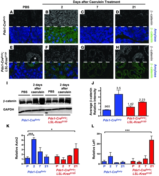
Cellular plasticity in adult organs is involved in both regeneration and carcinogenesis. WT mouse acinar cells rapidly regenerate following injury that mimics acute pancreatitis, a process characterized by transient reactivation of pathways involved in embryonic pancreatic development. In contrast, such injury promotes the development of pancreatic ductal adenocarcinoma (PDA) precursor lesions in mice expressing a constitutively active form of the GTPase, Kras, in the exocrine pancreas. The molecular environment that mediates acinar regeneration versus the development of PDA precursor lesions is poorly understood. Here, we used genetically engineered mice to demonstrate that mutant Kras promotes acinar-to-ductal metaplasia (ADM) and pancreatic cancer precursor lesion formation by blocking acinar regeneration following acute pancreatitis. Our results indicate that beta-catenin is required for efficient acinar regeneration. In addition, canonical beta-catenin signaling, a pathway known to regulate embryonic acinar development, is activated following acute pancreatitis. This regeneration-associated activation of beta-catenin signaling was not observed during the initiation of Kras-induced acinar-to-ductal reprogramming. Furthermore, stabilized beta-catenin signaling antagonized the ability of Kras to reprogram acini into PDA preneoplastic precursors. Therefore, these results suggest that beta-catenin signaling is a critical determinant of acinar plasticity and that it is inhibited during Kras-induced fate decisions that specify PDA precursors, highlighting the importance of temporal regulation of embryonic signaling pathways in the development of neoplastic cell fates.


Figures

Figure 1
Figure 1. Mutant Kras blocks acinar regeneration and promotes ADM/PanIN formation.
(AH) H&E staining of regeneration time course in Pdx1-CreEarly (AD) and Pdx1-CreEarly;LSL-KrasG12D (EH) mice. (E and M) Asterisks indicate spontaneous PanIN lesions in PBS-treated Pdx1-CreEarly;LSL-KrasG12D animals. Insets in B and F show morphologically similar duct-like cells 2 days after induction of acute pancreatitis. (IP) Amylase (red)/CK19 (green) immunofluorescent labeling. Note CK19 expression in spontaneous PanIN lesions in Pdx1-CreEarly;LSL-KrasG12D mice (asterisk, M). (J and N) Amylase is downregulated and CK19 is weakly expressed in transient duct-like cells in Pdx1-CreEarly mice (inset, J) while strongly expressed in duct-like cells in Pdx1-CreEarly;LSL-KrasG12D mice (inset, N). Rare amylase-positive cells are found in metaplastic epithelium (arrowheads, O). (QT) Amylase (red), CK19 (blue), YFP (green) immunofluorescent labeling in Elastase-CreERT;LSL-KrasG12D;R26R-EYFP mice. Without caerulein treatment, YFP expression is restricted to amylase-positive cells and restricted from CK19-positive cells. Arrowheads indicate autofluorescent erythrocytes (Q). Double-CK19/YFP–positive cells (cyan, indicating blue/green overlap) persist following caerulein treatment (RT). Original magnification, ×400 (AP; insets). Scale bars: 50 μm (QT).
Figure 2
Figure 2. Mutant Kras blocks acinar regeneration in favor of a persistently dedifferentiated state.
(AH) Clusterin and Sox9 (IP) expression during regeneration and Kras-induced ADM. (A and I) Clusterin expression is limited to some normal ducts in PBS-treated Pdx1-CreEarly mice, while Sox9 is restricted to ducts and centroacinar cells (arrowheads). (E and M) Acini in PBS-treated Pdx1-CreEarly;LSL-KrasG12D mice are negative for Clusterin and Sox9, while normal ducts and spontaneous PanINs are positive (asterisks). (B, F, J, and N) Damaged duct-like cells of both genotypes display clusterin- and Sox9-positive cells (insets). (C, D, K, and L) Clusterin and Sox9 are mainly restricted to duct cells (arrowheads) following regeneration in Pdx1-CreEarly pancreata. Rare clusterin-positive cells were observed 7 days following caerulein treatment (arrow, C). (G, H, O, and P) Clusterin and Sox9 remain strongly expressed in ADM and PanINs in Pdx1-CreEarly;LSL-KrasG12D mice. (Q) Schematic of failed regeneration of acini possessing mutant Kras (acini*) versus WT. WT acini transiently dedifferentiate and rapidly regenerate, while acini possessing mutant Kras are sensitized to persistent dedifferentiation and ADM/PanIN formation. Original magnification, ×400 (AP; insets).
Figure 3
Figure 3. Regeneration-associated reactivation of β-catenin signaling is inhibited during Kras-induced ductal reprogramming.
(AH) β-catenin (above dashed line, gray; below dashed line, green) and amylase (above dashed line, not shown; below dashed line, blue) immunofluorescent staining. (AD) Pdx1-CreEarly mice; (EH) Pdx1-CreEarly;LSL-KrasG12D mice. Asterisk in E marks spontaneous PanIN lesions. (I and J) Western blot analysis of β-catenin 2 days following caerulein treatment. Intensity, normalized to GAPDH, is quantified in J (bars represent mean ± SD). (K and L) Quantitative PCR for β-catenin target genes during WT acinar regeneration (blue bars) and ADM/PanIN (red bars). Values are relative to PBS-treated Pdx1-CreEarly mice and are presented as mean ± SD (n = 3). P, PBS treatment; 2, 7, 21 indicate days after caerulein treatment. *P < 0.05; ***P < 0.001. Original magnification, ×630 (AH).
Figure 4
Figure 4. β-catenin is required for efficient acinar regeneration.
(AF) H&E staining of regenerating control (p48Cre;β-cateninF/+) versus p48Cre;β-cateninF/F pancreas following caerulein treatment. f, fat accumulation (DF). Rare acini following caerulein treatment are marked with arrowheads (E and F). (GL) β-catenin (green), amylase (blue), and CK19 (red) immunofluorescent labeling. Acini in p48Cre;β-cateninF/F mice lack membrane β-catenin staining (compare insets in G and J). Arrowheads mark rare acini (K and L). Both β-catenin–negative (arrows) and –positive (hatch marks) CK19-labeled ducts are observed. i, islets. (M) Quantification of relative amylase area in control (blue bars) and p48Cre;β-cateninF/F (red bars) following caerulein pancreatitis. Bars represent mean ± SD. P, PBS treatment. 3 and 5 indicate days after caerulein treatment. *P < 0.05; **P < 0.01; ***P < 0.001. Original magnification, ×400 (AL, insets).
Figure 5
Figure 5. Stabilized β-catenin antagonizes Kras-induced ADM.
(AL) Characterization of 6-week-old PBS- and caerulein-treated p48Cre;β-cateninexon3/+;LSL-KrasG12D mice and caerulein-treated p48Cre;LSL-KrasG12D mice. (A, E, and I) H&E staining. Yellow lines separate areas of cells with acinar morphology (a), and ductal morphology (d). (B, F, and J) Amylase staining. (C, G, and K) Sox9 staining. (D, H, and L) Clusterin staining. (M) Quantification of relative amylase area 7 days following caerulein in p48Cre;β-cateninExon3/+;LSL-KrasG12D (red bars) and control p48Cre;LSL-KrasG12D (blue bars) mice. Bars represent mean ± SD. 7 indicates days after caerulein treatment. *P < 0.05; **P < 0.01; ***P < 0.001. Original magnification, ×400 (AL).
Figure 6
Figure 6. β-catenin signaling inhibits Kras-induced reprogramming of acini into PanINs.
(A, E, and I) H&E staining of tamoxifen-stimulated, caerulein- or PBS-treated ElaCreERT;β-cateninexon3/+;LSL-KrasG12D or ElaCreERT;LSL-KrasG12D mice. (B, F, and J) CK19/Alcian blue staining. Note that normal ducts in PBS-treated ElaCreERT;β-cateninexon3/+;LSL-KrasG12D mice are strongly CK19 positive but Alcian blue negative (B). (C, G, and K) β-catenin (green), DAPI (blue) immunofluorescent labeling. β-catenin accumulation is rare in PBS-treated ElaCreERT;β-cateninexon3/+;LSL-KrasG12D mice (asterisk, C). Nuclear localization is noted by cyan overlap of green/blue channels (K). (D, H, and L) phospho-p42/p44 staining. Original magnification, ×400 (A, B, DF, HJ, and L). Scale bars: 50 μm (C, G, and K).
Figure 7
Figure 7. β-catenin acts as a gatekeeper of Kras-induced reprogramming of acini into ductal PanINs.
Pattern of β-catenin signaling activity during acinar regeneration versus Kras-induced ADM/PanIN. β-catenin levels are kept below a critical threshold during the initiation of Kras-induced ductal reprogramming but increase as PanIN lesions form. Therefore, β-catenin antagonizes specification of a ductal state capable of forming PanINs, but likely contributes to PanIN progression and tumor growth.

References

    1. Jemal A, et al. Cancer statistics, 2008. CA Cancer J Clin. 2008;58(2):71–96. doi: 10.3322/CA.2007.0010. - DOI - PubMed
    1. Feldmann G, Beaty R, Hruban RH, Maitra A. Molecular genetics of pancreatic intraepithelial neoplasia. J Hepatobiliary Pancreat Surg. 2007;14(3):224–232. doi: 10.1007/s00534-006-1166-5. - DOI - PMC - PubMed
    1. De La O JP, et al. Notch and Kras reprogram pancreatic acinar cells to ductal intraepithelial neoplasia. Proc Natl Acad Sci U S A. 2008;105(48):18907–18912. doi: 10.1073/pnas.0810111105. - DOI - PMC - PubMed
    1. Habbe N, et al. Spontaneous induction of murine pancreatic intraepithelial neoplasia (mPanIN) by acinar cell targeting of oncogenic Kras in adult mice. Proc Natl Acad Sci U S A. 2008;105(48):18913–18918. doi: 10.1073/pnas.0810097105. - DOI - PMC - PubMed
    1. Guerra C, et al. Chronic pancreatitis is essential for induction of pancreatic ductal adenocarcinoma by K-Ras oncogenes in adult mice. Cancer Cell. 2007;11(3):291–302. doi: 10.1016/j.ccr.2007.01.012. - DOI - PubMed

Publication types

MeSH terms