. 2010 Feb;120(2):617-26.
doi: 10.1172/JCI40545. Epub 2010 Jan 11.

Constitutively active phosphatase inhibitor-1 improves cardiac contractility in young mice but is deleterious after catecholaminergic stress and with aging

Affiliations

Constitutively active phosphatase inhibitor-1 improves cardiac contractility in young mice but is deleterious after catecholaminergic stress and with aging

Katrin Wittköpper et al. J Clin Invest. 2010 Feb.

Abstract

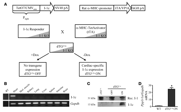
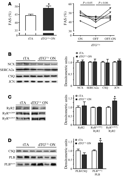
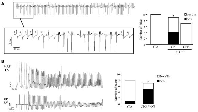
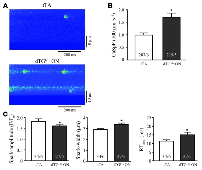
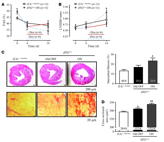
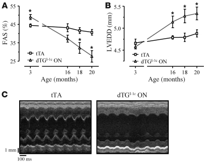
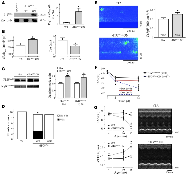
Phosphatase inhibitor-1 (I-1) is a distal amplifier element of beta-adrenergic signaling that functions by preventing dephosphorylation of downstream targets. I-1 is downregulated in human failing hearts, while overexpression of a constitutively active mutant form (I-1c) reverses contractile dysfunction in mouse failing hearts, suggesting that I-1c may be a candidate for gene therapy. We generated mice with conditional cardiomyocyte-restricted expression of I-1c (referred to herein as dTGI-1c mice) on an I-1-deficient background. Young adult dTGI-1c mice exhibited enhanced cardiac contractility but exaggerated contractile dysfunction and ventricular dilation upon catecholamine infusion. Telemetric ECG recordings revealed typical catecholamine-induced ventricular tachycardia and sudden death. Doxycycline feeding switched off expression of cardiomyocyte-restricted I-1c and reversed all abnormalities. Hearts from dTGI-1c mice showed hyperphosphorylation of phospholamban and the ryanodine receptor, and this was associated with an increased number of catecholamine-induced Ca2+ sparks in isolated myocytes. Aged dTGI-1c mice spontaneously developed a cardiomyopathic phenotype. These data were confirmed in a second independent transgenic mouse line, expressing a full-length I-1 mutant that could not be phosphorylated and thereby inactivated by PKC-alpha (I-1S67A). In conclusion, conditional expression of I-1c or I-1S67A enhanced steady-state phosphorylation of 2 key Ca2+-regulating sarcoplasmic reticulum enzymes. This was associated with increased contractile function in young animals but also with arrhythmias and cardiomyopathy after adrenergic stress and with aging. These data should be considered in the development of novel therapies for heart failure.


Figures

Figure 1
Figure 1. In vivo cardiac expression of I-1c.
(A) Generation and crossing strategy of a conditional mouse model (Tet-Off system) to express I-1c on a complete Ppp1r1a KO background. Dox, doxycycline; Ptight, Tet-responsive promoter; SV40, simian virus 40; VP16, herpes simplex virus protein 16. (B) RT-PCR analysis for I-1c and Gapdh from various organs of induced I-1c double-transgenic mice shows cardiomyocyte-restricted expression. -RT, minus RT control; Skel., skeletal. (C) Western blots from cardiac extracts demonstrate doxycycline-dependent I-1c expression and lack of leakiness in the single transgenic I-1c responder (sTGI-1c). Rec., recombinant. (D) I-1c mRNA analysis in dTGI-1c ON hearts revealed a 24.5-fold expression compared with Ppp1r1a mRNA from WT mice (n ≥ 4 each). *P < 0.05 versus WT.
Figure 2
Figure 2. I-1c enhances basal contractility and is associated with higher phosphorylation of PLB and cardiac RyR2.
(A) Echocardiographic assessment of FAS in single transgenic tTA mice and dTGI-1c ON mice at the age of 3 months (n = 13 each; left panel). *P < 0.05 versus tTA. Reassessment of FAS after doxycycline feeding (dTGI-1c OFF) and doxycycline withdrawal (dTGI-1c OFF-ON) indicates temporally controllable I-1c effects (n = 6; age, 3 months; right panel). (BD) Western blots and statistical analysis of total protein levels of NCX, SERCA2a, calsequestrin (CSQ), junctin (JCN), RyR2, and PLB and phosphorylation state of RyR2 (RyRSer2809 and RyRSer2815) and PLB (PLBSer16) in tTA and dTGI-1c ON hearts (n = 8 for each group). Samples were run on the same gel. *P < 0.05 versus tTA.
Figure 3
Figure 3. Higher susceptibility to arrhythmia in I-1c double-transgenic mice.
(A) Original telemetric recording of VTs in a dTGI-1c ON mouse (top panel). Number of tTA and double-transgenic mice that developed VTs (black bars) after arrhythmia provocation in freely moving mice. VTs were completely reversible after doxycycline feeding (dTGI-1c OFF). *P < 0.05 by χ2 test. P-waves are denoted by §.(B) Original recordings of ventricular arrhythmias in isolated Langendorff-perfused hearts of dTGI-1c ON and tTA mice, elicited during high-frequency pacing, with 80 or 100 ms cycle length. Shown are left ventricular monophasic action potentials (MAPs) and a bipolar electrogram of the endocardial right ventricular octapolar electrophysiological pacing catheter (EP RV). *P < 0.05 by χ2 test.
Figure 4
Figure 4. Isolated cardiomyocytes from dTGI-1c ON hearts show higher catecholamine-induced SR Ca2+ leak.
(A) Representative longitudinal line-scan images of tTA and dTGI-1c ON hearts. (B) Ca2+ spark frequency (CaSpF) in dTGI-1c ON versus tTA hearts (10 nM isoprenaline; *P < 0.05). (C) The Ca2+ spark characteristics, spark amplitude, spark width, and spark duration (time to 50% decay, RT50%), are shown. *P < 0.05 versus tTA. The numbers in the columns represent the numbers of myocytes and characterized sparks, respectively, from 5–6 hearts for each group.
Figure 5
Figure 5. Accelerated morphometric and functional deterioration after chronic catecholaminergic stress in I-1c double-transgenic mice.
(A) Echocardiographically determined FAS in dTGI-1c ON and tTA mice before (day 0) and after (days 4 and 14) isoprenaline infusion, respectively. Exaggerated decline in FAS in I-1c double-transgenic mice was stopped in a subgroup of I-1c double-transgenic mice fed with doxycycline for 10 days (red line). *P < 0.05 versus tTA+/–10d Dox; #P < 0.05 dTGI-1c ON day 4 versus day 0. (B) Left ventricular end-diastolic diameter (LVEDD). The dashed line represents I-1c double-transgenic mice kept in the ON state, and the red line represents I-1c double-transgenic mice fed with doxycycline for 10 days. *P < 0.05 versus dTGI-1c 10d OFF. (C) H&E-stained paraffin sections (top row; representative of 4–8 hearts each) demonstrate dilation in dTGI-1c ON hearts. Sirius red–stained paraffin sections (bottom row) are representative of 4–8 hearts each and demonstrate fibrosis in dTGI-1c hearts, confirmed by quantitative analysis of interstitial fibrosis in dTGI-1c hearts. The first number in each column indicates the number of analyzed areas (20–40 areas), and the second number indicates the number of analyzed hearts. Scale bars: 200 μm (top row); 20 μm (bottom row). *P < 0.05 versus tTA+/–10d Dox. (D) Myocyte cross-sectional area in tTA+/–10d Dox, dTGI-1c 10d OFF, and dTGI-1c ON hearts treated with isoprenaline for 14 days (the first number in each column indicates the number of analyzed cardiomyocytes (≥132), and the second number indicates the number of analyzed hearts). *P < 0.05 versus tTA+/–10d Dox; #P < 0.05 versus dTGI-1c 10d OFF.
Figure 6
Figure 6. Aging I-1c double-transgenic ON littermates show contractile dysfunction and left ventricular dilation.
(A) FAS in tTA and dTGI-1c ON littermates at the age of 18 and 20 months (n = 5 each). *P < 0.05 versus tTA. (B) LVEDD (n = 5 each). *P < 0.05 versus tTA. (C) Representative M-mode views from tTA and dTGI-1c ON mice.
Figure 7
Figure 7. Characterization of I-1S67A–expressing mice.
(A) Doxycycline-dependent I-1S67A expression and a lack of leakiness in single transgenic I-1S67A responders (I-1S67A). I-1S67A mRNA amount in dTGS67A ON mice is 8-fold higher compared with Ppp1r1a mRNA from WT mice (n = 5 each). *P < 0.05 versus WT. (B) I-1S67A enhances cardiac contractility in vivo (n = 6 for each group). *P < 0.05 versus tTA. (C) Phosphorylation state of PLB (PLBSer16) and RyR2 (RyRSer2815) in tTA and dTGS67A ON hearts (n ≥ 6 for each group). Samples were run on the same gel. *P < 0.05 versus tTA. (D) Number of tTA and dTGS67A ON mice that developed VTs (black bar) after arrhythmia provocation in freely moving mice. Doxycycline application completely repressed the development of VTs (dTGS67A OFF). *P < 0.05 by χ2 test. (E) Representative longitudinal line-scan images of tTA and dTGS67A ON mice and Ca2+ spark frequency, respectively (10 nM isoprenaline). The numbers in the columns represent the number of myocytes and hearts from each group. *P < 0.05 versus tTA. (F) Isoprenaline-infusion accelerated the decrease in contractility in dTGS67A ON versus tTA mice after 4 and 14 days. Exaggerated decline of contractility in dTGS67A ON mice could be stopped by doxycycline feeding from day 4 to 14 (red line). The dashed line represents the I-1c double-transgenic mice kept on water at any time. *P < 0.05 tTA versus dTGS67A ON on day 4; #P < 0.05 dTGS67A ON versus dTGS67A 10d OFF and tTA. (G) Aging I-1S67A double-transgenic mice show an exaggerated decrease in FAS and increase in LVEDD (n ≥ 5 each) compared with tTA mice at the age of 15 months. *P < 0.05 versus tTA. Representative M-mode views from tTA and dTGS67A ON mice. Experiments were done in parallel to the I-1c double-transgenic mice (CE).

References

    1. Bristow MR. beta-adrenergic receptor blockade in chronic heart failure. Circulation. 2000;101(5):558–569. - PubMed
    1. El-Armouche A, Eschenhagen T. beta-Adrenergic stimulation and myocardial function in the failing heart. Heart Fail Rev. 2008;14(4):225–241. doi: 10.1007/s10741-008-9132-8. - DOI - PubMed
    1. Hasenfuss G, Pieske B. Calcium cycling in congestive heart failure. J Mol Cell Cardiol. 2002;34(8):951–969. doi: 10.1006/jmcc.2002.2037. - DOI - PubMed
    1. Bers DM. Calcium cycling and signaling in cardiac myocytes. Annu Rev Physiol. 2008;70:23–49. doi: 10.1146/annurev.physiol.70.113006.100455. - DOI - PubMed
    1. Venetucci LA, Trafford AW, O’Neill SC, Eisner DA. The sarcoplasmic reticulum and arrhythmogenic calcium release. Cardiovasc Res. 2008;77(2):285–292. doi: 10.1093/cvr/cvm009. - DOI - PubMed

Publication types

MeSH terms

Substances