. 2010 Feb;120(2):545-58.
doi: 10.1172/JCI39319. Epub 2010 Jan 11.

Deleted in breast cancer-1 regulates SIRT1 activity and contributes to high-fat diet-induced liver steatosis in mice

Affiliations

Deleted in breast cancer-1 regulates SIRT1 activity and contributes to high-fat diet-induced liver steatosis in mice

Carlos Escande et al. J Clin Invest. 2010 Feb.

Abstract

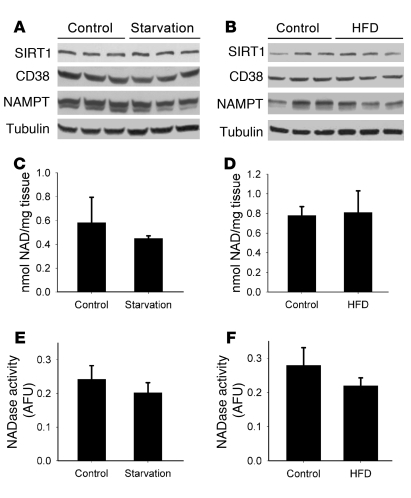
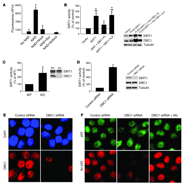
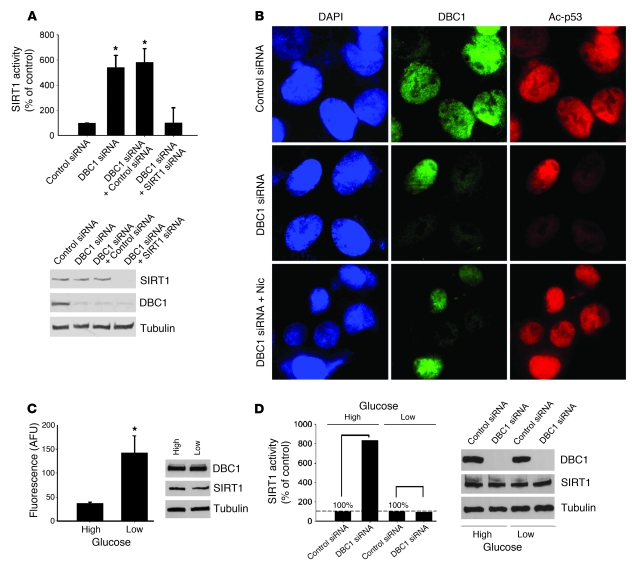
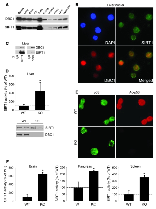
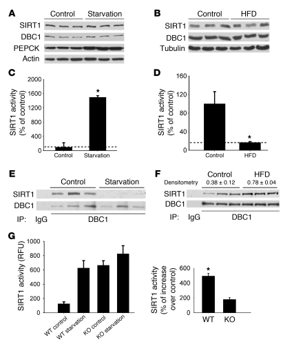
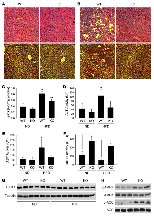
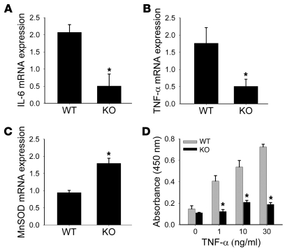
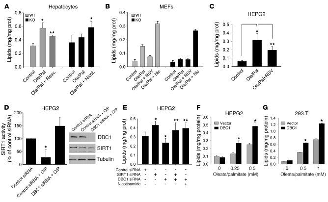
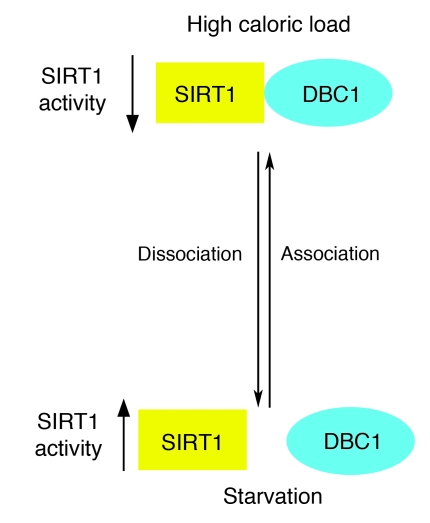
The enzyme sirtuin 1 (SIRT1) is a critical regulator of many cellular functions, including energy metabolism. However, the precise mechanisms that modulate SIRT1 activity remain unknown. As SIRT1 activity in vitro was recently found to be negatively regulated by interaction with the deleted in breast cancer-1 (DBC1) protein, we set out to investigate whether DBC1 regulates SIRT1 activity in vivo. We found that DBC1 and SIRT1 colocalized and interacted, and that DBC1 modulated SIRT1 activity, in multiple cell lines and tissues. In mouse liver, increased SIRT1 activity, concomitant with decreased DBC1-SIRT1 interaction, was detected after 24 hours of starvation, whereas decreased SIRT1 activity and increased interaction with DBC1 was observed with high-fat diet (HFD) feeding. Consistent with the hypothesis that DBC1 is crucial for HFD-induced inhibition of SIRT1 and for the development of experimental liver steatosis, genetic deletion of Dbc1 in mice led to increased SIRT1 activity in several tissues, including liver. Furthermore, DBC1-deficient mice were protected from HFD-induced liver steatosis and inflammation, despite the development of obesity. These observations define what we believe to be a new role for DBC1 as an in vivo regulator of SIRT1 activity and liver steatosis. We therefore propose that the DBC1-SIRT1 interaction may serve as a new target for therapies aimed at nonalcoholic liver steatosis.


Figures

Figure 1
Figure 1. SIRT1 protein levels and NAD+ do not change in the mouse liver in response to starvation or HFD.
Mice were put under 24 hours of starvation (A, C, and E) or fed HFD for 4 weeks (B, D, and F). Control groups consisted of mice fed a normal chow diet ad libitum (n = 3). (A and B) Western blots were performed for liver SIRT1, CD38, NAMPT, and tubulin. Each lane represents 1 different mouse liver. (C and D) Total NAD+ was isolated and measured from liver extracts after starvation (P = 0.37) and HFD (P = 0.42). (E and F) CD38 NADase activity was measured from liver extracts from starved (P = 0.32) and HFD-fed mice (P = 0.37). All experiments were performed with at least 6 mice in each group. Data are mean ± SD.
Figure 2
Figure 2. DBC1 regulates NAD-dependent SIRT1 activity.
(A) Isolated rat liver nuclei extracts were incubated in the absence or presence of 200 μM NAD+, or with NAD+ plus nicotinamide (2 mM), suramin (100 μM), or sirtinol (100 μM), and SIRT1 activity was measured. (B) Flag-SIRT1, full-length DBC1 (myc-DBC1-FL), and DBC1 with the leucine zipper deleted (myc-DBC1-ΔLZ) were overexpressed in 293T cells. At 24 hours after transfection, cell lysates were obtained, and SIRT1 activity was measured. NAD-dependent SIRT1 activity was expressed as a percentage of control (empty vector) activity. Protein overexpression was confirmed by Western blot. *P < 0.05 versus control; **P < 0.05 versus myc-DBC1-ΔLZ, t test. (C) MEFs derived from WT or Dbc1 KO mice were analyzed for NAD-dependent SIRT1 activity. Expression levels of SIRT1 and DBC1 were analyzed by Western blot. (D) DBC1 was knocked down in INS cells by siRNA transfection. SIRT1 activity was measured from cell lysates 72 hours after the first transfection. DBC1 knockdown was confirmed by Western blot. (E) INS cells were transfected with DBC1 siRNA, fixed after 72 hours, and stained for DBC1. Nuclei were stained with DAPI. Original magnification, ×600. (F) Endogenous p53 acetylation was analyzed by immunofluorescence after DBC1 siRNA transfection. Cells were treated with control siRNA, DBC1 siRNA, or DBC1 siRNA plus 5 mM nicotinamide (added 16 hours before fixation). Original magnification, ×600. Data are mean ± SD (n = 3). *P < 0.05, t test.
Figure 3
Figure 3. DBC1 controls glucose-dependent SIRT1 activity in human hepatocytes.
(A) HEPG2 cells were transfected with DBC1 siRNA, DBC1 siRNA plus control siRNA, and DBC1 siRNA plus SIRT1 siRNA. NAD-dependent SIRT1 activity was measured, and expressed as a percentage of control (nonspecific siRNA). The efficiency of transfection was confirmed by Western blot. (B) HEPG2 cells were transfected with DBC1 siRNA and DBC1; 5 mM nicotinamide was added 16 hours before fixation. Endogenous p53 acetylation levels were studied by immunofluorescence. Original magnification, ×600. (C) HEPG2 cells were cultured in serum-free media for 24 hours and then incubated for 24 hours in low (5 mM) or high (30 mM) glucose. SIRT1 activity was expressed in AFUs. n = 3. *P < 0.05, t test. (D) SIRT1 activity in HEPG2 cells transfected with DBC1 siRNA and then treated with high and low glucose as described in C. SIRT1 activity measured with DBC1 siRNA was normalized to the respective controls (nonspecific siRNA transfection) with high and low glucose. Data are shown as percent increase in SIRT1 activity over the respective controls (with high or low glucose, set as 100%). The fluorescence values in the control siRNA condition corresponded to 73 and 154 AFUs for cells in high and low glucose, respectively. Shown is 1 representative experiment of 3. The siRNA transfection efficiency was confirmed by Western blot. Except where indicated, data are mean ± SD.
Figure 4
Figure 4. SIRT1 and DBC1 interact in mouse tissue, and Dbc1 KO mice show increased endogenous SIRT1 activity in vivo.
(A) Western blots showing the expression of SIRT1 and DBC1 in different mouse tissues. (B) Isolated mouse liver nuclei immunofluorescence for SIRT1 and DBC1, showing that both proteins colocalized inside the nucleus. Original magnification, ×400. (C) Coimmunoprecipitation of SIRT1 and DBC1 from isolated nuclear extracts from liver. Shown is DBC1-SIRT1 coimmunoprecipitation using SIRT1 (left) and DBC1 (right) antibodies. Normal rabbit IgG was used as control for the immunoprecipitation. (D) Endogenous NAD-dependent SIRT1 activity was measured in isolated nuclear extracts from the livers of WT and Dbc1 KO mice. SIRT1 activity is expressed as a percentage of WT activity. Western blots of SIRT1 and DBC1 from these nuclear fractions served as control for protein levels. (E) Endogenous p53 acetylation was analyzed in isolated liver nuclei. Total p53 antibody was used as control. Original magnification, ×400. (F) Endogenous NAD-dependent SIRT1 activity was measured in nuclear extracts from brain, pancreas, and spleen of WT and Dbc1 KO mice. SIRT1 activity is expressed as a percentage of WT activity. Data are mean ± SD. *P < 0.05, t test.
Figure 5
Figure 5. SIRT1 activity and DBC1-SIRT1 interaction correlate under different metabolic conditions in the liver.
(AF) Mice were starved for 24 hours (A, C, and E) or placed on a HFD for 4 weeks (B, D, and F). Control mice were fed a normal chow diet. (A and B) Western blot analysis showed that SIRT1 and DBC1 protein levels did not change in the liver after starvation or HFD. Each lane represents a different mouse liver. (C and D) SIRT1 activity was measured from liver nuclear extracts. Data were normalized to the activity measured in the normal chow diet (n = 6). (E and F) Coimmunoprecipitation from liver nuclear extracts using DBC1 antibody. Normal rabbit IgG was used as control for the immunoprecipitation (n = 3). DBC1 and SIRT1 band intensities were calculated by densitometry as described in Methods. The ratio between SIRT1 and DBC1 values was used to calculate changes in interaction. (G) WT and Dbc1 KO mice were put on starvation for 24 hours, and liver SIRT1 activity was measured from nuclear extracts. Shown is 1 representative experiment of 3. SIRT1 activity in WT and Dbc1 KO mice after starvation was also normalized to that of the respective fed states, shown at right as percent increase over control (set as 100%). Data are mean ± SD. *P < 0.05, t test.
Figure 6
Figure 6. Dbc1 KO mice are protected against HFD-induced liver steatosis.
(A and B) WT and Dbc1 KO mice were fed normal chow (A) or put on HFD for 20 weeks (B). Liver sections were stained with H&E (top) or Oil Red O–hematoxylin (bottom). Original magnification, ×200. (C) Lipid content from liver homogenates of mice fed normal chow diet (ND) or HFD. *P < 0.05, **P < 0.05, ANOVA. (D and E) ALT (D) and AST (E) activities were measured in plasma from mice fed normal chow or HFD. *P < 0.05, ANOVA. Dbc1 KO mice showed no significant increase in the activity of either protein compared with the groups fed normal chow. (F) SIRT1 activity in liver nuclear extracts from mice fed normal chow or HFD. Statistically significant differences (P < 0.05, ANOVA) are shown by brackets. RFU, relative fluorescence units. (G) Western blots for SIRT1 from WT and Dbc1 KO mouse livers in mice fed normal chow or HFD for 20 weeks. Each lane represents a different mouse liver. (H) Western blots for phosphorylated ACC (Ser80), ACC, phosphorylated AMPK (Thr172), and AMPK from WT and Dbc1 KO mice livers after 20 weeks of HFD. Each lane represents a different mouse liver. Data are mean ± SD.
Figure 7
Figure 7. Dbc1 KO mice and cells are protected against inflammation.
(AC) Semiquantitative RT-PCR from mouse livers after 20 weeks of HFD for IL-6 (A), TNF-α (B), and MnSOD (C) mRNA. Total levels were normalized to 18s rRNA expression levels. (D) WT and Dbc1 KO–derived MEFs were treated with different doses of TNF-α for 6 hours. NF-κB activation (p65) was measured from nuclear cell extracts. *P < 0.05, t test. Data are mean ± SD.
Figure 8
Figure 8. DBC1 controls oleate/palmitate-induced cellular steatosis through a SIRT1-dependent mechanism.
(A) Lipid accumulation levels in primary hepatocytes isolated from WT and Dbc1 KO mice. An average of 3 different experiments is shown. *P < 0.05, **P < 0.1 versus control, t test. (B) Lipid accumulation in MEFs derived from WT and Dbc1 KO mice. Shown is 1 representative experiment of 4. (C) Lipid accumulation in HEPG2 cells. *P < 0.05, **P < 0.1, t test. n = 3. (D) Lipid accumulation in HEPG2 cells. Cells were transfected with control or DBC1 siRNA and then incubated with oleate/palmitate. SIRT1 activity was measured; values were normalized to those from control siRNA transfection. Protein levels were evaluated by Western blot. *P < 0.05, t test. n = 3. (E) Lipid accumulation in HEPG2 cells. Cells were transfected with control, SIRT1, and DBC1 siRNAs and then incubated with oleate/palmitate. *P < 0.05, **P < 0.1 versus control siRNA, t test. n = 4. (F and G) Effect of DBC1 overexpression on lipid accumulation in HEPG2 (F) and 293T (G) cells. Cells were transfected with empty vector or myc-DBC1 for 48 hours. During the last 16 hours of transfection, cells were incubated with different concentrations of oleate/palmitate (2:1 ratio). *P < 0.05, t test. n = 3. Data are mean ± SD.
Figure 9
Figure 9. Working model for the regulation of DBC1-SIRT1 interactions under different metabolic conditions.

References

    1. Guarente L. Sirtuins as potential targets for metabolic syndrome. Nature. 2006;444(7121):868–874. doi: 10.1038/nature05486. - DOI - PubMed
    1. Guarente L. Sirtuins in aging and disease. Cold Spring Harb Symp Quant Biol. 2007;72:483–488. doi: 10.1101/sqb.2007.72.024. - DOI - PubMed
    1. Ghosh HS. The anti-aging, metabolism potential of SIRT1. Curr Opin Investig Drugs. 2008;9(10):1095–1102. - PubMed
    1. Pfluger PT, Herranz D, Velasco-Miguel S, Serrano M, Tschop MH. Sirt1 protects against high-fat diet-induced metabolic damage. Proc Natl Acad Sci U S A. 2008;105(28):9793–9798. doi: 10.1073/pnas.0802917105. - DOI - PMC - PubMed
    1. Deng XQ, Chen LL, Li NX. The expression of SIRT1 in nonalcoholic fatty liver disease induced by high-fat diet in rats. Liver Int. 2007;27(5):708–715. doi: 10.1111/j.1478-3231.2007.01497.x. - DOI - PubMed

Publication types