. 2010 Jan 8;6(1):e1000715.
doi: 10.1371/journal.ppat.1000715.

Staphylococcus aureus panton-valentine leukocidin is a very potent cytotoxic factor for human neutrophils

Affiliations

Staphylococcus aureus panton-valentine leukocidin is a very potent cytotoxic factor for human neutrophils

Bettina Löffler et al. PLoS Pathog. .

Abstract

The role of the pore-forming Staphylococcus aureus toxin Panton-Valentine leukocidin (PVL) in severe necrotizing diseases is debated due to conflicting data from epidemiological studies of community-associated methicillin-resistant S. aureus (CA-MRSA) infections and various murine disease-models. In this study, we used neutrophils isolated from different species to evaluate the cytotoxic effect of PVL in comparison to other staphylococcal cytolytic components. Furthermore, to study the impact of PVL we expressed it heterologously in a non-virulent staphylococcal species and examined pvl-positive and pvl-negative clinical isolates as well as the strain USA300 and its pvl-negative mutant. We demonstrate that PVL induces rapid activation and cell death in human and rabbit neutrophils, but not in murine or simian cells. By contrast, the phenol-soluble modulins (PSMs), a newly identified group of cytolytic staphylococcal components, lack species-specificity. In general, after phagocytosis of bacteria different pvl-positive and pvl-negative staphylococcal strains, expressing a variety of other virulence factors (such as surface proteins), induced cell death in neutrophils, which is most likely associated with the physiological clearing function of these cells. However, the release of PVL by staphylococcal strains caused rapid and premature cell death, which is different from the physiological (and programmed) cell death of neutrophils following phagocytosis and degradation of virulent bacteria. Taken together, our results question the value of infection-models in mice and non-human primates to elucidate the impact of PVL. Our data clearly demonstrate that PVL acts differentially on neutrophils of various species and suggests that PVL has an important cytotoxic role in human neutrophils, which has major implications for the pathogenesis of CA-MRSA infections.


Conflict of interest statement

The authors have declared that no competing interests exist.

Figures
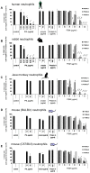
Figure 1
Figure 1. The cytolytic effect of purified S.aureus virulence factors on neutrophils from different species.
Neutrophils from different species, including human (A), rabbit (B), Java monkey (C), BALB/c mice (D), C57/BL6 mice (E) were freshly isolated and 1×10⁶ 0.5 ml−1 cells were incubated with increasing doses of PVL, α-toxin, protein A or PSMs (PSMα1, PSMα2, PSMα3), respectively. PVL: 0.02–0.2 µg/ml (0.5–5 nM); α-toxin: 5, 10 µg/ml (150, 300 nM); protein A: 10, 100 µg/ml (0.238, 2.38 µM); PSMs: 10–60 µg/ml (4–24 µM). Neutrophils were stimulated for 1 h and then cells were washed, stained with annexin V and propidium iodide (taking another hour) and then cell death was measured by flow cytometry. The values represent the mean ± SEM of at least three independent experiments. * P≤0.05, ** P≤0.01, *** P≤0.001 (independent t-test comparing the rate of intact cells between control and stimulated cells). Taking of blood samples from humans and animals were approved by the local ethics committee.
Figure 2
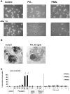
Figure 2. Differences between PVL- and PSMs-induced cell death.
Human neutrophils were freshly isolated and stimulated with staphylococcal components as described in figure 1. Neutrophils were stimulated for 1 h with PVL (80 ng/ml) or PSMs (60 µg/ml) and cells were analyzed by light microscopy with a live cell imaging system (A). Neutrophils were stimulated for 1 h with PVL (40 ng/ml) and processed for electron microscopy (B). Cells were stimulated for 10 min and an oxidative burst reaction was determined by a burst-test (Orpegen Pharma). The values represent the mean ± SEM of at least three independent experiments. * P≤0.05, ** P≤0.01, *** P≤0.001 (independent t-test comparing the rate of burst reaction between control and stimulated cells; C).
Figure 3
Figure 3. The impact of PVL expression on human neutrophil survival.
Human neutrophils were freshly isolated and 1×10⁶ 0.5 ml−1 cells were incubated with live bacteria, which were grown in overnight cultures and used for stimulating cells at an multiplicity of infection (MOI 10–200) as indicated. In these experiments we used heterologous expression strains of TM300 and Cowan I (A, E, F), the wild-type strain USA300 and its knock-out mutant USA300ΔPVL (B) and pvl-positive (C) and pvl-negative (D) clinical isolates from invasive diseases. After 1 h of incubation with bacteria the cells were washed, stained with annexin V and propidium iodide (taking another hour) and then cell death was measured by flow cytometry. The values represent the mean ± SEM of at least three independent experiments.
Figure 4
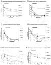
Figure 4. Time-dependent effect of purified PVL vs. live bacteria on neutrophil cell death induction.
Human neutrophils were freshly isolated and 1×10⁶ 0.5 ml−1 cells were stimulated with purified PVL (A) or with live bacteria of wild-type strains at an MOI of 100 (B). Cell death was determined every 15 min. For this, cells were washed, rapidly (for 5 min) stained with PI and cell death was instantly determined by flow cytometry. The values represent the mean ± SEM of at least three independent experiments.
Figure 5
Figure 5. The cytotoxic effect of bacterial culture supernatants is dependent on PVL expression.
Human neutrophils were freshly isolated and 1×10⁶ 0.5 ml−1 cells were incubated with bacterial supernatants, which were prepared from overnight cultures of different strains and used for stimulating cells (10%). In these experiments we used bacterial supernatants of the heterologous expression strain TM300+PVL and of the wild-type strain USA300 and its knock-out mutant USA300ΔPVL (A); furthermore we used bacterial supernatants of pvl-positive and pvl-negative clinical isolates from invasive diseases (B). The presence of the pvl-gene in the indicated strains and the amount of PVL production in the bacterial supernatants is given semi-quantitatively as listed in table 1 and demonstrated in Figure S1C. After 30 min of incubation of the cells with bacterial supernatants, cells were washed, rapidly (for 5 min) stained with PI and cell death was instantly determined by flow cytometry. The values represent the mean ± SEM of at least three independent experiments. *** P≤0.001 (independent t-test comparing the rate of intact cells after stimulation with supernatants of USA300 and USA300ΔPVL).

References

    1. Lowy FD. Staphylococcus aureus infections. N Engl J Med. 1998;339:520–532. - PubMed
    1. Gordon RJ, Lowy FD. Pathogenesis of methicillin-resistant Staphylococcus aureus infection. Clin Infect Dis. 2008;46(Suppl 5):S350–S359. - PMC - PubMed
    1. King MD, Humphrey BJ, Wang YF, Kourbatova EV, Ray SM, et al. Emergence of community-acquired methicillin-resistant Staphylococcus aureus USA 300 clone as the predominant cause of skin and soft-tissue infections. Ann Intern Med. 2006;144:309–317. - PubMed
    1. Gillet Y, Issartel B, Vanhems P, Fournet JC, Lina G, et al. Association between Staphylococcus aureus strains carrying gene for Panton-Valentine leukocidin and highly lethal necrotising pneumonia in young immunocompetent patients. Lancet. 2002;359:753–759. - PubMed
    1. Rubinstein E, Kollef MH, Nathwani D. Pneumonia caused by methicillin-resistant Staphylococcus aureus. Clin Infect Dis. 2008;46(Suppl 5):S378–S385. - PubMed

Publication types

LinkOut - more resources