Localisation and mislocalisation of the interferon-inducible immunity-related GTPase, Irgm1 (LRG-47) in mouse cells
- PMID: 20072621
- PMCID: PMC2799677
- DOI: 10.1371/journal.pone.0008648
Localisation and mislocalisation of the interferon-inducible immunity-related GTPase, Irgm1 (LRG-47) in mouse cells
Abstract
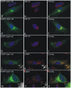
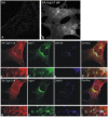
Irgm1 (LRG-47) is an interferon-inducible Golgi membrane associated GTPase of the mouse whose disruption causes susceptibility to many different intracellular pathogens. Irgm1 has been variously interpreted as a regulator of homologous effector GTPases of the IRG family, a regulator of phagosome maturation and as an initiator of autophagy in interferon-induced cells. We find that endogenous Irgm1 localises to late endosomal and lysosomal compartments in addition to the Golgi membranes. The targeting motif known to be required for Golgi localisation is surprisingly also required for endolysosomal localisation. However, unlike Golgi localisation, localisation to the endolysosomal system also requires the functional integrity of the nucleotide binding site, and thus probably reflects transient activation. Golgi localisation is lost when Irgm1 is tagged at either N- or C-termini with EGFP, while localisation to the endolysosomal system is relatively favoured. N-terminally tagged Irgm1 localises predominantly to early endosomes, while C-terminally tagged Irgm1 localises to late endosomes and lysosomes. Both these anomalous distributions are reversed by inactivation of the nucleotide binding site, and the tagged proteins both revert to Golgi membrane localisation. Irgm1 is the first IRG protein to be found associated with the endolysosomal membrane system in addition to either Golgi (Irgm1 and Irgm2) or ER (Irgm3) membranes, and we interpret the result to be in favour of a regulatory function of IRGM proteins at cellular membrane systems. In future analyses it should be borne in mind that tagging of Irgm1 leads to loss of Golgi localisation and enhanced localisation on endolysosomal membranes, probably as a result of constitutive activation.
Conflict of interest statement
Figures


Similar articles
-
Loss of the interferon-γ-inducible regulatory immunity-related GTPase (IRG), Irgm1, causes activation of effector IRG proteins on lysosomes, damaging lysosomal function and predicting the dramatic susceptibility of Irgm1-deficient mice to infection.BMC Biol. 2016 Apr 20;14:33. doi: 10.1186/s12915-016-0255-4. BMC Biol. 2016. PMID: 27098192 Free PMC article.
-
Irgm1 (LRG-47), a regulator of cell-autonomous immunity, does not localize to mycobacterial or listerial phagosomes in IFN-γ-induced mouse cells.J Immunol. 2013 Aug 15;191(4):1765-74. doi: 10.4049/jimmunol.1300641. Epub 2013 Jul 10. J Immunol. 2013. PMID: 23842753 Free PMC article.
-
Palmitoylation of the immunity related GTPase, Irgm1: impact on membrane localization and ability to promote mitochondrial fission.PLoS One. 2014 Apr 21;9(4):e95021. doi: 10.1371/journal.pone.0095021. eCollection 2014. PLoS One. 2014. PMID: 24751652 Free PMC article.
-
The immunity-related GTPases in mammals: a fast-evolving cell-autonomous resistance system against intracellular pathogens.Mamm Genome. 2011 Feb;22(1-2):43-54. doi: 10.1007/s00335-010-9293-3. Epub 2010 Oct 30. Mamm Genome. 2011. PMID: 21052678 Free PMC article. Review.
-
The IRG proteins: a function in search of a mechanism.Immunobiology. 2008;213(3-4):367-75. doi: 10.1016/j.imbio.2007.11.005. Epub 2007 Dec 31. Immunobiology. 2008. PMID: 18406381 Review.
Cited by
-
The GTPase activity of murine guanylate-binding protein 2 (mGBP2) controls the intracellular localization and recruitment to the parasitophorous vacuole of Toxoplasma gondii.J Biol Chem. 2012 Aug 10;287(33):27452-66. doi: 10.1074/jbc.M112.379636. Epub 2012 Jun 22. J Biol Chem. 2012. PMID: 22730319 Free PMC article.
-
Loss of the interferon-γ-inducible regulatory immunity-related GTPase (IRG), Irgm1, causes activation of effector IRG proteins on lysosomes, damaging lysosomal function and predicting the dramatic susceptibility of Irgm1-deficient mice to infection.BMC Biol. 2016 Apr 20;14:33. doi: 10.1186/s12915-016-0255-4. BMC Biol. 2016. PMID: 27098192 Free PMC article.
-
Innate immunity to Toxoplasma gondii infection.Nat Rev Immunol. 2014 Feb;14(2):109-21. doi: 10.1038/nri3598. Nat Rev Immunol. 2014. PMID: 24457485 Review.
-
Irgm1 protects hematopoietic stem cells by negative regulation of IFN signaling.Blood. 2011 Aug 11;118(6):1525-33. doi: 10.1182/blood-2011-01-328682. Epub 2011 Jun 1. Blood. 2011. PMID: 21633090 Free PMC article.
-
Host interferon-γ inducible protein contributes to Brucella survival.Front Cell Infect Microbiol. 2012 Apr 27;2:55. doi: 10.3389/fcimb.2012.00055. eCollection 2012. Front Cell Infect Microbiol. 2012. PMID: 22919646 Free PMC article.
References
-
- Martens S, Howard J. The Interferon-Inducible GTPases. Annu Rev Cell Dev Biol. 2006;22:559–589. - PubMed
-
- Taylor GA. IRG proteins: key mediators of interferon-regulated host resistance to intracellular pathogens. Cell Microbiol. 2007;9:1099–1107. - PubMed
-
- Zhao YO, Rohde C, Lilue JT, Konen-Waisman S, Khaminets A, et al. Toxoplasma gondii and the Immunity-Related GTPase (IRG) resistance system in mice: a review. Mem Inst Oswaldo Cruz. 2009;104:234–240. - PubMed
-
- Coers J, Bernstein-Hanley I, Grotsky D, Parvanova I, Howard JC, et al. Chlamydia muridarum evades growth restriction by the IFNγamma-inducible host resistance factor Irgb10. J Immunol. 2008;180:6237–6245. - PubMed
Publication types
MeSH terms
Substances
Grants and funding
LinkOut - more resources
Full Text Sources
Molecular Biology Databases
Miscellaneous

