. 2008 Dec;1(2-4):73-84.
doi: 10.1007/s12177-008-9011-5. Epub 2008 Nov 18.

Identification of the molecular signatures integral to regenerating photoreceptors in the retina of the zebra fish

Identification of the molecular signatures integral to regenerating photoreceptors in the retina of the zebra fish

Sonya E L Craig et al. J Ocul Biol Dis Infor. 2008 Dec.

Abstract

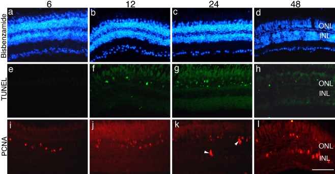
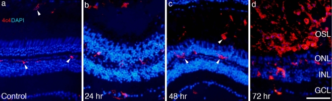
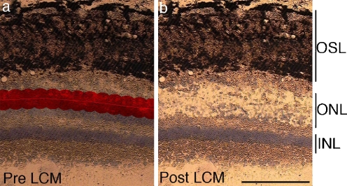
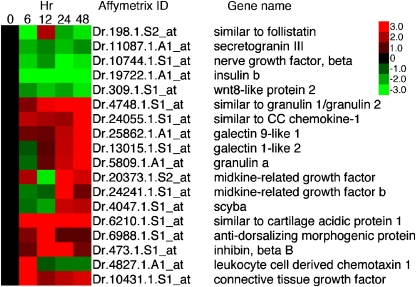
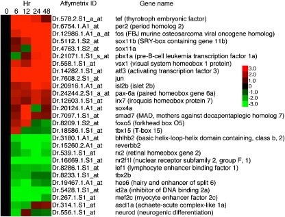
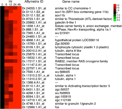
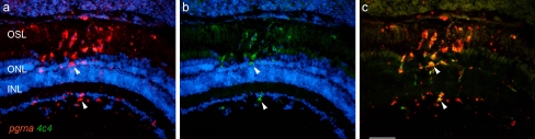
Investigating neuronal and photoreceptor regeneration in the retina of zebra fish has begun to yield insights into both the cellular and molecular means by which this lower vertebrate is able to repair its central nervous system. However, knowledge about the signaling molecules in the local microenvironment of a retinal injury and the transcriptional events they activate during neuronal death and regeneration is still lacking. To identify genes involved in photoreceptor regeneration, we combined light-induced photoreceptor lesions, laser-capture microdissection of the outer nuclear layer (ONL) and analysis of gene expression to characterize transcriptional changes for cells in the ONL as photoreceptors die and are regenerated. Using this approach, we were able to characterize aspects of the molecular signature of injured and dying photoreceptors, cone photoreceptor progenitors, and microglia within the ONL. We validated changes in gene expression and characterized the cellular expression for three novel, extracellular signaling molecules that we hypothesize are involved in regulating regenerative events in the retina.

Electronic supplementary material: The online version of this article (doi:10.1007/s12177-008-9011-5) contains supplementary material, which is available to authorized users.

Keywords: Laser-capture microdissection; Microarray; Microglia; Regenerative neurogenesis; Retinal stem cells.


Figures

Fig. 1
Fig. 1
Light induces photoreceptor-specific cell death and triggers proliferation. Animals were exposed to constant light and retinal sections were prepared from animals after 6 h (a, e, i), 12 h (b, f, j), 24 h (c, g, k), and 48 h (d, h, l). Sections were stained with bisbenzimide to label nuclear layers (ad). The same retinal sections illustrated in ad were labeled with TUNEL (arrowheads, eh). Adjacent sections were immunolabeled with antibodies against PCNA (arrowheads, il). Scale bar = 50 μm. ONL, outer nuclear layer; INL, inner nuclear layer
Fig. 2
Fig. 2
As photoreceptors die, resident microglia migrate into the outer nuclear and outer segment layers. Retinal sections were taken from control (a) and experimental retinas exposed to constant light for 24 h (b), 48 h (c), and 72 h (d) and immunolabeled with the microglia-specific antibody, 4C4 (arrowheads), and counterstained with 4′,6-diamidino-2-phenylindole. Scale bar = 50 μm. OSL, outer segment layer; ONL, outer nuclear layer; INL, inner nuclear layer; GCL, ganglion cell layer
Fig. 3
Fig. 3
Laser-capture microdissection of cells from the ONL. A video image of a section from a control retina is illustrated in a and b. The ONL is highlighted using the LCM robot, identifying the area selected for dissection (a). Panel b illustrates the retinal section in a after LCM. Scale bar = 100 μm. OSL, outer segment layer; ONL, outer nuclear layer; INL, inner nuclear layer
Fig. 4
Fig. 4
Heat map representing the hierarchical clustering of 1,937 genes observed to change in expression in the ONL over the course of photoreceptor injury and regeneration. Uncentered Pearson’s correlation and centroid linkage of genes were used to cluster transcriptional changes. Three clustering differences were observed: genes with expression that increased, genes with expression that decreased, and genes that showed differential expression at the 12-h time point
Fig. 5
Fig. 5
Heat map of selected phototransduction genes that change over the course of the light injury. The majority of photoreceptor-specific genes decreased their levels of expression, consistent with photoreceptor loss due to cell death
Fig. 6
Fig. 6
Heat map of selected stress response genes that change in expression over the course of light injury. The majority of these genes showed marked increase in expression during the early phases of the light-induced injury
Fig. 7
Fig. 7
Heat map of selected secreted proteins that change over the course of the light injury
Fig. 8
Fig. 8
Heat map of selected transcription factors that change over the course of light injury
Fig. 9
Fig. 9
Heat map of selected genes that showed a marked increase in transcription at the 48-h time point
Fig. 10
Fig. 10
Expression of Lgals9l1 mRNA in microglia in the ONL. Panels ac illustrate a retinal section from an animal exposed to constant light for 12 h, then labeled by in situ hybridization with probes against lgals9l1 mRNA (a), immunolabeled for microglia with 4C4 antibody (b). Panel c is the digital overlay of a and b. Panels df illustrate a retina section from an animal exposed to constant light for 48 h, then labeled by in situ hybridization with probes against lgals9l1 mRNA (d), immunolabeled for microglia with 4C4 antibody (e). Panel f is the digital overlay of d and e. Arrowheads indicate the same cells in ac and df. ONL, outer nuclear layer; INL, inner nuclear layer. Scale bar = 50 μm
Fig. 11
Fig. 11
Lgals1l2 protein is expressed by proliferating Müller glial stem cells and their progeny and microglia in the ONL of injured retina. Panels ac illustrate a retinal section from a transgenic (gfap:GFP)mi2001 fish exposed to constant light for 48 h followed by immunolabeling for Lgals1l2 (a) and GFP (b). Panel c is a digital overlay of a and b. Panels df illustrate a retinal section from an animal exposed to constant light for 48 h and immunolabeled for Lgals1l2 (d) and 4C4 (e). Panel f is a digital overlay of d and e. Arrowheads indicate the same cells in ac and df. Scale bar = 50 μm. ONL, outer nuclear layer; INL, inner nuclear layer
Fig. 12
Fig. 12
Progranulin-a is expressed by microglia at the site of photoreceptor injury. Panel a illustrates an in situ hybridization showing pgrna at the site of photoreceptor injury. Panel b is the same section as in a but immunostained with the 4c4 antibody. Panel c is the digital overlay of panels a and b. Arrowheads identify the same cells in ac. Scale bar = 50 μm. OSL, outer segment layer; ONL, outer nuclear layer; INL, inner nuclear layer
Fig. 13
Fig. 13
Heat map highlighting selected genes that increased expression at the 12-h time point
Fig. 14
Fig. 14
Heat map highlighting selected genes that decreased in expression at the 12-h time point

Similar articles

Cited by

References

    1. Benjamini Y, Yekutieli DJ. Am Stat Assoc. 2005;100:71–80. doi: 10.1198/016214504000001907. - DOI
    1. Bernardos RL, Raymond PA. GFAP transgenic zebra fish. Gene Expr Patterns. 2006;6:1007–1013. doi: 10.1016/j.modgep.2006.04.006. - DOI - PubMed
    1. Bernardos RL, Barthel LK, Meyers JR, Raymond PA. Late-stage neuronal progenitors in the retina are radial Müller glia that function as retinal stem cells. J Neurosci. 2007;27:7028–7040. doi: 10.1523/JNEUROSCI.1624-07.2007. - DOI - PMC - PubMed
    1. Cadieux B, Chitramuthu BP, Baranowski D, Bennett HP. The zebra fish progranulin gene family and antisense transcripts. BMC Genomics. 2005;6:156. doi: 10.1186/1471-2164-6-156. - DOI - PMC - PubMed
    1. Camby I, Le Mercier M, Lefranc F, Kiss R. Galectin-1: a small protein with major functions. Glycobiology. 2006;16:137R–157R. doi: 10.1093/glycob/cwl025. - DOI - PubMed