. 2008 Dec;1(2-4):85-93.
doi: 10.1007/s12177-008-9013-3. Epub 2008 Nov 4.

Systemic 7-methylxanthine in retarding axial eye growth and myopia progression: a 36-month pilot study

Systemic 7-methylxanthine in retarding axial eye growth and myopia progression: a 36-month pilot study

Klaus Trier et al. J Ocul Biol Dis Infor. 2008 Dec.

Abstract

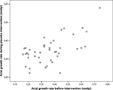
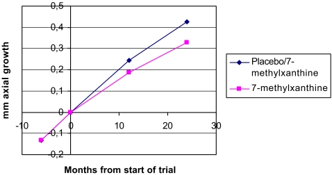
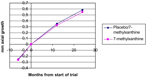
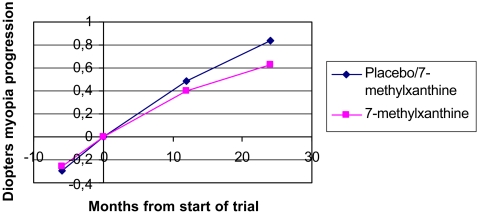
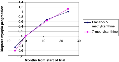
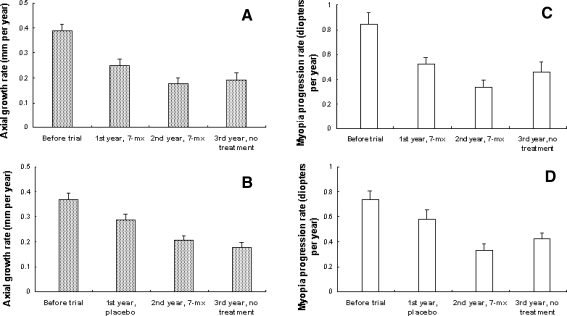
The adenosine antagonist 7-methylxanthine (7-mx) works against myopia in animal models. In a clinical trial, 68 myopic children (mean age 11.3 years) received either placebo or 7-mx tablets for 12 months. All participants subsequently received 7-mx for another 12 months, after which treatment was stopped. Axial length was measured with Zeiss IOL-Master and cycloplegic refraction with Nikon Retinomax at -6, 0, 12, 24, and 36 months. Axial growth was reduced among children treated with 7-mx for 24 months compared with those only treated for the last 12 months. Myopia progression and axial eye growth slowed down in periods with 7-mx treatment, but when the treatment was stopped, both myopia progression and axial eye growth continued with invariable speed. The results indicate that 7-mx reduces eye elongation and myopia progression in childhood myopia. The treatment is safe and without side effects and may be continued until 18-20 years of age when myopia progression normally stops.

Keywords: 7-methylxanthine; Adenosine receptor antagonist; Clinical trial; Myopia.


Figures

Fig. 1
Fig. 1
Correlation between axial length growth-rate during the 12-months placebo controlled trial and the observation period before intervention, shown as mean values for right and left eye in each of the participants in the placebo group (p < 0.01)
Fig. 2
Fig. 2
Axial growth in children with a base-line axial growth rate of 0.075–0.190 mm per 6 months (n = 40). In the period 12–24 months, both groups received 7-methylxanthine. The difference at 24 months was −0.098 mm (95% Cl −0.211 and 0.013 mm, p = 0.048)
Fig. 3
Fig. 3
Axial growth in children with a base-line axial growth rate of 0.200–0.390 mm per 6 months (n = 28). In the period 12–24 months, both groups received 7-methylxanthine. The difference at 24 months was −0.037 mm (95% Cl −0.195 and 0.120 mm, p = 0.667)
Fig. 4
Fig. 4
Myopia progression in children with a base-line axial growth rate of 0.075–0.190 mm per 6 months (n = 40). In the period 12–24 months, both groups received 7-methylxanthine. The difference at 24 months was −0.217 diopters (95% Cl −0.478 and 0.044 diopters, p = 0.141)
Fig. 5
Fig. 5
Myopia progression in children with a base-line axial growth rate of 0.200–0.390 mm per 6 months (n = 28). In the period 12–24 months, both groups received 7-methylxanthine. The difference at 24 months was 0.121 diopters (95% Cl −0.267 and 0.508 diopters, p = 0.541)
Fig. 6
Fig. 6
a, b Axial growth rate before trial, during treatment with 7-mx or placebo in the first year, during treatment with 7-mx in the second year, and after discontinuation of treatment. In the first and second year, axial growth rate is significantly lower than in the precedent period, but in the third year, after discontinuation of treatment, axial growth rate is not different from the precedent year. c, d Myopia progression rate in the same periods. The myopia progression rate is significantly reduced compared with the precedent period during treatment with 7-mx, but unchanged in periods with placebo treatment or no treatment

References

    1. McBrien NA, Gentle A. Role of sclera in the development and pathological complications of myopia. Prog Ret Eye Res. 2003;22:307–338. doi: 10.1016/S1350-9462(02)00063-0. - DOI - PubMed
    1. Guggenheim JA, McBrien NA. Form-deprivation myopia induces activation of scleral matrix metalloproteinase-2 in tree shrew. Invest Ophthalmol Vis Sci. 1996;37:1380–1395. - PubMed
    1. Jobling A, Nguyen M, Gentle A, et al. Isoform-specific changes in scleral TGF-β expression and the regulation of collagen synthesis during myopia progression. J Biol Chem. 2004;279:18121–18126. doi: 10.1074/jbc.M400381200. - DOI - PubMed
    1. Jones LA, Mitchell GL, Mutti DO, et al. Comparison of ocular component growth curves among refractive error groups in children. Invest Ophthalmol Vis Sci. 2005;46:2317–2327. doi: 10.1167/iovs.04-0945. - DOI - PubMed
    1. Thorn F, Gwiazda J, Held R. Myopia progression is specified by a double exponential growth function. Opt Vis Sci. 2005;82:E286. doi: 10.1097/01.OPX.0000159370.66540.34. - DOI - PubMed

LinkOut - more resources