. 2009 Jan 1:41:347-374.
doi: 10.1146/annurev.fluid.010908.165136.

Fluid and Solute Transport in Bone: Flow-Induced Mechanotransduction

Affiliations

Fluid and Solute Transport in Bone: Flow-Induced Mechanotransduction

Susannah P Fritton et al. Annu Rev Fluid Mech. .

Abstract

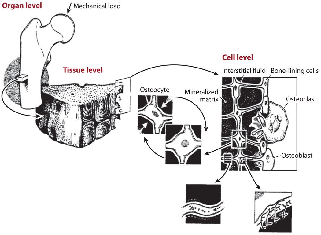
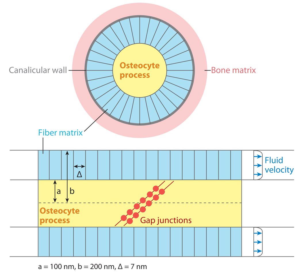
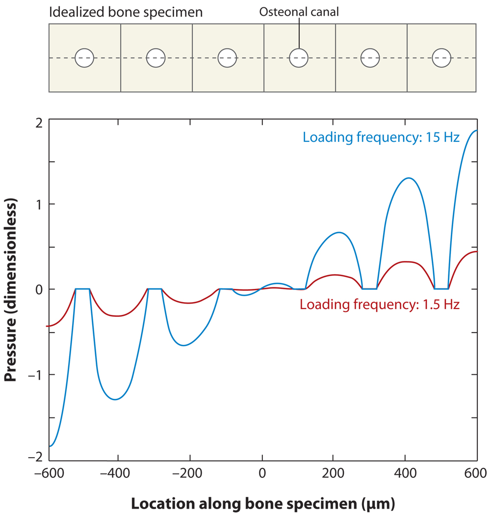
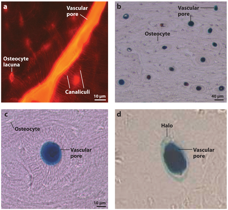
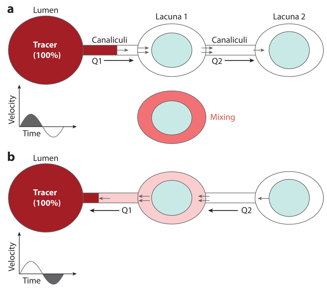
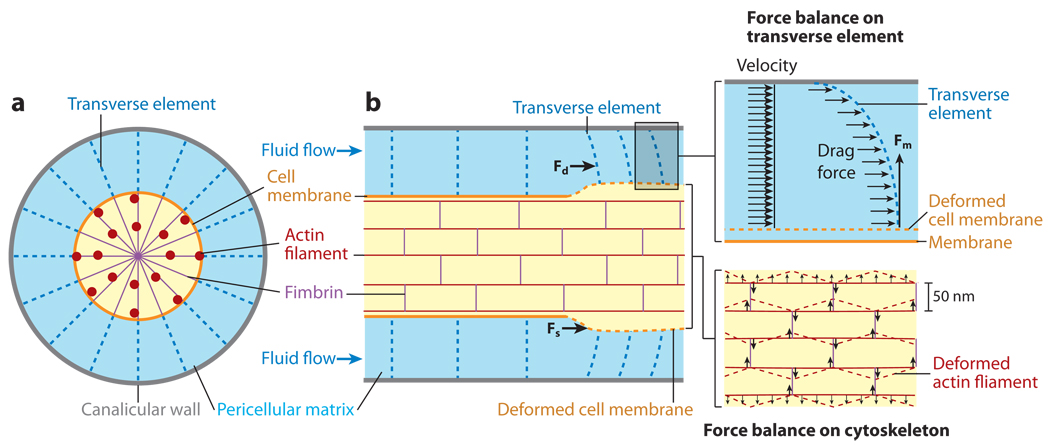
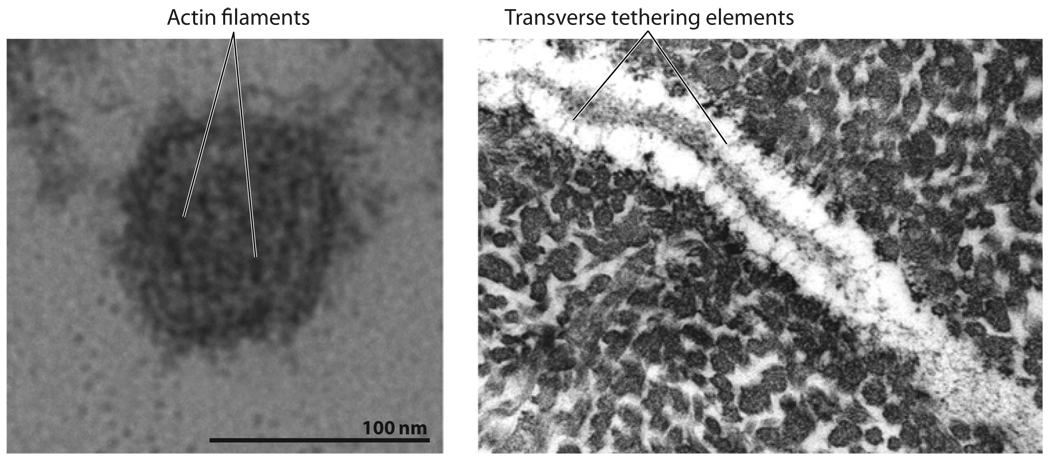
Much recent evidence suggests that bone cells sense their mechanical environment via interstitial fluid flow. In this review, we summarize theoretical and experimental approaches to quantify fluid and solute transport in bone, starting with the early investigations of fluid shear stress applied to bone cells. The pathways of bone interstitial fluid and solute movement are high-lighted based on recent theoretical models, as well as a new generation of tracer experiments that have clarified and refined the structure and function of the osteocyte pericellular matrix. Then we trace how the fluid-flow models for mechanotransduction have evolved as new ultrastructural features of the osteocyte lacunar-canalicular porosity have been identified and how more recent in vitro fluid-flow and cell-stretch experiments have helped elucidate at the molecular level the possible pathways for cellular excitation in bone.


Figures

Figure 1
Figure 1
Mechanical loading applied at the whole bone level is transmitted through the bone tissue to the cellular level and causes movement of the interstitial fluid surrounding osteocytes in the mineralized matrix. The osteocytes are distributed throughout bone tissue and connect to each other and to bone-lining cells and osteoblasts on the bone surfaces. Figure adapted from Rubin et al. 1990. Reprinted with permission from Elsevier.
Figure 2
Figure 2
Osteocytes are housed in lacunae; their cell processes connect with one another through canaliculi. During cyclic mechanical loading, interstitial fluid from the canaliculi flows into and out of the vascular porosity surrounding bone capillaries. Figure adapted and used with permission from Kelly PJ. 1983. Pathways of transport in bone. In Handbook of Physiology: The Cardiovascular System. Peripheral Circulation and Organ Blood Flow, ed. JT Shephard, FM Abboud, sect. 2, pt. 1, chapt. 12, pp. 371–96. Bethesda, MD: Am. Physiol. Soc.
Figure 3
Figure 3
Schematic illustration of an osteocyte process and canaliculus, showing a cross-section view (top) and a longitudinal segment (bottom) with the ends of two cell processes connecting via gap junctions. The fluid annulus surrounding the osteocyte is filled with a pericellular matrix with pore size ∆. Figure adapted from Weinbaum et al. 1994. Reprinted with permission from Elsevier.
Figure 4
Figure 4
Pressure distribution across a portion of an idealized bone specimen of thickness 1.2 mm (shown on top), mimicking Starkebaum et al.’s (1979) bending experiments. The circles represent osteonal canals, and the cusps around the canals demonstrate higher pressure gradients near the vascular pores, which are further amplified at higher loading frequencies. Figure adapted from Wang et al. 1999. Reprinted with permission from Elsevier.
Figure 5
Figure 5
Tracer labeling in the rat tibia mid-diaphysis. (a) Reactive red (~1-nm diameter) labeling shows vascular pore, osteocyte lacunae, and canaliculi staining. (b,c) Ferritin (~12-nm diameter) was found in vascular pores without labeling the surrounding osteocyte lacunae. Figure adapted from Wang et al. 2004. (d ) By varying the histological processing methods, ferritin halos surrounding vascular pores can be produced. Figure adapted from Ciani et al. 2005. Reprinted with permission from Elsevier.
Figure 6
Figure 6
Schematic of Wang et al.’s (2000) proposed mixing model. (a) For cyclic loading, if the fluid displacement is large enough to pass from the vascular lumen (the tracer source) through the canaliculi to the first osteocyte lacuna, an instant mixing process occurs in the lacuna. (b) Upon the reversal of cyclic loading, even though the fluid returns to the vascular pore, net solute transport to the lacuna occurs. Figure from Wang et al. 2000 with kind permission from Springer Science + Business Media.
Figure 7
Figure 7
You et al.’s (2001) strain-amplification model illustrating the osteocyte process in cross section (a) and longitudinal section (b). Actin filaments span the process, which is attached to the canalicular wall via transverse elements. Applied loading results in interstitial fluid flow through the pericellular matrix, producing a drag force on the tethering fibers. Figure reprinted from You et al. 2001, with permission from Elsevier.
Figure 8
Figure 8
(Left panel) Electron microscopy (EM) of a cross section of osteocyte process from mouse bone showing the central actin filament bundle. (Right panel) EM of a longitudinal section of osteocyte process from mouse bone showing tethering fibers. Figure taken from You et al. 2004 with permission.
Figure 9
Figure 9
Schematic of Han et al.’s (2004) strain-amplification model, illustrating greatly refined actin filament and cross-filament arrangements. (a) Cross section of osteocyte process. (b) Fimbrin cross-linking arrangement. (c) Longitudinal section of osteocyte process. (d ) Double helix of cross-filaments. Figure taken from Han et al. 2004, Proc. Natl. Acad. Sci. USA 101:16689–94. Copyright (2004) National Academy of Sciences, U.S.A.
Figure 10
Figure 10
(a) Electron micrographs illustrating the canalicular protrusions and transverse tethering elements of the osteocyte process. (b) Schematic of Wang et al.’s (2007) integrin-based strain-amplification model. The conical canalicular protrusions can be seen both in cross section (left) and longitudinal section (right). Figure adapted from Wang et al. 2007, Proc. Natl. Acad. Sci. USA 104:15941–46. Copyright (2007) National Academy of Sciences, U.S.A.
Figure 11
Figure 11
Cellular axial strains as a function of applied tissue-level strains and loading frequency. Physiological loading is marked as loading at 1 Hz, the approximate frequency of walking. Vibration loading applied by a low-magnitude oscillating platform is also indicated. For vibration loading shown by Rubin et al. (2004) to inhibit bone loss in humans, the osteocyte axial strain is predicted to be greater than the 0.5% strain (5000 microstrain) required to elicit intracellular signaling in vitro. Figure adapted from Wang et al. 2007, Proc. Natl. Acad. Sci. USA 104:15941–46. Copyright (2007) National Academy of Sciences, U.S.A.

References

    1. Ajubi NE, Klein-Nulend J, Alblas MJ, Burger EH, Nijweide PJ. Signal transduction pathways involved in fluid flow-induced PGE2 production by cultured osteocytes. Am. J. Physiol. 1999;276:E171–E178. - PubMed
    1. Ajubi NE, Klein-Nulend J, Nijweide PJ, Vrijheid-Lammers T, Alblas MJ, Burger EH. Pulsating fluid flow increases prostaglandin production by cultured chicken osteocytes—a cytoskeleton-dependent process. Biochem. Biophys. Res. Commun. 1996;225:62–68. - PubMed
    1. Anderson EJ, Kaliyamoorthy S, Iwan J, Alexander D, Knothe Tate ML. Nano-microscale models of periosteocytic flow show differences in stresses imparted to cell body and processes. Ann. Biomed. Eng. 2005;33:52–62. - PubMed
    1. Ayasaka N, Kondo T, Goto T, Kido MA, Nagata E, Tanaka T. Differences in the transport systems between cementocytes and osteocytes in rats using microperoxidase as a tracer. Arch. Oral Biol. 1992;37:363–369. - PubMed
    1. Bakker A, Klein-Nulend J, Burger E. Shear stress inhibits while disuse promotes osteocyte apoptosis. Biochem. Biophys. Res. Commun. 2004;320:1163–1168. - PubMed

LinkOut - more resources