

doi: 10.1007/978-1-60761-411-1_11.
Multiphoton redox ratio imaging for metabolic monitoring in vivo
Affiliations
- PMID: 20072916
- PMCID: PMC2874879
- DOI: 10.1007/978-1-60761-411-1_11
Item in Clipboard
Multiphoton redox ratio imaging for metabolic monitoring in vivo
Methods Mol Biol.
2010.
Abstract
Metabolic monitoring at the cellular level in live tissues is important for understanding cell function, disease processes, and potential therapies. Multiphoton imaging of the relative amounts of NADH and FAD (the primary electron donor and acceptor, respectively, in the electron transport chain) provides a noninvasive method for monitoring cellular metabolic activity with high resolution in three dimensions in vivo. NADH and FAD are endogenous tissue fluorophores, and thus this method does not require exogenous stains or tissue excision. We describe the principles and protocols of multiphoton redox ratio imaging in vivo.
Figures
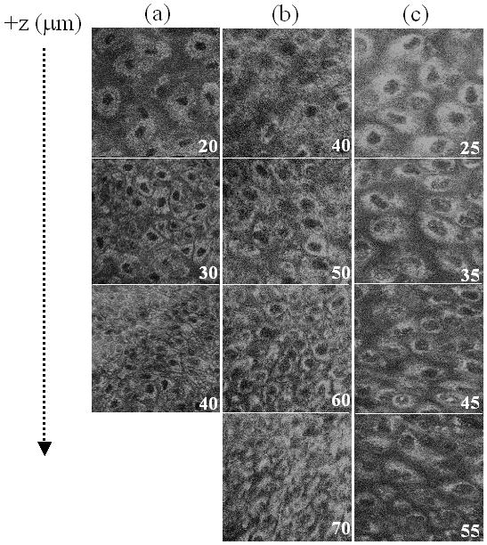
Representative in vivo three-dimensional multiphoton images of the redox ratio (fluorescence intensity of FAD/NADH) from tissues diagnosed as normal (a), low-grade precancer (b), and high-grade precancer (c) in the 7,12-dimethylbenz(a)anthracene (DMBA)-treated hamster cheek pouch model of oral cancer. The numbers in the corner of each image indicate the depth below the tissue surface in microns, and each image is 100 × 100 μm. From ref [15], © 2007, National Academy of Sciences, U.S.A.
References
-
- Chance B, Schoener B, Oshino R, Itshak F, Nakase Y. Oxidation-reduction ratio studies of mitochondria in freeze-trapped samples. NADH and flavoprotein fluorescence signals. J Biol Chem. 1979;254:4764–71. - PubMed
-
- Drezek R, Brookner C, Pavlova I, Boiko I, Malpica A, Lotan R, Follen M, Richards-Kortum R. Autofluorescence microscopy of fresh cervical-tissue sections reveals alterations in tissue biochemistry with dysplasia. Photochem Photobiol. 2001;73:636–41. - PubMed
-
- Gulledge CJ, Dewhirst MW. Tumor oxygenation: a matter of supply and demand. Anticancer Res. 1996;16:741–9. - PubMed
-
- Ramanujam N, Kortum RR, Thomsen S, Jansen AM, Follen M, Chance B. Low temperature fluorescence imaging of freeze-trapped human cervical tissues. Opt Express. 2001;8:335–343. - PubMed
Publication types
MeSH terms
Substances
Grants and funding
LinkOut - more resources
Full Text Sources
Other Literature Sources
