. 2010 Mar 31;28(3):620-32.
doi: 10.1002/stem.301.

Development and function of myeloid-derived suppressor cells generated from mouse embryonic and hematopoietic stem cells

Affiliations

Development and function of myeloid-derived suppressor cells generated from mouse embryonic and hematopoietic stem cells

Zuping Zhou et al. Stem Cells. .

Abstract

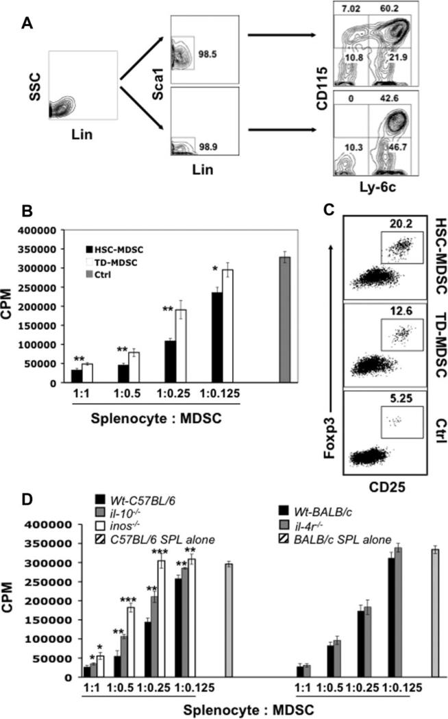
Emerging evidence suggests that myeloid-derived suppressor cells (MDSCs) have great potential as a novel immune intervention modality in the fields of transplantation and autoimmune diseases. Thus far, efforts to develop MDSC-based therapeutic strategies have been hampered by the lack of a reliable source of MDSCs. Here we show that functional MDSCs can be efficiently generated from mouse embryonic stem (ES) cells and bone marrow hematopoietic stem (HS) cells. In vitro-derived MDSCs encompass two homogenous subpopulations: CD115(+)Ly-6C(+) and CD115(+)Ly-6C(-) cells. The CD115(+)Ly-6C(+) subset is equivalent to the monocytic Gr-1(+)CD115(+)F4/80(+) MDSCs found in tumor-bearing mice. In contrast, the CD115(+)Ly-6C(-) cells, a previously unreported population of MDSCs, resemble the granulocyte/macrophage progenitors developmentally. In vitro, ES- and HS-MDSCs exhibit robust suppression against T-cell proliferation induced by polyclonal stimuli or alloantigens via multiple mechanisms involving nitric oxide synthase-mediated NO production and interleukin (IL)-10. Impressively, they display even stronger suppressive activity and significantly enhance ability to induce CD4(+)CD25(+)Foxp3(+) regulatory T-cell development compared with tumor-derived MDSCs. Furthermore, adoptive transfer of ES-MDSCs can effectively prevent alloreactive T-cell-mediated lethal graft-versus-host disease, leading to nearly 82% long-term survival among treated mice. The successful in vitro generation of MDSCs may represent a critical step toward potential clinical application of MDSCs.


Figures

Figure 1
Figure 1
Directed differentiation of HoxB4 ES cells into myeloid-derived suppressor cells. (A): Kinetics of MDSC development. HoxB4 ES cells were first grown in suspension to form embryonic bodies (EB), and cells dissociated from day-6 EB were plated onto semiconfluent (irradiated) OP9 stromal cells and cultured in medium one (M1, see details in Methods). After 48 hours, M1 medium was removed and replaced with medium two (M2, see details in Methods). Floating and loosely attached cells recovered on different days after coculture in M2 were stained and analyzed by fluorescence-activated cell sorting. Data shown are representative of at least five experiments with reproducible results. (B): Opposing roles of M-CSF and GM-CSF in the generation of ES-derived CD115+ cells. Data shown are representative of three experiments with reproducible results. (C): Dose-dependent effect of M-CSF on the generation of ES-derived CD115+ cells. Error bars represent standard deviations (n = 3, triplicates). For experiments in (B) and (C), HoxB4 ES cells were differentiated using the same procedure as described in (A) except for the last step, where cells were cultured for 10 days in M2 or M2 supplanted with M-CSF or GM-CSF (B) or M2 plus various concentration of M-CSF (C). (D): Efficiency of myeloid-derived suppressor cell production: at the indicated number of dates in M2+M-CSF, floating and loosely adherent cells were retrieved and cell numbers were determined based on cell count and percentage of individual populations. Error bars represent standard deviations (n = 3, independent experiments). Abbreviations: Ctrl, control; D, days; GM-CSF, granulocyte-macrophage colony-stimulating factor; M-CSF, macrophage colony stimulating factor.
Figure 2
Figure 2
Potent suppressive capacity of ES-MDSCs. (A): Inhibition of polyclonally stimulated T-cell proliferation by ES-derived CD115+ cells (CD115+ versus control, **p <.001; ***p <.0001). (B): Inhibition of alloantigen-stimulated T-cell proliferation by ES-derived CD115+ cells (CD115+ versus control, ***p <.0001). (C): Suppression of T-cell proliferation by CD115+Ly-6C+ and CD115+Ly-6C populations (CD115+Ly-6C versus CD115+Ly-6C+, *p <.05). (D): A comparison of suppressive activity of ES-derived CD115+ cells versus purified CD115+ cells isolated from tumor-bearing mice (TD-CD115+) (ES-CD115+ versus TD-CD115+, **p <.001). Various numbers of ES-derived cells or TD-CD115+ cells were coincubated with 129SvEv splenocytes in the presence of anti-CD3/anti-CD28 (A, C, D) or irradiated BALB/c splenocytes (B). [3H]-Thymidine was pulsed for the final 8 hours of a 3-day (A, C, D) or a 4-day (B) culture. Data shown are representatives of at least three experiments with consistent results. Abbreviations: Ctrl, Control (splenocyte alone); ES, embryonic stem; MDSC, myeloid-derived suppressor cell; TD, tumor-derived.
Figure 3
Figure 3
Various pathways mediating suppressor function of ES-MDSCs. (A): Treg development induced by ES-MDSCs. Data shown are representative dot plots and numbers inserted are mean ± SD from three reproducible experiments. (B): NO production by ES-MDSCs. In A and B, 1 × 10⁶ of ES-derived CD115+ and CD115 cells or TD-CD115+ cells were cocultured with 4 × 10⁶ 129SvEv splenocytes for 5 days in the presence of anti-CD3/anti-CD28. CD4+CD25+Foxp3+ cells were analyzed by intracellular staining. NO levels in the supernatants were determined per manufacturer's protocol (n = 3 triplicate, ES-CD115+ vs. Ctrl or ES-CD115 , **p <.0001; ES-CD115+ vs. TD-CD115+, *p <.05). Data are representative of two experiments with consistent results. (C): Expression of iNOS, arginase 1, IL-10, and TGF-β by ES-MDSCs. ES-derived CD115+ and CD115 cells and TD-CD115+ cells were cultured for 24 hours in the presence or absence of IFN-γ or IL-13. We assessed mRNA expression by reverse transcription polymerase chain reaction. Abbreviations: Arg.1, arginase 1; Ctrl, control; ES, embryonic stem; GAPDH, glyceraldehyde 3-phosphate dehydrogenase; IL, interleukin; iNOS, inducible nitric oxide synthase; NO, nitric oxide; TD, tumor-derived IFN-γ, interferon-gamma; TGF-ß, transforming growth factor-beta.
Figure 4
Figure 4
Morphologic, phenotypic, and developmental characterization of ES-MDSCs. (A): Morphology of ES-derived MDSCs. Differentiated cells were sorted by fluorescence-activated cell sorting (FACS), cytospun, and stained by May-Giemsa (original magnification: 1,000). (B): Surface phenotype of various subsets differentiated from HoxB4 ESs. Cells recovered were directly stained with a panel of surface markers and analyzed by flow cytometry. (C): Distinct potentials of CD115+Ly-6C (upper profiles) and CD115+Ly-6C+ (lower profiles) cells. FACS-sorted subsets of MDSCs were seeded into gelatinized plates and cultured in medium supplemented with the appropriate cytokine mix (see details in Methods) for various lengths of time. Differentiated cells were collected and analyzed by FACS for the expression of CD115 and Ly-6C. (D): Colony forming activity of FACS-sorted CD115+Ly-6C and CD115+Ly-6C+ populations in methylcellulose. CD115+Ly-6C cells, which had higher clonogenic activity than CD115+Ly-6C+ cells (upper histogram, *p <.001), gave rise to M, GM, G colonies, whereas the CD115+Ly-6C+ population generated only M colonies (lower histogram). M, macrophage colonies; GM, macrophage and granulocyte mixed colonies; G, granulocyte colonies. (E): May-Grünwald Giemsa staining of pooled colonies developed from CD115+Ly-6C cells (upper panel, arrows and arrowheads indicated macrophages and granulocytes, respectively) and CD115+Ly-6C+ cells (lower panel). Original magnification: ×400. Cells used in (AD) were obtained on day 10 after differentiation on OP9 in M2 MCSF. Abbreviations: D, day; G, granulocyte colony stimulating factor; GM, granulocyte macrophage colony stimulating factor; M, macrophage stimulating+factor; No., number.
Figure 5
Figure 5
Prevention of allo-HSCT-associated GVHD by ES-MDSCs. (A): Survival curve of recipient mice. Lethally irradiated (8 Gy, TBI) BALB/c mice were left untreated (ν, n = 6) or transplanted via tail vein injection with 129SvEv T-cell-depleted bone marrow cells (TCDBM, 5 × 10⁶/mouse) alone (v, n = 6), or TCDBM plus purified 129SvEv splenic T-cells (T, 5 × 10⁵/mouse) (σ, n = 10), or TCDBM plus T and ES-CD115 (2 × 10⁶/mouse) (î, n = 4), or TCDBM plus T and ES-MDSCs (2 × 10⁶/mouse) (λ, n = 11). Two additional treatments of ES-MDSCs (2 × 10⁶/mouse, each) were given to ES-MDSC recipients on days 4 and 10 after the initial transplantation. Data shown are combined results from two independent experiments. (B): Changes of mean body weight in treated mice (values are mean ± SD. v, n = 6; σ, n= 3–10; λ, n= 9–11 mice). (C): Sections of livers stained with hematoxylin and eosin and harvested on day 23 from indicated groups. Representative micrographs are shown. (D): Kinetics of chimerism in the recipient mice. Spleens (SP), livers, and lymph nodes (LN) were recovered from the recipients (n = 3) on days 4, 7, and 14 after transplantation. Donor-derived T-cells (H-2Kb+) were identified by flow cytometry. Representative dot plots of peripheral T-cell chimerism from one of reproducible experiments are presented. The overall allogeneic chimerism was not apparently affected but the frequencies of donor T-cell subsets were increased in the recipients treated with MDSCs. Abbreviations: BM, bone marrow; ES, embryonic stem; LN, lymph nodes; MDSC myeloid-derived suppressor cells; SP, spleens; T, T cells; TCDBM, T-cell-depleted bone marrow.
Figure 6
Figure 6
Migration pattern of MDSC in GVHD. Irradiated BALB/c mice were injected, via tail vein, with 5 × 10⁶ TCDBM cells and 0.5 × 10⁶ donor T-cells with (n = 5) or without (baseline, n = 4) 1 × 10⁷ PKH26 labeled MDSCs. Mice were killed on day 3 after transfer. Single cell suspensions were prepared from spleen (SP), bone marrow (BM), lymph nodes (LN), liver, and lung and stained with anti-Ly-6C-FITC or isotype control followed by flow cytometric analysis. (A): Absolute numbers of transferred MDSCs in various organs. Significantly higher numbers of transferred MDSCs were found in the spleen (p = .01) and liver (p = .0016). (B): Fold changes of transferred MDSCs in various organs when compared with the baseline (without MDSC transfer). Abbreviations: BM, bone marrow; CT, control; LN, lymph nodes; SP, spleen.
Figure 7
Figure 7
Generation of myeloid-derived suppressor cells from marrow hematopoietic stem/progenitor cells. (A): In vitro differentiation of bone marrow hematopoietic stem/progenitor cells into MDSCs (HSMDSC). BM cells isolated from B6 mice were depleted of lineage positive cells (left panel: after depletion) and then FACS-sorted into Sca1+ and Sca1 Cells (middle panel). Purified cells were differentiated using similar culture condition used in the derivation of ES-MDSCs. (B): Comparison of suppressive activity between HS-MDSC and TD-MDSC. HS-MDSCs or TDMDSCs were coincubated with B6 splenocytes at different ratios in the presence of anti-CD3/anti-CD28 and [3H]-thymidine was pulsed for the final 8 hours of a 3-day culture. (C): Enhanced Treg induction by HS-MDSCs. MDSCs were coincubated for 5 days with B6 splenocytes at a 1:4 ratio in the presence of anti-CD3/anti-CD28. Foxp3 expression was analyzed via intra-cellular staining. (D): The role of IL-10, iNOS, and IL-4 in the suppression mediated by HS-MDSC. HS-MDSCs developed from Lin BM cells of indicated strains of mice were coincubated with B6 (for il10−/− and inos−/− MDSCs) or BALB/c (for il-4r−/− MDSCs, BALB/c background) splenocytes at different ratios in the presence of anti-CD3/anti-CD28. [3H]-Thymi-dine was pulsed for the final 8 hours of a 3-day culture (il-10−/− MDSCs or inos−/− MDSCs versus Wt B6 MDSCs, *p <.05; **p <.01; ***p <.001). Abbreviations: BM, bone marrow; CPM, counts per minute; Ctrl, control; FACS, fluorescence-activated cell sorter; HSC, hematopoietic stem cell; Lin, lineage; MDSC myeloid-derived suppressor cells; TD-MDSC, MDSC cells generated from tumor-bearing mice; Wt, wild-type.

References

    1. Serafini P, Borrello I, Bronte V. Myeloid suppressor cells in cancer: Recruitment, phenotype, properties, and mechanisms of immune suppression. Semin Cancer Biol. 2006;16:53–65. - PubMed
    1. Huang B, Pan PY, Li Q, et al. Gr-1+CD115+ immature myeloid suppressor cells mediate the development of tumor-induced T regulatory cells and T-cell anergy in tumor-bearing host. Cancer Res. 2006;66:1123–1131. - PubMed
    1. Kusmartsev SA, Li Y, Chen SH. Gr-1+ myeloid cells derived from tumor-bearing mice inhibit primary T cell activation induced through CD3/CD28 costimulation. J Immunol. 2000;165:779–785. - PubMed
    1. Rodriguez PC, Hernandez CP, Quiceno D, et al. Arginase I in myeloid suppressor cells is induced by COX-2 in lung carcinoma. J Exp Med. 2005;202:931–939. - PMC - PubMed
    1. Rodriguez PC, Quiceno DG, Zabaleta J, et al. Arginase I production in the tumor microenvironment by mature myeloid cells inhibits T-cell receptor expression and antigen-specific T-cell responses. Cancer Res. 2004;64:5839–5849. - PubMed

Publication types

MeSH terms