. 2010 Jan 14:9:7.
doi: 10.1186/1476-4598-9-7.

Cell aggregation induces phosphorylation of PECAM-1 and Pyk2 and promotes tumor cell anchorage-independent growth

Affiliations

Cell aggregation induces phosphorylation of PECAM-1 and Pyk2 and promotes tumor cell anchorage-independent growth

Xing Zhang et al. Mol Cancer. .

Abstract

Background: Apoptosis caused by inadequate or inappropriate cell-matrix interactions is defined as anoikis. Although transformed cells are known to be anoikis-resistant, the underlying mechanisms have not been well understood. We investigated the mechanisms of anoikis resistance of tumor cells.

Results: We observed that cell aggregation in suspension promoted cell survival and proliferation. We demonstrated a correlation between tumor cell aggregation in suspension and cell growth in soft agar. Analysis of tyrosine kinase-mediated cell survival and growth signaling pathways revealed increased levels of tyrosine-phosphorylation of PECAM-1 and Pyk2 in cell aggregates. We also showed that PECAM-1 and Pyk2 physically interact with each other, and that PECAM-1 carrying a deletion of exons 11-16 could no longer bind to Pyk2. Furthermore, RNA interference-mediated reduction of Pyk2 and PECAM-1 protein levels reduced cell aggregation and inhibited the growth of tumor cells in soft agar.

Conclusions: The data demonstrated that Pyk2 and PECAM-1 were critical mediators of both anchorage-independent growth and anoikis resistance in tumor cells.


Figures
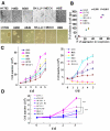
Figure 1
Figure 1
Cell aggregation correlated with cell growth in suspension culture and in soft agar. A, Upper panel: Cell aggregation in polyHEMA-coated dishes (× 100). H1792, H460, A549, SK-LU-1, MDCK, and HBE cells were cultured on 60-mm polyHEMA-coated dishes (2.5 × 10⁵/dish) for 8 hours and photographed. Lower panel: Colony-formation of cells in soft agar. 4 × 10⁴ cells were seeded into 60 mm soft agar plates and incubated for 2 weeks and photographed. B, Correlation between cell aggregation and soft agar growth. The numbers of clonies formed in soft agar after 14 d incubation were plotted against the mean numbers of cell aggregates per 10 fields formed in polyHEMA-coated dishes. Pearson's correlation coefficient was used. N: Number C, Proliferation of tumor cells and normal cells in regular (left) and polyHEMA-coated (right) dishes. Cells as indicated from regular cell culture dishes were plated into regular or polyHEMA-coated dishes (4 × 10⁴/dish) for 1, 2, 3, 4 or 5 d. At the end of each incubation viable cells were counted using trypan blue exclusion. D, Cell aggregation promoted tumor cell proliferation in suspension cultures. 2.5 × 10⁴ tumor cells as indicated from regular cell culture dishes were plated into polyHEMA-coated or methyl cellulose plates for 1, 2, or 3 d. At the end of each incubation viable cells were counted using trypan blue exclusion. Data from at least three separate experiments were analyzed using Student's t test. "***": P < 0.001. S: Suspension culture in polyHEMA-coated dishes MS: Suspension culture in methyl cellulose dishes.
Figure 2
Figure 2
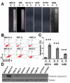
Detachment from ECM induced apoptosis in normal epithelial MDCK and HBE cells. A, DNA laddering analysis of cell death. Normal cells and tumor cells from regular cell culture dishes (1 × 10⁷ ) were plated into 100-mm polyHEMA-coated dishes for 24 h and harvested. Cytosolic DNA was extracted and analyzed by agarose gel electrophoresis. B and C Annexin V staining analysis of cell death. 5 × 10⁵ MDCK, HBE, and H460 cells from regular cell culture dishes were plated into 60-mm polyHEMA-coated dishes for 24 h, harvested, fixed, and assessed by Annexin V/propidium iodide (AV/PI) DNA staining and flow cytometry. B, Percent of early apoptotic cells determined by flow cytometry using AV/PI binding. Quadrant D1: PI/AV +/-, necrotic; D2: PI/AV +/+, late apoptotic/necrotic; D3: PI/AV -/-, live cells; D4: PI/AV -/+, early apoptotic cells. C, Histograms of flow cytometry data. Data from at least three separate experiments were analyzed using Student's t test. "***": P < 0.001. A: Attached culture; S: Suspension culture in polyHEMA-coated dishes. D, Immunoblotting analysis of cleaved caspase-3. After the indicated time periods cells were lysed and total protein was extracted separated by SDS-PAGE, and analyzed by immunoblotting with the indicated antibodies. Tubulin was used as a loading control. A: Attached culture; S: Suspension culture in polyHEMA-coated dishes. "***": P < 0.001.
Figure 3
Figure 3
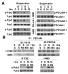
Induction of phosphorylation of Pyk2 and PECAM by cell aggregation. A, Phosphorylation of Pyk2 and PECAM in high versus low cell density cultures. Different concentrations of HBE, SK-LU-1, or H460 cells (H: 5.0 × 10⁵ cells L: 2.5 × 10⁵ cells) were cultured in 60-mm polyHEMA-coated dishes for 0, 6, or 15 h. Cell lysates were resolved by SDS-PAGE and analyzed by immunoblotting with anti-phospho-Pyk2 (Tyr881), or anti-Pyk2 antibody (left). Equal amounts of whole-cell extracts were immunoprecipitated with anti-PECAM antibody followed by immunoblotting with 4G10 antibody (Right). B. Phosphorylation of Pyk2 and PECAM in regular suspension versus methyl-cellulose suspension cultures. H1792 and H460 cells were cultured in polyHEMA-coated dishes (S) or methyl-cellulose dishes (MS) for 6 or 15 h, harvested, and lysed with RIPA buffer. A, Equal amounts of whole-cell extracts were immunoprecipitated with anti-PECAM antibody followed by immunoblotting with anti-phosphotyrosine 4G10 antibody (Upper). Equal amounts of whole-cell extracts were immunoblotted with anti-pPyk2 (Tyr881) or anti-Pyk2 antibody (Lower).
Figure 4
Figure 4
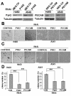
Knock-down of PECAM-1 or Pyk2 decreased cell aggregation and colony formation in soft agar. A, H460 (left) and A549 (right) cells were infected with pRetrosuper/Pyk2, pRetrosuper/PECAM-1, or scrambled (negative control) RNAi retrovirus. The cells were then selected with puromycin for 48 h. RNAi-infected cell lysates were analyzed for the level of protein expression by immunoblotting analysis using the antibodies indicated. Anti-tubulin antibody was used to show equal protein loading. B, The morphology of aggregates of H460 cells (left) and A549 (right) infected with PECAM or Pyk2 RNAi vectors. The RNAi-infected cells were selected with puromycin for 48 h. Cells were harvested cultured on 100-mm polyHEMA-coated dishes for 8 h, and photographed by phase-contrast microscope (× 40). C and D Function of PECAM-1 or Pyk2 in promoting soft agar growth of H460 (left) and A549 (right) cells. C. RNAi-infected cells were harvested and plated in soft agar grown for 2 wk at 37°C, and assayed for colony formation. D, Graphical representation of microscopy data expressed as the percentage of colonies. Data from at least three separate experiments were analyzed using Student's t test. "***": P < 0.001
Figure 5
Figure 5
PECAM-1 interacted with Pyk2 physically in vivo. A and B Binding of endogenous PECAM with endogenous Pyk2 in H460 (A) and A549 (B) cells. Confluent H460 and A549 cells from regular cell culture dishes were plated onto 100-mm polyHEMA-coated dishes for 3 or 15 h. Cells were harvested and lysed. Equal amounts of whole-cell extracts were immunoprecipitated with anti-PECAM-1 antibody, anti-Pyk2 antibody, or anti-Flag antibody followed by immunoblotting with anti-Pyk2 antibody or anti-PECAM-1 antibody. C, In vivo interaction of Pyk2 and PECAM-1 in 293T cells. 293T cells (1 × 10⁶/100-mm dish) were co-transfected with pCDNA3.1 empty vector, PECAM FL construct, PECAM Mu (Δ11-16) construct, PYK2 construct, or a combination as indicated. After treatment for 48 h, equal amounts of whole-cell extracts were immunoprecipitated with anti-PECAM antibody and immunoblotted for Pyk2 and PECAM (upper) or immunoprecipitated with anti-Pyk2 antibody and immunoblotted for PECAM and Pyk2 (middle). Western blot analysis of equal amounts of cell extracts was also performed in parallel (lower). FL: Full-length; Mu: Mutant (Δ11-16).

Similar articles

Cited by

References

    1. Frisch SM, Francis H. Disruption of epithelial cell-matrix interactions induces apoptosis. J Cell Biol. 1994;24:619–626. doi: 10.1083/jcb.124.4.619. - DOI - PMC - PubMed
    1. Grossmann J. Molecular mechanisms of "detachment-induced apoptosis--Anoikis". Apoptosis. 2002;7:247–260. doi: 10.1023/A:1015312119693. - DOI - PubMed
    1. Rennebeck G, Martelli M, Kyprianou N. Anoikis and survival connections in the tumor microenvironment: is there a role in prostate cancer metastasis? Cancer Res. 2005;65:11230–11235. doi: 10.1158/0008-5472.CAN-05-2763. - DOI - PMC - PubMed
    1. Jiang K, Sun J, Cheng J, Djeu JY, Wei S, Sebti S. Akt mediates Ras downregulation of RhoB a suppressor of transformation invasion, and metastasis. Mol Cell Biol. 2004;24:5565–5576. doi: 10.1128/MCB.24.12.5565-5576.2004. - DOI - PMC - PubMed
    1. Steuer AF, Ting RC. Formation of larger cell aggregates by transformed cells: an in vitro index of cell transformation. J Natl Cancer Inst. 1976;56:1279–1280. - PubMed

Publication types

MeSH terms

Substances

LinkOut - more resources