

Novel application of low pH-dependent fluorescent dyes to examine colitis
- PMID: 20074376
- PMCID: PMC2824706
- DOI: 10.1186/1471-230X-10-4
Novel application of low pH-dependent fluorescent dyes to examine colitis
Abstract
Background: Endoscopy capable of fluorescence observation provides histological information on gastrointestinal lesions. We explored the novel application of low pH-dependent fluorescent dyes for fluorescence observation of crypt structure and inflammatory cell infiltration in the colon.
Methods: Low pH-dependent fluorescent dyes were applied to the colonic mucosa of normal mice for observation under fluorescence stereomicroscopy system. We also examined mouse models of colitis, which were induced by trinitrobenzenesulfonic acid, dextran sulfate sodium or interleukin-10 deficiency.
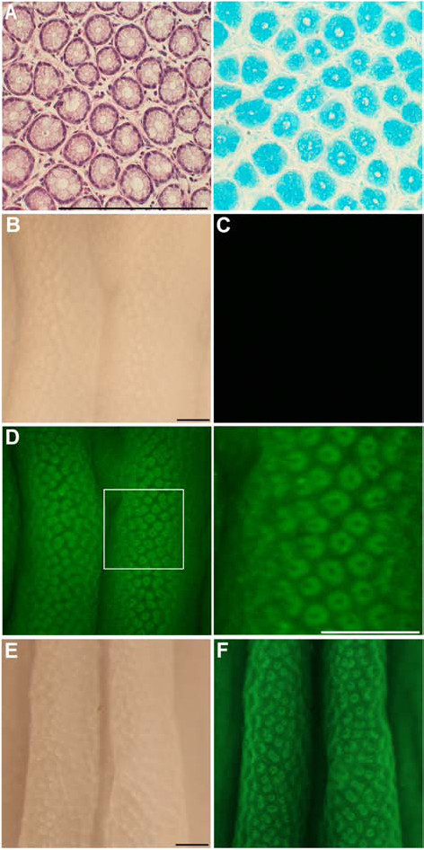
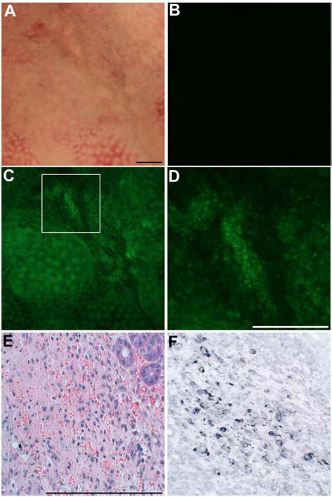
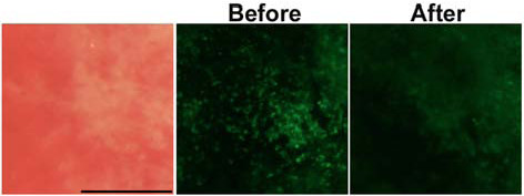
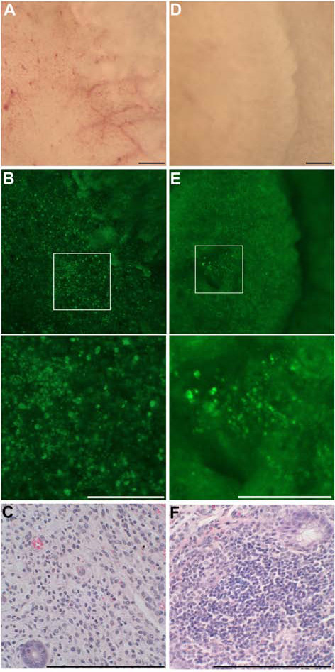
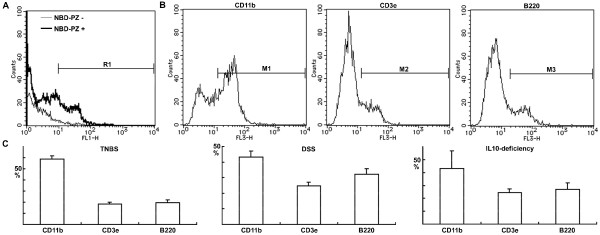
Results: Topical application of low pH-dependent fluorescent dyes revealed crypts as ring-shaped fluorescent stains by visualizing the mucin granules of goblet cells. Because of the minimal fluorescence intensity of the low pH-dependent fluorescent dyes in phosphate-buffered saline, it was not necessary to wash the mucosa before the fluorescence observation. 4-Nitro-7-piperazino-2,1,3-benzoxadiazole (NBD-PZ) was quicker to achieve complete staining (three minutes) than LysoSensor Green DND-153 and DND-189 (20 minutes). In each type of colitis, NBD-PZ revealed the destruction of the crypts as the disappearance of the ring-shaped fluorescent stains and the infiltration of inflammatory cells as the aggregation of punctate fluorescent stains through visualization of lysosomes.
Conclusions: Low pH-dependent fluorescent dyes, especially NBD-PZ, are suitable for topical application to the colonic mucosa and have characteristics that allow for the histological examination of colitis.
Figures
Similar articles
-
Novel mouse model of colitis characterized by hapten-protein visualization.Biotechniques. 2010 Sep;49(3):641-8. doi: 10.2144/000113496. Biotechniques. 2010. PMID: 20854265
-
Novel application of 4-nitro-7-(1-piperazinyl)-2,1,3-benzoxadiazole to visualize lysosomes in live cells.Biotechniques. 2008 Oct;45(4):465, 467-8. doi: 10.2144/000112912. Biotechniques. 2008. PMID: 18855774
-
Design and synthesis of a hydrophilic fluorescent derivatization reagent for carboxylic acids, 4-N-(4-N-aminoethyl)piperazino-7-nitro-2,1,3-benzoxadiazole (NBD-PZ-NH2), and its application to capillary electrophoresis with laser-induced fluorescence detection.Biomed Chromatogr. 2002 Dec;16(8):523-8. doi: 10.1002/bmc.200. Biomed Chromatogr. 2002. PMID: 12474216
-
Confocal endomicroscopy in patients with ulcerative colitis.J Gastroenterol Hepatol. 2008 Dec;23 Suppl 2:S286-90. doi: 10.1111/j.1440-1746.2008.05559.x. J Gastroenterol Hepatol. 2008. PMID: 19120913
-
Fluorogenic and fluorescent labeling reagents with a benzofurazan skeleton.Biomed Chromatogr. 2001 Aug;15(5):295-318. doi: 10.1002/bmc.75. Biomed Chromatogr. 2001. PMID: 11507712 Review.
Cited by
-
Endogenous Bioelectric Signaling Networks: Exploiting Voltage Gradients for Control of Growth and Form.Annu Rev Biomed Eng. 2017 Jun 21;19:353-387. doi: 10.1146/annurev-bioeng-071114-040647. Annu Rev Biomed Eng. 2017. PMID: 28633567 Free PMC article. Review.
-
Acidification of uterine epithelium during embryo implantation in mice.Biol Reprod. 2017 Jan 1;96(1):232-243. doi: 10.1095/biolreprod.116.144451. Biol Reprod. 2017. PMID: 28395338 Free PMC article.
-
Endogenous voltage gradients as mediators of cell-cell communication: strategies for investigating bioelectrical signals during pattern formation.Cell Tissue Res. 2013 Apr;352(1):95-122. doi: 10.1007/s00441-012-1329-4. Epub 2012 Feb 17. Cell Tissue Res. 2013. PMID: 22350846 Free PMC article. Review.
-
The presence of blastocyst within the uteri facilitates lumenal epithelium transformation for implantation via upregulating lysosome proteostasis activity.Autophagy. 2024 Jan;20(1):58-75. doi: 10.1080/15548627.2023.2247747. Epub 2023 Aug 22. Autophagy. 2024. PMID: 37584546 Free PMC article.
-
Regulation of cell behavior and tissue patterning by bioelectrical signals: challenges and opportunities for biomedical engineering.Annu Rev Biomed Eng. 2012;14:295-323. doi: 10.1146/annurev-bioeng-071811-150114. Annu Rev Biomed Eng. 2012. PMID: 22809139 Free PMC article. Review.
References
-
- Kiesslich R, Burg J, Vieth M, Gnaendiger J, Enders M, Delaney P, Polglase A, McLaren W, Janell D, Thomas S. et al.Confocal laser endoscopy for diagnosing intraepithelial neoplasias and colorectal cancer in vivo. Gastroenterology. 2004;127(3):706–713. doi: 10.1053/j.gastro.2004.06.050. - DOI - PubMed
-
- Kiesslich R, Goetz M, Lammersdorf K, Schneider C, Burg J, Stolte M, Vieth M, Nafe B, Galle PR, Neurath MF. Chromoscopy-guided endomicroscopy increases the diagnostic yield of intraepithelial neoplasia in ulcerative colitis. Gastroenterology. 2007;132(3):874–882. doi: 10.1053/j.gastro.2007.01.048. - DOI - PubMed
Publication types
MeSH terms
Substances
LinkOut - more resources
Full Text Sources
Other Literature Sources
