. 2010 Jan 8;140(1):88-98.
doi: 10.1016/j.cell.2009.12.024.

Mutations in potassium channel Kir2.6 cause susceptibility to thyrotoxic hypokalemic periodic paralysis

Affiliations

Mutations in potassium channel Kir2.6 cause susceptibility to thyrotoxic hypokalemic periodic paralysis

Devon P Ryan et al. Cell. .

Abstract

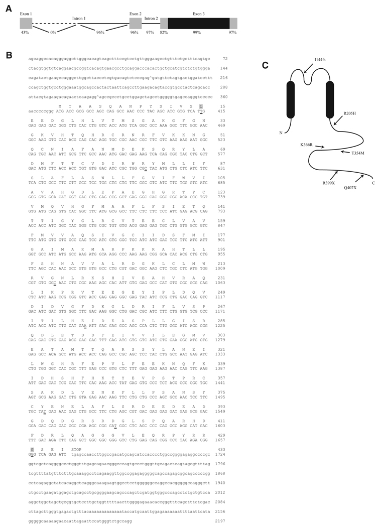
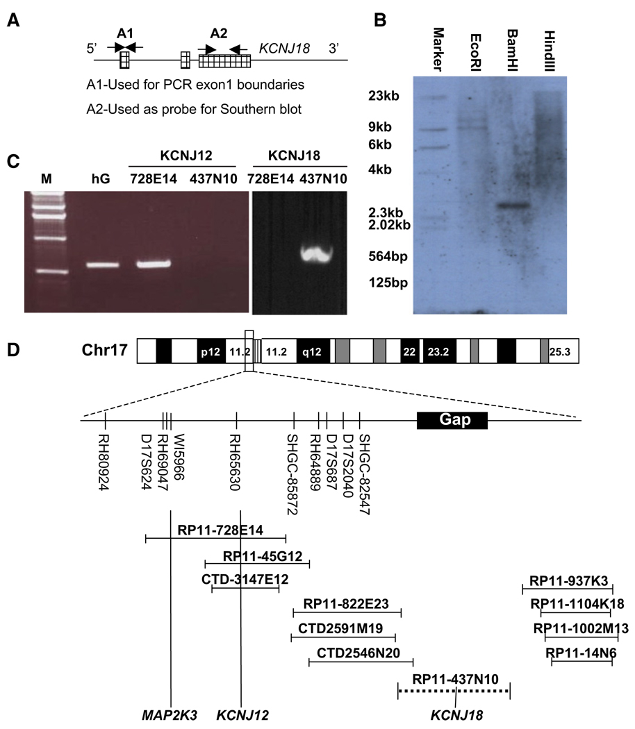
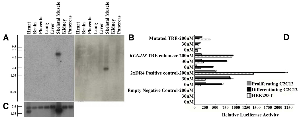
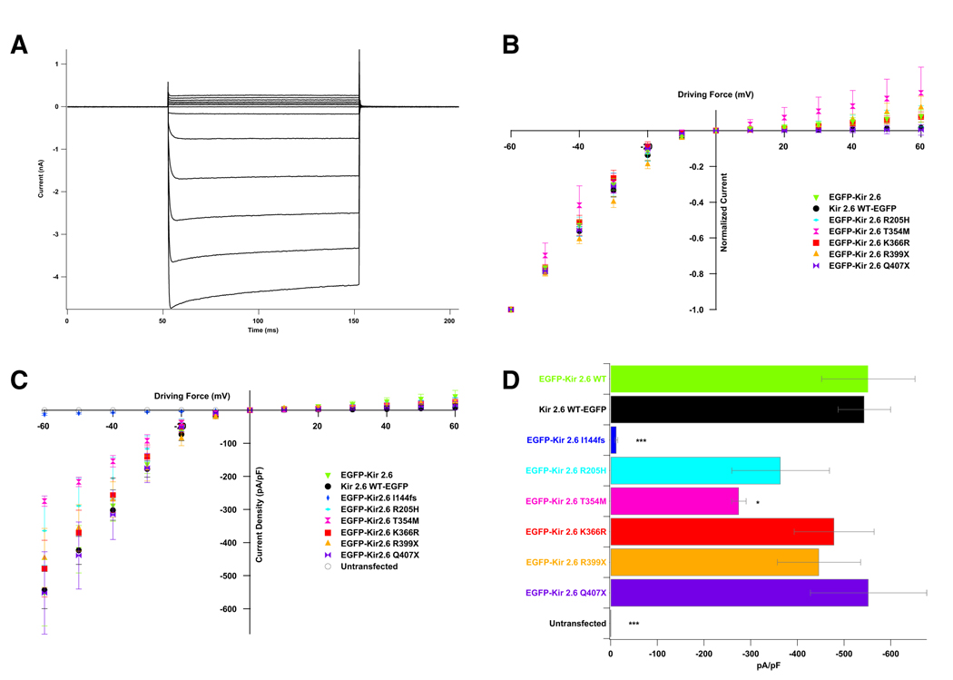
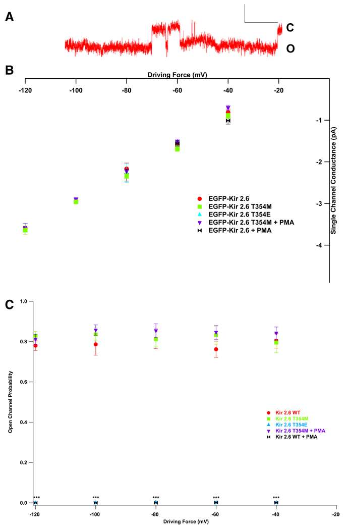
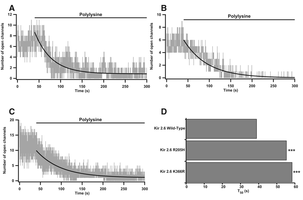
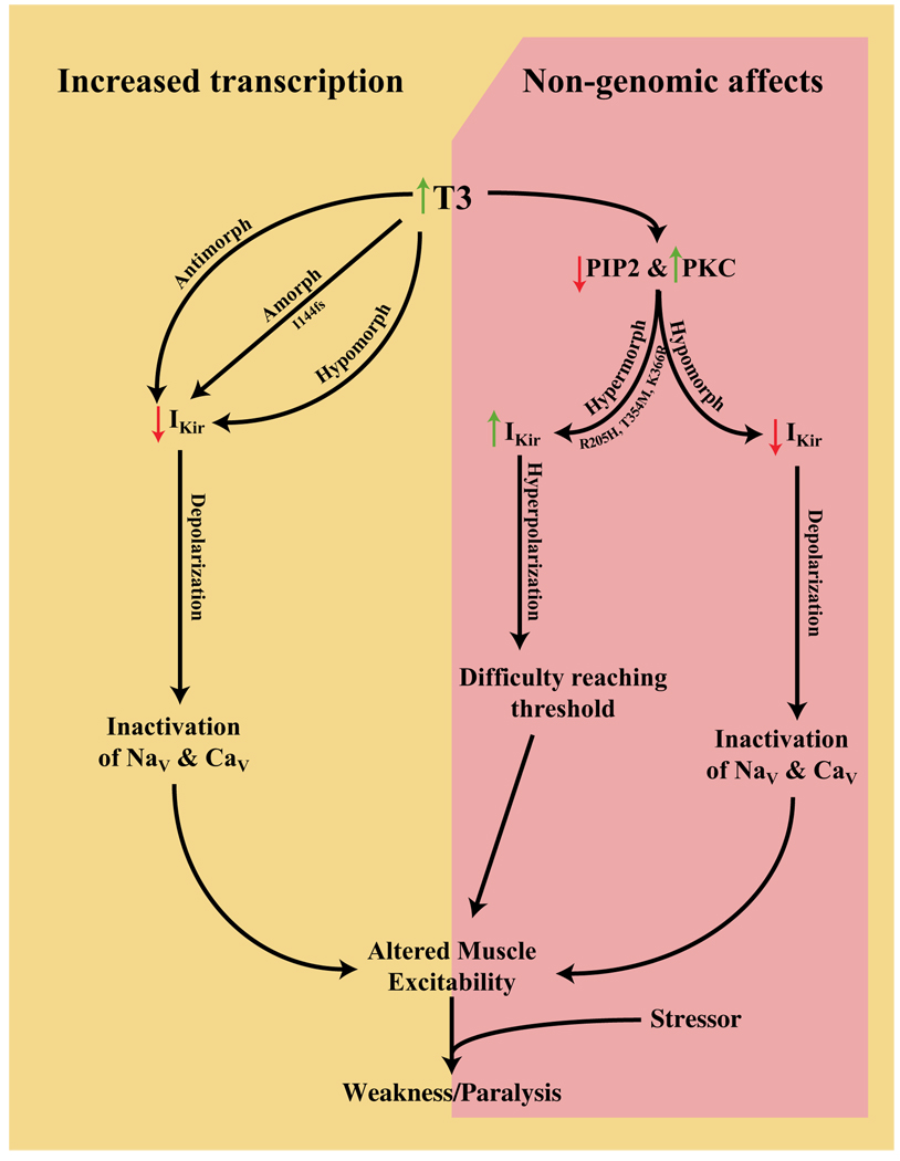
Thyrotoxic hypokalemic periodic paralysis (TPP) is characterized by acute attacks of weakness, hypokalemia, and thyrotoxicosis of various etiologies. These transient attacks resemble those of patients with familial hypokalemic periodic paralysis (hypoKPP) and resolve with treatment of the underlying hyperthyroidism. Because of the phenotypic similarity of these conditions, we hypothesized that TPP might also be a channelopathy. While sequencing candidate genes, we identified a previously unreported gene (not present in human sequence databases) that encodes an inwardly rectifying potassium (Kir) channel, Kir2.6. This channel, nearly identical to Kir2.2, is expressed in skeletal muscle and is transcriptionally regulated by thyroid hormone. Expression of Kir2.6 in mammalian cells revealed normal Kir currents in whole-cell and single-channel recordings. Kir2.6 mutations were present in up to 33% of the unrelated TPP patients in our collection. Some of these mutations clearly alter a variety of Kir2.6 properties, all altering muscle membrane excitability leading to paralysis.


Figures

Figure 1
Figure 1. Structure and Sequence of KCNJ18 and Kir2.6
(A) KCNJ18 shares a high degree of identity with KCNJ12 in both exons (boxes) and introns (lines). The coding region of both KCNJ18 and KCNJ12 is contained within exon 3 (black region). The first intron of KCNJ12 is longer than that of KCNJ18, causing 0% identity in this nonoverlapping region (dotted line). (B) KCNJ18 sequence-exon boundaries are denoted with a caret (^). Coding sequence is capitalized with the corresponding amino acid above. Underlined nucleotides denote differences between KCNJ18 and KCNJ12, with nonsynonymous differences having a gray background. (C) Diagram of Kir2.6 with the relative locations of TPP associated mutations. See also Figure S1.
Figure 2
Figure 2. Genomic Characterization of KCNJ18
(A) Both KCNJ12 and KCNJ18 have three recognized exons (hatched regions). Probes constructed from exon 1 (A1, unique for each gene) were used for northern blot and genome walking to determine exon 1 boundaries. (B) Radiolabeled A2 primers from exon 3 were used as a probe for Southern blot upon BamH1-digested KCNJ18 containing BAC DNA, (C) KCNJ12 specific primers were able to yield PCR products from control human genomic (hG) DNA and BAC RP11-728E14 (containing KCNJ12) but not RP11-437N10 (containing KCNJ18). (D) From this BAC end sequencing, we place KCNJ18 centromeric to KCNJ12 on chromosome 17 in a previously identified sequencing gap and not overlapping with KCNJ12. See also Figure S2.
Figure 3
Figure 3. Expression Pattern and Transcriptional Regulation of KCNJ18
(A and B) The A1 probe set (Figure 2A) was used to specifically probe multiple tissue northern blot membranes for KCNJ18 (A) and KCNJ12 (B) mRNAs, which are found specifically in skeletal muscle. (C) β-actin was used as a loading control. (D) The KCNJ18 regulatory region can function as an enhancer for luciferase expression in HEK293T and either proliferating or differentiated C2C12 cells. A DR4-positive control, an empty vector control, and a mutated KCNJ18 TRE control were also tested. Luciferase expression by the WT KCNJ18 is enhanced in a T3 dose-dependent manner from 0 nM T3 (hypothyroid) through 200 nM (hyperthyroid) T3 after compensating for the internal renilla control. Error bars indicate the mean ± standard error.
Figure 4
Figure 4. KCNJ18 Encodes an Inwardly Rectifying Potassium Channel Whose Conductance Properties Are Altered by Some TPP Mutations
(A) When expressed in 293T cells, Kir2.6 produces stereotypical Kir currents. Voltage steps were performed from the resting membrane potential (0 mV driving force) to between −60 and +60 mV in increments of 10 mV. (B) Normalization of these values to maximal current allows for comparison of rectification between WT and mutant channels. TPP mutations do not lead to altered rectification. (C) Current density can instead be measured by normalizing currents to cellular capacitance. (D) Both the I144fs and T354M mutations lead to decreased current density, most easily seen at −60 mV. p values were calculated with a t test versus EGFP-Kir2.6 WT. There are five to 12 cells per data point. Error bars indicate the mean ± standard error. See also Figure S3.
Figure 5
Figure 5. TPP Mutations in Kir2.6 Alter Single-Channel Response to PKC
(A) Kir2.6 produces stereotypical single-channel currents. Scale bars represent 200 ms by 2 pA. Openings (O) are down, and closings (C) are up. (B) Wild-type and mutant single-channel conductance is unaltered by PKC activation or mimicking constitutive phosphorylation at T354 (T354E). (C) Open probability, however, is decreased in WT but not T354M mutant channels by the activation of PKC or T354E. p values were calculated a t test versus EGFP-Kir2.6 WT. There are five to nine patches per data point, each with one to three (typically two or three) channels.
Figure 6
Figure 6. Kir2.6-PIP2 Interactions Are Altered by TPP Mutations
(A–C) Inside-out patches were perfused with isotonic solution for 40 s prior to perfusion with polylysine. Wild-type (A), R205H (B), and K366R (C) openings decreased after polylysine perfusion. (D) Curve fits of idealized channel openings (smooth black curves in A–C), were used to derive the T50 value. Both the R205H and the K366R mutant channels have an altered interaction with PIP2 and take significantly longer to reach half-maximal opening than WT channels. There are five to ten cells and nine to 20 total channels per channel type. p < 0.001 in accordance to a statistical model. See also Figure S4.
Figure 7
Figure 7. Model of TPP Pathophysiology
Amorphic and antimorphic alleles would cause decreased K+ currents leading to depolarization and gradual transitioning of voltage-gated channels to their inactivated states. Hypermorphic alleles, causing hyperpolarization, conversely, would cause difficulty reaching threshold. These opposite shifts in membrane excitability, when coupled with a stressor event, are both predicted to lead to the weakness and paralysis observed in TPP. How the genomic and nongenomic effects of thyrotoxicosis on other ion channels interact with these proposed Kir current alterations remains to be elucidated.

Comment in

References

    1. Abraham MR, Jahangir A, Alekseev AE, Terzic A. Channelopathies of inwardly rectifying potassium channels. FASEB J. 1999;13:1901–1910. - PubMed
    1. Bailey JA, Yavor AM, Massa HF, Trask BJ, Eichler EE. Segmental duplications: organization and impact within the current human genome project assembly. Genome Res. 2001;11:1005–1017. - PMC - PubMed
    1. Bassett JH, Harvey CB, Williams GR. Mechanisms of thyroid hormone receptor-specific nuclear and extra nuclear actions. Mol. Cell. Endocrinol. 2003;213:1–11. - PubMed
    1. Bulman DE, Scoggan KA, van Oene MD, Nicolle MW, Hahn AF, Tollar LL, Ebers GC. A novel sodium channel mutation in a family with hypokalemic periodic paralysis. Neurology. 1999;53:1932–1936. - PubMed
    1. Donaldson MR, Jensen JL, Tristani-Firouzi M, Tawil R, Bendahhou S, Suarez WA, Cobo AM, Poza JJ, Behr E, Wagstaff J, et al. PIP2 binding residues of Kir2.1 are common targets of mutations causing Andersen syndrome. Neurology. 2003;60:1811–1816. - PubMed

Publication types

MeSH terms