. 2010 Apr 1;316(6):1070-81.
doi: 10.1016/j.yexcr.2010.01.006. Epub 2010 Jan 11.

Isoform- and dose-sensitive feedback interactions between paired box 6 gene and delta-catenin in cell differentiation and death

Affiliations

Isoform- and dose-sensitive feedback interactions between paired box 6 gene and delta-catenin in cell differentiation and death

Jiao Zhang et al. Exp Cell Res. .

Abstract

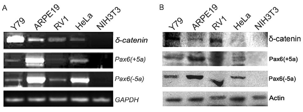
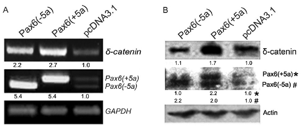
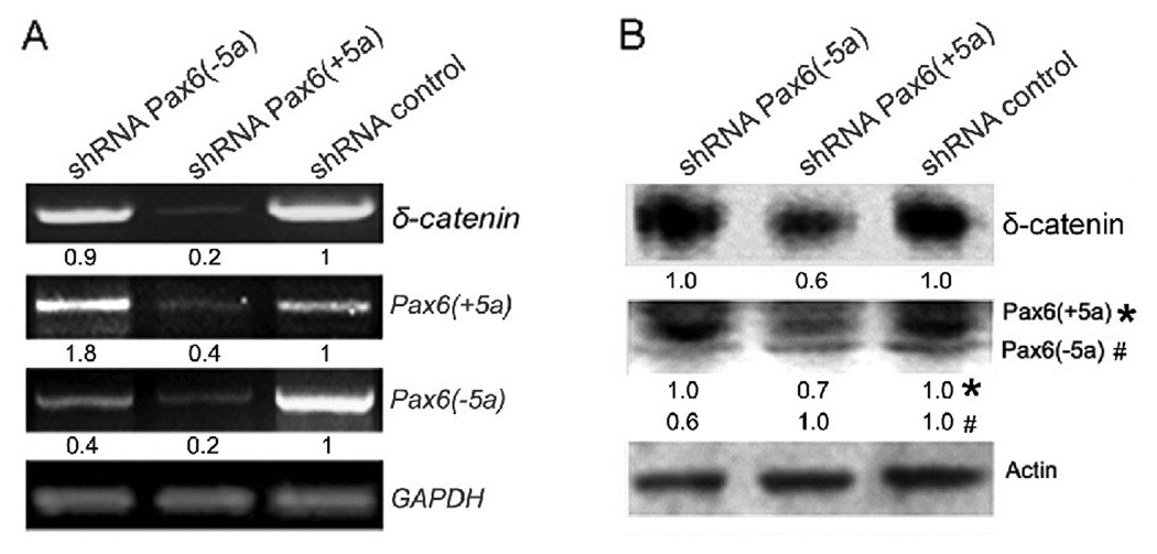
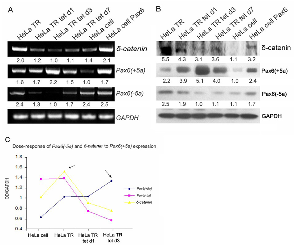
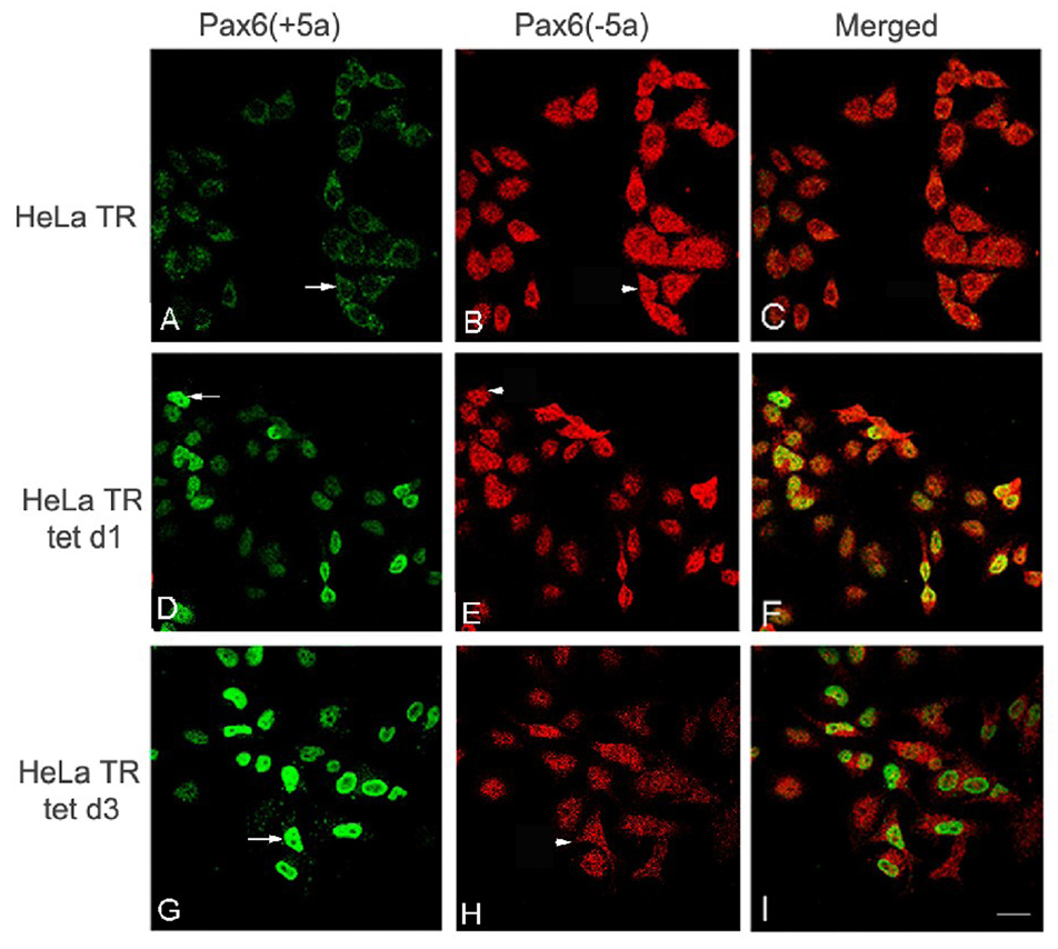
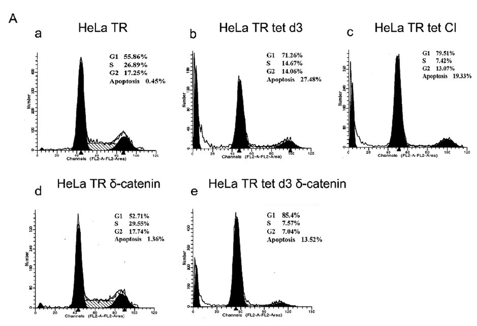
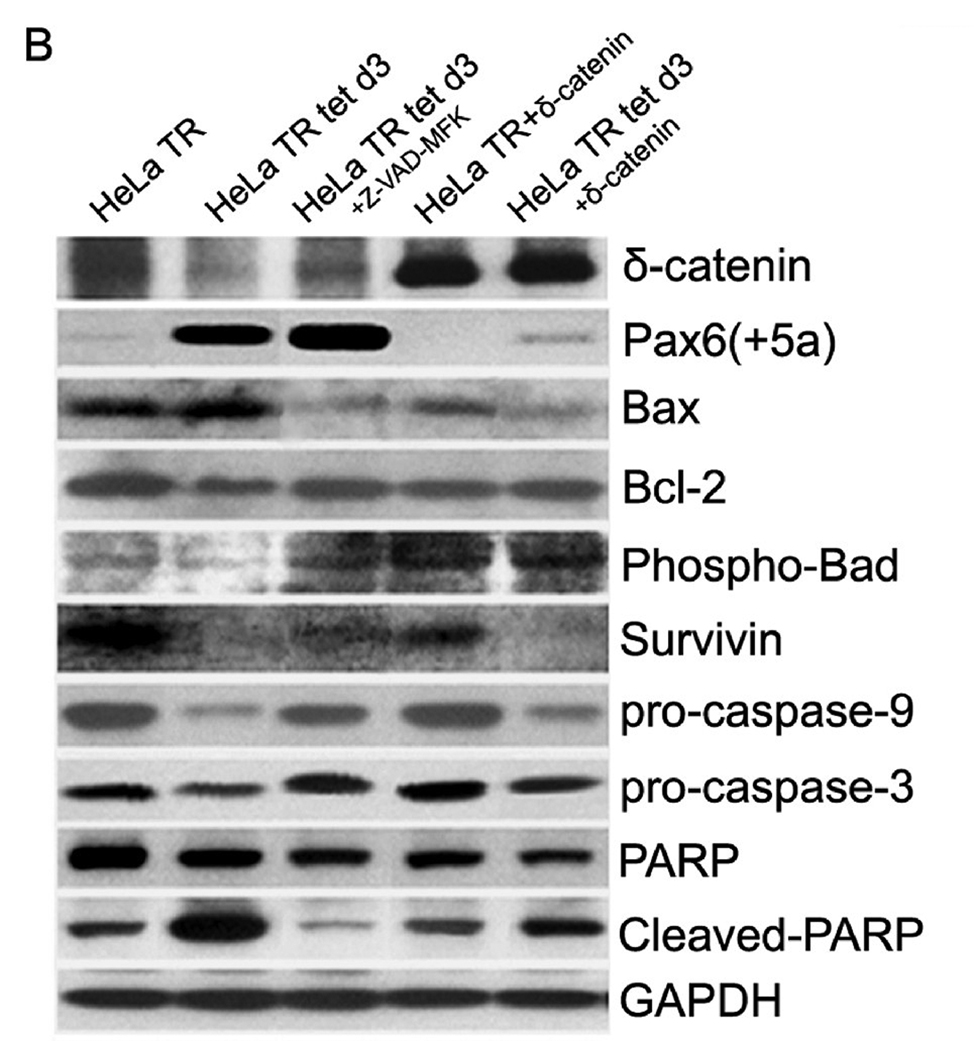
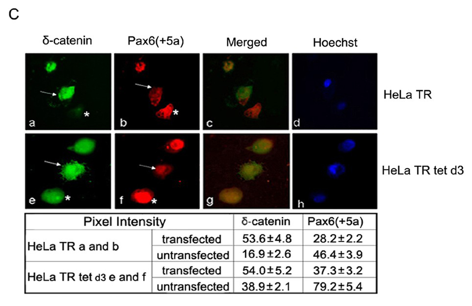
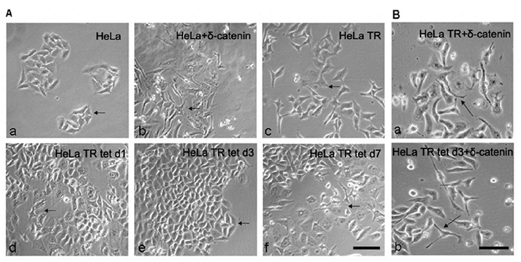
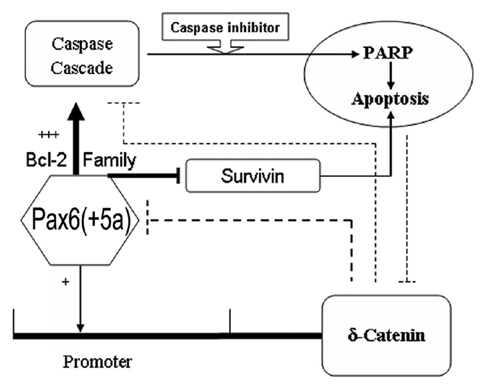
Pax6, a mammalian homolog of the Drosophila paired box gene family member expressed in stem and progenitor cells, resides at the top of the genetic hierarchy in controlling cell fates and morphogenesis. While Pax6 activation can lead to mitotic arrest, premature neurogenesis, and apoptosis, the underlying molecular mechanisms have not been resolved. Here we report that either Pax6(+5a) or Pax6(-5a) was sufficient to promote, whereas their knockdown reduced the expression of delta-catenin (CTNND2), a neural specific member of the armadillo/beta-catenin superfamily. Pax6(+5a) elicited stronger effects on delta-catenin than Pax6(-5a). Inducible Pax6(+5a) expression demonstrated a biphasic and dose-dependent regulation of delta-catenin expression and cell fates. A moderate upregulation of Pax6(+5a) promoted delta-catenin expression and induced neurite-like cellular protrusions, but increasing expression of Pax6(+5a) reversed these processes. Furthermore, sustained high expression of Pax6(+5a) triggered apoptosis as determined by the reduction of phospho-Bad, Bcl-2, survivin and procaspases, as well as the increases in Bax and cleaved poly(ADP-ribose) polymerase. Importantly, re-introducing delta-catenin by ectopic expression elicited a feedback suppression on Pax6(+5a) expression and reduced Pax6(+5a) induced apoptosis. Therefore, delta-catenin expression is not only controlled by Pax6, but it also provides a feedback suppression mechanism for their functional interactions with important implications in cellular morphogenesis, apoptosis, and cancer.


Conflict of interest statement

Conflict of Interest

The authors declare that they have no conflict of interest.

Figures

Fig. 1
Fig. 1. Pax6(+5a), Pax6(−5a) and δ-catenin expression in cell lines
A: RT-PCR showing Pax6(+5a), Pax6(−5a), and δ-catenin mRNA expression in Y79, ARPE-19, CWR22Rv-1 (Rv-1), HeLa and NIH3T3 cells. GAPDH is used as internal mRNA control. B: Western blots showing Pax6(+5a), Pax6(−5a), and δ-catenin protein expression in Y79, ARPE-19, CWR22Rv-1 (Rv-1), HeLa and NIH3T3 cells. Actin is used as protein loading control.
Fig. 2
Fig. 2. Overexpression of either Pax6(+5a) or Pax6(−5a) is sufficient to promote δ-catenin expression
A. RT-PCR showing Pax6(+5a), Pax6(−5a), and δ-catenin mRNA expression in Y79 cells transiently transfected with Pax6(+5a), Pax6(−5a), or pcDNA3.1 as vector control. GAPDH is used as internal mRNA control. B. Western blots showing Pax6(+5a), Pax6(−5a), and δ-catenin protein expression in Y79 cells transiently transfected with Pax6(+5a), Pax6(−5a), or pcDNA3.1. Note: semi-quantitative measurements of the RT-PCR and Western blot are shown underneath each lane. Actin is used as protein loading control.
Fig. 3
Fig. 3. Knockdown of Pax6(+5a) or Pax6(−5a) suppresses δ-catenin expression
Pax6 isoform specific shRNA is designed to knockdown Pax6(+5a) or Pax6(−5a) respectively in Y79 cells to explore either Pax6 isoform’s function on δ-catenin expression. A: RT-PCR showing Pax6(+5a), Pax6(−5a), and δ-catenin mRNA expression in Y79 cells after knockdown of Pax6(+5a) or Pax6(−5a) respectively. GAPDH is used as internal mRNA control. B: Western blots showing Pax6(+5a), Pax6(−5a), and δ-catenin protein expression in Y79 cells after knockdown of Pax6(+5a) or Pax6(−5a) respectively. Note: semi-quantitative measurements of the RT-PCR and Western blot are shown underneath each lane. The results showed that knockdown of Pax6(+5a) elicits the stronger effects on δ-catenin while the effects of Pax6(−5a) is more moderate. Actin is used as protein loading control.
Fig. 4
Fig. 4. Dose responses of Pax6(−5a) and δ-catenin expression to induced overexpression of Pax6(+5a)
A. RT-PCR showing Pax6(+5a), Pax6(−5a), and δ-catenin expression in time course in HeLa Tat-TetR-Pax6 cells with (HeLa tet) or without (HeLa TR) induction of Pax6(+5a) overexpression as well as parental HeLa cells (HeLa) and HeLa cells transiently transfected with Pax6(+5a)(HeLa Pax6). Note: a moderate upregulation of Pax6(+5a) promotes δ-catenin expression, whereas a sustained high expression of Pax6(+5a) inhibits δ-catenin expression. GAPDH is used as internal mRNA control. B: Western blots showing Pax6(+5a), Pax6(−5a), and δ-catenin protein expression in HeLa cell and HeLa Tat-TetR-Pax6 cells with or without induction of Pax6(+5a) overexpression. Note: semi-quantitative measurements of the RT-PCR and Western blot are shown underneath each lane in A and B. Actin is used as protein loading control. C. Dose-response curve of Pax6(−5a) and δ-catenin to Pax6(+5a) expression shows that the regulation of δ-catenin expression by Pax6 is isoform- and dose-dependent. With the increasing Pax6(+5a) expression, Pax6(−5a) expression decreases gradually. δ-Catenin expression shows a parabolic curve, which reaches a peak when Pax6(+5a) is moderately upregulated, and then downregulates with the increasing Pax6(+5a) expression. Note: HeLa TR: HeLa Tat-TetR-Pax6 cells without doxycycline induction of Pax6(+5a) overexpression; HeLa Tat tet d1 and d3 indicates day1 and day 3 after HeLa Tat-TetR-Pax6 cells are induced to overexpress Pax6(+5a) by doxycycline treatment. HeLa Tat tet d7 indicates that HeLa Tat-TetR-Pax6 cells are induced to overexpress Pax6(+5a) by doxycycline treatment for 3 days, then treatment was stopped and the cells were grown to day 7 before analysis. HeLa: parental HeLa cells without any treatment or transfection. HeLa cell Pax6: HeLa cells transiently transfected with Pax6(+5a).
Fig. 5
Fig. 5. Induced overexpression of Pax6(+5a) is accompanied by the reduction of Pax6(−5a)
Immunofluorescent laser confocal light microscopy shows the time course of Pax6(+5a) and Pax6(−5a) expression in HeLa Tat-TetR-Pax6 cells double labeled using anti-Pax6(+5a) and anti-Pax6(−5a). A–C: Pax6(+5a) and Pax6(−5a) expression in HeLa Tat-TetR-Pax6 cells without (HeLa TR) induction of Pax6(+5a) overexpression. D–F: HeLa Tat-TetR-Pax6 cells at day 1 (HeLa tet d1) with induction of Pax6(+5a) overexpression. G–I: HeLa Tat-TetR-Pax6 cells at day 3 (HeLa tet d3) with induction of Pax6(+5a) overexpression. Note: compared with A, many cells in D show moderately increased Pax6(+5a) expression and show dramatically increased expression in G (arrows). On the other hand, Pax6(−5a) shows slightly decreased expression in cells in D but more pronounced decrease in expression in H when compared to A (arrowheads). A, D, and G: Pax6(+5a); B, E, and H: Pax6(−5a); C, F, and I: Merged Images. Bar: 25 µm.
Fig. 6
Fig. 6. Mitotic and apoptotic changes in HeLa Tat-TetR-Pax6 cell line
A. Cell cycle kinetics analysis. HeLa Tat-TetR-Pax6 cells after doxycycline induction is treated respectively with caspase inhibitor (Z-VAD-FMK) or transfected with δ-catenin. At day3, cells in each group are collected and analyzed by flow cytometry with propidium iodide (PI) staining. a: HeLa Tat-TetR-Pax6 cells without doxycycline induction (HeLa TR); b: HeLa Tat-TetR-Pax6 cell 3 days after doxycycline induction (HeLa TR tet d3); c: HeLa Tat-TetR-Pax6 cell with Pax6(+5a) overexpression in the presence of Z-VAD-FMK (HeLa TR tet CI); d: Overexpression of δ-catenin in HeLa Tat-TetR-Pax6 cells without Pax6(+5a) overexpression (HeLa TR δ-catenin); e: Overexpression of δ-catenin in HeLa Tat-TetR-Pax6 cells with Pax6(+5a) overexpression (HeLa TR tet δ-catenin). B. The effects of Pax6 and δ-catenin expression on apoptotic protein expression. Western blots show protein expression profiles of Pax6(+5a), δ-catenin, PARP, cleaved-PARP, procaspase-9, procaspase-3, Bax, Bcl-2, Phospho-Bad and survivin. Note that δ-catenin expression in HeLa TR tet d3 cells is reduced with high Pax6(+5a) expression when compared to that in HeLa TR cells. Blocking caspase activation using caspase inhibitor (HeLa TR tet d3+Z-VAD-FMK) reduces the inhibitory effects of high Pax6(+5a) expression on δ-catenin expression. Pax6(+5a) and δ-catenin expression is both increased when compared to that of no-caspase inhibitor treatment. On the other hand, overexpression of δ-catenin (HeLa TR tet d3+δ-catenin) also reduces Pax6(+5a) expression and the apoptotic protein expression induced by Pax6(+5a) overexpression. The results show that overexpression of δ-catenin reduces Pax6(+5a) expression in HeLa Tat-TetR-Pax6 cells, which provides a negative feedback between Pax6 and δ-catenin expression. C. Double immunofluorescent light microscopy showing the effects of δ-catenin overexpression on Pax6(+5a) expression. a–d: Pax6(+5a) (red) and δ-catenin (green) expression in HeLa TR cells; c: Merged image of a and b; d: Nuclei staining. e–h: Pax6(+5a) (red) and δ-catenin (green) expression in HeLa tet d3 cells. g: Merged image of e and f; h: Nuclei staining. Note: arrows indicate δ-catenin transfected cells; asterisks indicate untransfected cells. The arrows show that the expression of Pax6(+5a) (red) is reduced in cells transfected with δ-catenin (green). The table in C shows the morphometric density of pixel values in cells stained by immunofluorescence light microscopy. Bar: 20 µm.
Fig. 7
Fig. 7. Morphological changes of HeLa and HeLa Tat-TetR-Pax6 cells influenced by Pax6 and δ-catenin expression
A. A moderate upregulation of Pax6(+5a) promotes, whereas its sustained high expression suppresses the extension of cellular protrusions and processes. a: Parental HeLa cells without any treatments as negative control. Arrows: epithelial cell clusters. b: HeLa cells transiently transfected with Pax6(+5a) as positive control. Arrows: cells extending processes. c: HeLa TR cells without doxycycline induction [with moderate level of Pax6(+5a) expression]. Arrows: cells extending processes. d: HeLa TR tet d1 cells at day 1 after doxycycline induction [with increasing Pax6(+5a) expression]. Arrow: cells still showing processes; Arrowheads: cells no longer extending processes. e: HeLa TR tet d3 cells at day 3 after doxycycline induction [with increasingly high Pax6(+5a) expression]. Arrowheads: cells no longer extending processes. f: HeLa TR tet d7 cells with doxycycline induction [with the highest Pax6(+5a) expression] for 3 days and followed by in the absence of induction until day 7. Arrows: cells extending processes. Bar: 50 µm. B. δ-Catenin overexpression promotes the extension of cellular protrusions and processes regardless whether Pax6 is overexpressed. a: HeLa TR cells without doxycycline induction transfected with δ-catenin; b: HeLa TR tet d3 cells with a 3-day doxycycline induction transfected with δ-catenin. Arrows indicate the extension of cellular processes. Bar: 30 µm.
Fig. 8
Fig. 8. Schematic illustration of the model highlighting putative Pax6 and δ-catenin feedback regulation and their roles in apoptosis
Note: +: indicates moderate expression of Pax6(+5a); +++: indicates higher Pax6(+5a) overexpression; →: indicates promote or upregulate □□ : indicates inhibit or downregulate

Similar articles

Cited by

References

    1. Walther C, Gruss P. Pax-6, a murine paired box gene, is expressed in the developing CNS. Development. 1991;113:1435–1449. - PubMed
    1. Gehring WJ, Ikeo K. Pax 6: mastering eye morphogenesis and eye evolution. Trends Genet. 1999;15:371–377. - PubMed
    1. Wawersik S, Maas RL. Vertebrate eye development as modeled in Drosophila. Hum Mol Genet. 2000;9:917–925. - PubMed
    1. Halder G, Callaerts P, Gehring WJ. Induction of ectopic eyes by targeted expression of the eyeless gene in Drosophila. Science. 1995;267:1788–1792. - PubMed
    1. Tang HK, Singh S, Saunders GF. Dissection of the transactivation function of the transcription factor encoded by the eye developmental gene PAX6. J Biol Chem. 1998;273:7210–7221. - PubMed

Publication types

MeSH terms