

Inwardly rectifying potassium channel Kir4.1 is responsible for the native inward potassium conductance of satellite glial cells in sensory ganglia
- PMID: 20074622
- PMCID: PMC2823846
- DOI: 10.1016/j.neuroscience.2010.01.005
Inwardly rectifying potassium channel Kir4.1 is responsible for the native inward potassium conductance of satellite glial cells in sensory ganglia
Abstract
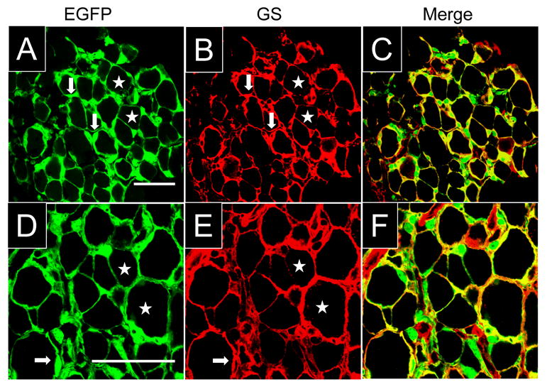
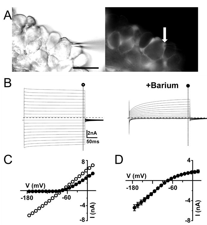
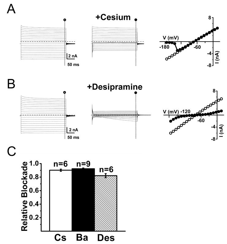
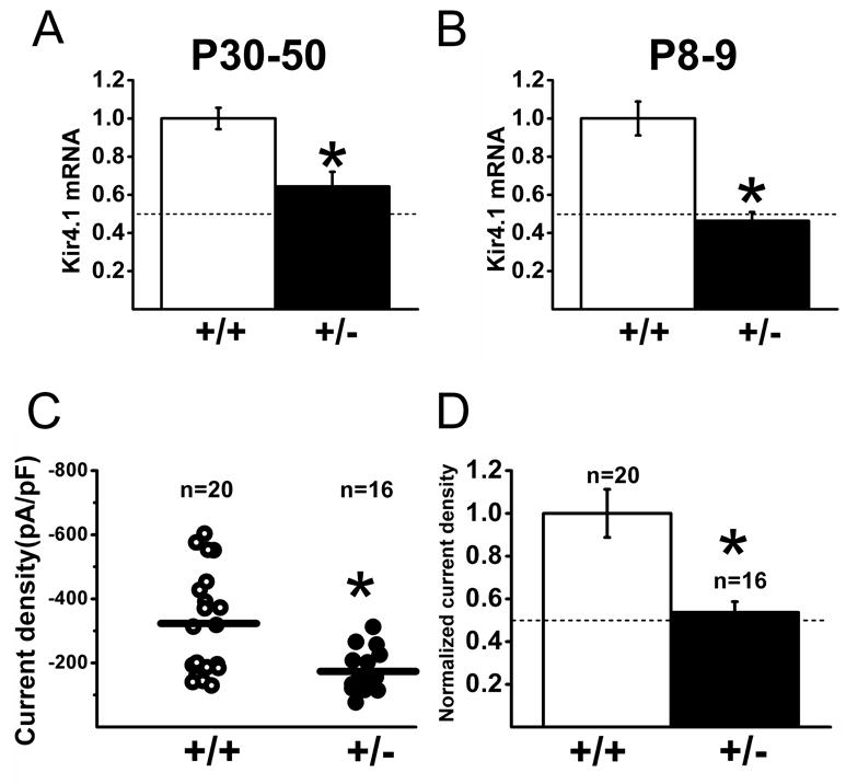
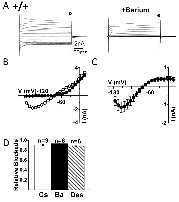
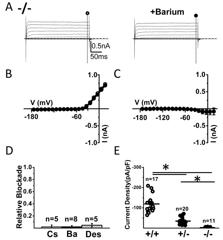
Satellite glial cells (SGCs) surround primary afferent neurons in sensory ganglia, and increasing evidence has implicated the K(+) channels of SGCs in affecting or regulating sensory ganglion excitability. The inwardly rectifying K(+) (Kir) channel Kir4.1 is highly expressed in several types of glial cells in the central nervous system (CNS) where it has been implicated in extracellular K(+) concentration buffering. Upon neuronal activity, the extracellular K(+) concentration increases, and if not corrected, causes neuronal depolarization and uncontrolled changes in neuronal excitability. Recently, it has been demonstrated that knockdown of Kir4.1 expression in trigeminal ganglia leads to neuronal hyperexcitability in this ganglia and heightened nociception. Thus, we investigated the contribution of Kir4.1 to the membrane K(+) conductance of SGCs in neonatal and adult mouse trigeminal and dorsal root ganglia. Whole cell patch clamp recordings were performed in conjunction with immunocytochemistry and quantitative transcript analysis in various mouse lines. We found that in wild-type mice, the inward K(+) conductance of SGCs is blocked almost completely with extracellular barium, cesium and desipramine, consistent with a conductance mediated by Kir channels. We then utilized mouse lines in which genetic ablation led to partial or complete loss of Kir4.1 expression to assess the role of this channel subunit in SGCs. The inward K(+) currents of SGCs in Kir4.1+/- mice were decreased by about half while these currents were almost completely absent in Kir4.1-/- mice. These findings in combination with previous reports support the notion that Kir4.1 is the principal Kir channel type in SGCs. Therefore Kir4.1 emerges as a key regulator of SGC function and possibly neuronal excitability in sensory ganglia.
Copyright (c) 2010 IBRO. Published by Elsevier Ltd. All rights reserved.
Figures
Similar articles
-
Peripheral inflammation suppresses inward rectifying potassium currents of satellite glial cells in the trigeminal ganglia.Pain. 2011 Sep;152(9):2147-2156. doi: 10.1016/j.pain.2011.05.023. Epub 2011 Jun 15. Pain. 2011. PMID: 21680091
-
Activation of GABA(B) receptors potentiates inward rectifying potassium currents in satellite glial cells from rat trigeminal ganglia: in vivo patch-clamp analysis.Neuroscience. 2015 Mar 12;288:51-8. doi: 10.1016/j.neuroscience.2014.12.024. Epub 2014 Dec 24. Neuroscience. 2015. PMID: 25542421
-
Silencing the Kir4.1 potassium channel subunit in satellite glial cells of the rat trigeminal ganglion results in pain-like behavior in the absence of nerve injury.J Neurosci. 2008 Apr 16;28(16):4161-71. doi: 10.1523/JNEUROSCI.5053-07.2008. J Neurosci. 2008. PMID: 18417695 Free PMC article.
-
Inwardly rectifying potassium channels (Kir) in central nervous system glia: a special role for Kir4.1 in glial functions.J Cell Mol Med. 2006 Jan-Mar;10(1):33-44. doi: 10.1111/j.1582-4934.2006.tb00289.x. J Cell Mol Med. 2006. PMID: 16563220 Free PMC article. Review.
-
The role of an inwardly rectifying K(+) channel (Kir4.1) in the inner ear and hearing loss.Neuroscience. 2014 Apr 18;265:137-46. doi: 10.1016/j.neuroscience.2014.01.036. Epub 2014 Jan 28. Neuroscience. 2014. PMID: 24480364 Free PMC article. Review.
Cited by
-
pH modulation of glial glutamate transporters regulates synaptic transmission in the nucleus of the solitary tract.J Neurophysiol. 2013 Jul;110(2):368-77. doi: 10.1152/jn.01074.2012. Epub 2013 Apr 24. J Neurophysiol. 2013. PMID: 23615553 Free PMC article.
-
Intrinsic oscillatory activity arising within the electrically coupled AII amacrine-ON cone bipolar cell network is driven by voltage-gated Na+ channels.J Physiol. 2012 May 15;590(10):2501-17. doi: 10.1113/jphysiol.2011.225060. Epub 2012 Mar 5. J Physiol. 2012. PMID: 22393249 Free PMC article.
-
Store-operated calcium entry in the satellite glial cells of rat sympathetic ganglia.Korean J Physiol Pharmacol. 2024 Jan 1;28(1):93-103. doi: 10.4196/kjpp.2024.28.1.93. Korean J Physiol Pharmacol. 2024. PMID: 38154968 Free PMC article.
-
Desipramine induces anti-inflammatory dorsal root ganglion transcriptional signatures in the murine spared nerve injury model.Neurobiol Pain. 2024 Mar 20;15:100153. doi: 10.1016/j.ynpai.2024.100153. eCollection 2024 Jan-Jun. Neurobiol Pain. 2024. PMID: 38549875 Free PMC article.
-
Molecular mechanisms of EAST/SeSAME syndrome mutations in Kir4.1 (KCNJ10).J Biol Chem. 2010 Nov 12;285(46):36040-8. doi: 10.1074/jbc.M110.163170. Epub 2010 Aug 31. J Biol Chem. 2010. PMID: 20807765 Free PMC article.
References
-
- Bockenhauer D, Feather S, Stanescu HC, Bandulik S, Zdebik AA, Reichold M, Tobin J, Lieberer E, Sterner C, Landoure G, Arora R, Sirimanna T, Thompson D, Cross JH, van't Hoff W, Al Masri O, Tullus K, Yeung S, Anikster Y, Klootwijk E, Hubank M, Dillon MJ, Heitzmann D, Arcos-Burgos M, Knepper MA, Dobbie A, Gahl WA, Warth R, Sheridan E, Kleta R. Epilepsy, ataxia, sensorineural deafness, tubulopathy, and KCNJ10 mutations. The New England journal of medicine. 2009;360:1960–1970. - PMC - PubMed
-
- Cherkas PS, Huang TY, Pannicke T, Tal M, Reichenbach A, Hanani M. The effects of axotomy on neurons and satellite glial cells in mouse trigeminal ganglion. Pain. 2004;110:290–298. - PubMed
-
- Doupnik CA, Davidson N, Lester HA. The inward rectifier potassium channel family. Curr Opin Neurobiol. 1995;5:268–277. - PubMed
-
- Dublin P, Hanani M. Satellite glial cells in sensory ganglia: their possible contribution to inflammatory pain. Brain Behav Immun. 2007;21:592–598. - PubMed
Publication types
MeSH terms
Substances
Grants and funding
LinkOut - more resources
Full Text Sources
Medical
Molecular Biology Databases
