

T-cell antagonism by short half-life pMHC ligands can be mediated by an efficient trapping of T-cell polarization toward the APC
- PMID: 20075022
- PMCID: PMC2806700
- DOI: 10.1073/pnas.0911258107
T-cell antagonism by short half-life pMHC ligands can be mediated by an efficient trapping of T-cell polarization toward the APC
Abstract
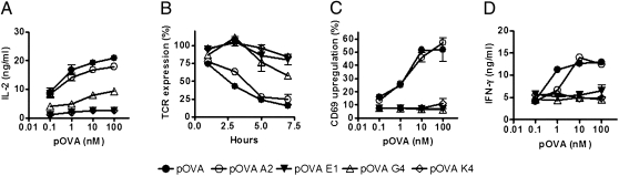
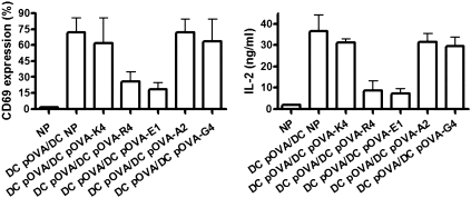
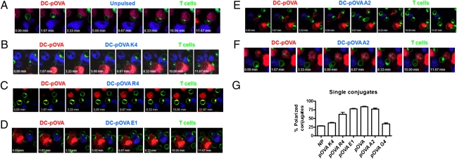
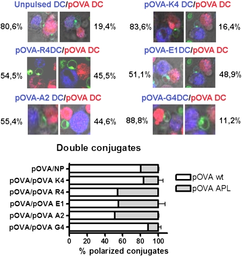
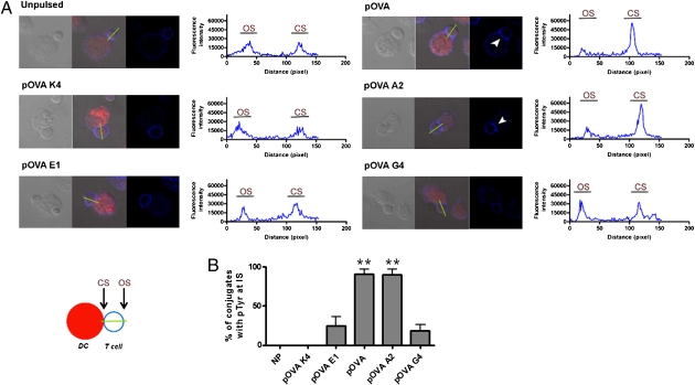
T-cell activation results from productive T-cell receptor (TCR) engagement by a cognate peptide-MHC (pMHC) complex on the antigen presenting cell (APC) surface, a process leading to the polarization of the T-cell secretory machinery toward the APC interface. We have previously shown that the half-life of the TCR/pMHC interaction and the density of pMHC on the APC are two parameters determining T-cell activation. However, whether the half-life of the TCR/pMHC interaction can modulate the efficiency of T-cell secretory machinery polarization toward an APC still remains unclear. Here, by using altered peptide ligands conferring different half-lives to the TCR/pMHC interaction, we have tested how this parameter can control T-cell polarization. We observed that only TCR/pMHC interactions with intermediate half-lives can promote the assembly of synapses that lead to T-cell activation. Strikingly, intermediate half-life interactions can be competed out by short half-life interactions, which can efficiently promote T-cell polarization and antagonize T-cell activation that was induced by activating intermediate half-life interactions. However, short TCR/pMHC interactions fail at promoting phosphorylation of signaling molecules at the T-cell-APC contact interface, which are needed for T-cell activation. Our data suggest that although intermediate half-life pMHC ligands promote assembly of activating synapses, this process can be inhibited by short half-life antagonistic pMHC ligands, which promote the assembly of non activating synapses.
Conflict of interest statement
The authors declare no conflict of interest.
Figures

Similar articles
-
The half-life of the T-cell receptor/peptide-major histocompatibility complex interaction can modulate T-cell activation in response to bacterial challenge.Immunology. 2007 Jun;121(2):227-37. doi: 10.1111/j.1365-2567.2007.02561.x. Epub 2007 Feb 20. Immunology. 2007. PMID: 17313485 Free PMC article.
-
Modulation of T cell function by TCR/pMHC binding kinetics.Immunobiology. 2006;211(1-2):47-64. doi: 10.1016/j.imbio.2005.09.003. Epub 2006 Jan 4. Immunobiology. 2006. PMID: 16446170 Review.
-
T cell receptor binding kinetics required for T cell activation depend on the density of cognate ligand on the antigen-presenting cell.Proc Natl Acad Sci U S A. 2005 Mar 29;102(13):4824-9. doi: 10.1073/pnas.0500922102. Epub 2005 Mar 16. Proc Natl Acad Sci U S A. 2005. PMID: 15772168 Free PMC article.
-
T-cell receptor triggering is critically dependent on the dimensions of its peptide-MHC ligand.Nature. 2005 Jul 28;436(7050):578-82. doi: 10.1038/nature03843. Nature. 2005. PMID: 16049493
-
Diversity in immunological synapse structure.Immunology. 2010 Dec;131(4):466-72. doi: 10.1111/j.1365-2567.2010.03366.x. Epub 2010 Oct 29. Immunology. 2010. PMID: 21039474 Free PMC article. Review.
Cited by
-
Conformational States Control Lck Switching between Free and Confined Diffusion Modes in T Cells.Biophys J. 2020 Mar 24;118(6):1489-1501. doi: 10.1016/j.bpj.2020.01.041. Epub 2020 Feb 11. Biophys J. 2020. PMID: 32097620 Free PMC article.
-
Identifying Individual T Cell Receptors of Optimal Avidity for Tumor Antigens.Front Immunol. 2015 Nov 18;6:582. doi: 10.3389/fimmu.2015.00582. eCollection 2015. Front Immunol. 2015. PMID: 26635796 Free PMC article. Review.
-
Cross-TCR Antagonism Revealed by Optogenetically Tuning the Half-Life of the TCR Ligand Binding.Int J Mol Sci. 2021 May 6;22(9):4920. doi: 10.3390/ijms22094920. Int J Mol Sci. 2021. PMID: 34066527 Free PMC article.
-
Opposite effects of endogenous peptide-MHC class I on T cell activity in the presence and absence of CD8.J Immunol. 2011 May 1;186(9):5193-200. doi: 10.4049/jimmunol.1003755. Epub 2011 Mar 30. J Immunol. 2011. PMID: 21451107 Free PMC article.
-
Human metapneumovirus keeps dendritic cells from priming antigen-specific naive T cells.Immunology. 2013 Jul;139(3):366-76. doi: 10.1111/imm.12083. Immunology. 2013. PMID: 23374037 Free PMC article.
References
-
- Carreño LJ, González PA, Kalergis AM. Modulation of T cell function by TCR/pMHC binding kinetics. Immunobiology. 2006;211:47–64. - PubMed
-
- Kalergis AM. Modulation of T cell immunity by TCR/pMHC dwell time and activating/inhibitory receptor pairs on the antigen-presenting cell. Curr Pharm Des. 2003;9:233–244. - PubMed
-
- González PA, Carreño LJ, Figueroa CA, Kalergis AM. Modulation of immunological synapse by membrane-bound and soluble ligands. Cytokine Growth Factor Rev. 2007;18:19–31. - PubMed
-
- Grakoui A, et al. The immunological synapse: A molecular machine controlling T cell activation. Science. 1999;285:221–227. - PubMed
-
- Huppa JB, Davis MM. T-cell-antigen recognition and the immunological synapse. Nat Rev Immunol. 2003;3:973–983. - PubMed
Publication types
MeSH terms
Substances
LinkOut - more resources
Full Text Sources
Research Materials
