. 2010 Jan;31(1):E12-24.

Reporting standards for endovascular repair of saccular intracranial cerebral aneurysms

Affiliations

Reporting standards for endovascular repair of saccular intracranial cerebral aneurysms

P M Meyers et al. AJNR Am J Neuroradiol. 2010 Jan.

Abstract

Background and purpose: The goal of this article is to provide consensus recommendations for reporting standards, terminology, and written definitions when reporting on the radiological evaluation and endovascular treatment of intracranial, cerebral aneurysms. These criteria can be used to design clinical trials, to provide uniformity of definitions for appropriate selection and stratification of patients, and to allow analysis and meta-analysis of reported data.

Methods: This article was written under the auspices of the Joint Writing Group of the Technology Assessment Committee, Society of NeuroInterventional Surgery, Society of Interventional Radiology; Joint Section on Cerebrovascular Neurosurgery of the American Association of Neurological Surgeons and Congress of Neurological Surgeons; and Section of Stroke and Interventional Neurology of the American Academy of Neurology. A computerized search of the National Library of Medicine database of literature (PubMed) from January 1991 to December 2007 was conducted with the goal to identify published endovascular cerebrovascular interventional data about the assessment and endovascular treatment of cerebral aneurysms useful as benchmarks for quality assessment. We sought to identify those risk adjustment variables that affect the likelihood of success and complications. This article offers the rationale for different clinical and technical considerations that may be important during the design of clinical trials for endovascular treatment of cerebral aneurysms. Included in this guidance article are suggestions for uniform reporting standards for such trials. These definitions and standards are primarily intended for research purposes; however, they should also be helpful in clinical practice and applicable to all publications.

Conclusions: The evaluation and treatment of brain aneurysms often involve multiple medical specialties. Recent reviews by the American Heart Association have surveyed the medical literature to develop guidelines for the clinical management of ruptured and unruptured cerebral aneurysms. Despite efforts to synthesize existing knowledge on cerebral aneurysm evaluation and treatment, significant inconsistencies remain in nomenclature and definition for research and reporting purposes. These operational definitions were selected by consensus of a multidisciplinary writing group to provide consistency for reporting on imaging in clinical trials and observational studies involving cerebral aneurysms. These definitions should help different groups to publish results that are directly comparable.


Figures
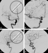
Fig 1.
Fig 1.
Arteriographic projections used to assess aneurysm dimensions. Posterior-anterior Townes and direct lateral projections demonstrate the presence of the right carotid–ophthalmic aneurysm, but these projections do not allow for optimal assessment of aneurysm size, dome-to-neck ratio, or artery-to-neck ratio. Magnified posterior-anterior Waters and lateral oblique projections in this particular case provide better planar imaging to assess the aneurysm for surgical or endovascular treatment.
Fig 2.
Fig 2.
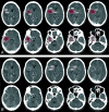
CT brain scan with intravenous iodinated contrast of a patient with a partially thrombosed, giant right middle cerebral aneurysm. A, 5-mm contiguous axial images through the aneurysm demonstrate a ring-enhancing mass with a maximal dimension of 4.8 cm and a total volume of 48 mL. B, The patent component of the aneurysm measures only 14 mm in maximal dimension with a volume of 1.33 mL. The opacified component of the aneurysm at catheter arteriography represents 2.8% of the total aneurysm volume.
Fig 3.
Fig 3.
Diagram of measurements to determine aneurysm dimensions without geometric magnification using 1-cm measuring rings attached to the patient's head. All measurements are made on radiographic images using x-rays. Tube side magnification factor (T), film-side magnification factor (F), and the uncorrected lesion size (L) are measured on the radiograph. The distance from the tube side of the head to the lesion (D) and the width of the head (H) are measured from radiographs in the orthogonal plane. The magnification factor (M) at the level of the lesion is derived by the following formula: M=(DH)×F+(1DH)×T. Due to inherent geometric magnification in radiographic images, the actual aneurysm size is calculated by dividing the apparent aneurysm size as follows: Size=LM.
Fig 4.
Fig 4.
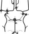
Common location of cerebral aneurysms surrounding the circle of Willis. (A) Middle cerebral (proximal to bifurcation, bifurcation, distal to bifurcation); (B) carotid terminus; (C) anterior choroidal; (D) superior hypophyseal; (E) anterior communicating (proximal to communicating artery, at communicating artery); (F) posterior communicating; (G) ophthalmic; (H) basilar artery (terminus, trunk); (I) superior cerebellar; (J) V4 segment, vertebral; (K) posterior inferior cerebellar; (L) pericallosal artery.
Fig 5.
Fig 5.
Consensus grading scale for endovascular aneurysm occlusion is applied to orthogonal images obtained in optimal projections to assess aneurysm dimensions: Grade 0, complete aneurysm occlusion; Grade 1, ≥90% aneurysm occlusion; Grade 2, 70% to 89% aneurysm occlusion; Grade 3, 50% to 69% aneurysm occlusion; Grade 4, 25% to 49% aneurysm occlusion; Grade 5, <25% aneurysm occlusion.
Fig 6.
Fig 6.
Modification to consensus grading scale for endovascular aneurysm occlusion describing interstitial opacification within coil mass. A, Left internal carotid arteriography in right frontal oblique projection during the arterial phase of injection shows coil occlusion of a 12×11×14 mm left posterior communicating artery aneurysm. B, Because there is a small ventral neck remnant and residual opacification within the aneurysm sac (colored areas), this represents consensus Grade 1 occlusion with “I” for persistent interstitial opacification within the coil mass.

Republished from

Similar articles

  • Reporting standards for endovascular repair of saccular intracranial cerebral aneurysms.
    Meyers PM, Schumacher HC, Higashida RT, Derdeyn CP, Nesbit GM, Sacks D, Wechsler LR, Bederson JB, Lavine SD, Rasmussen P; American Society of Interventional Neuroradiology; Society of Interventional Radiology. Meyers PM, et al. J Vasc Interv Radiol. 2009 Jul;20(7 Suppl):S435-50. doi: 10.1016/j.jvir.2009.03.004. J Vasc Interv Radiol. 2009. PMID: 19560031
  • Reporting standards for endovascular repair of saccular intracranial cerebral aneurysms.
    Meyers PM, Schumacher HC, Higashida RT, Derdeyn CP, Nesbit GM, Sacks D, Wechsler LR, Bederson JB, Lavine SD, Rasmussen P. Meyers PM, et al. Stroke. 2009 May;40(5):e366-79. doi: 10.1161/STROKEAHA.108.527572. Epub 2009 Feb 26. Stroke. 2009. PMID: 19246711 Review.
  • Reporting standards for endovascular repair of saccular intracranial cerebral aneurysms.
    Meyers PM, Schumacher HC, Higashida RT, Derdeyn CP, Nesbit GM, Sacks D, Wechsler LR, Bederson JB, Lavine SD, Rasmussen P; Joint Writing Group of the Technology Assessment Committee, Society of Neurolnterventional Surgery, Society of Interventional Radiology; Joint Section on Cerebrovascular Neurosurgery of the American Association of Neurological Surgeons and Congress of Neurological Surgeons; Section of Stroke and Interventional Neurology of the American Academy of Neurology. Meyers PM, et al. J Neurointerv Surg. 2010 Dec;2(4):312-23. doi: 10.1136/jnis.2010.002337. Epub 2010 Sep 21. J Neurointerv Surg. 2010. PMID: 21990640
  • Reporting standards for angioplasty and stent-assisted angioplasty for intracranial atherosclerosis.
    Schumacher HC, Meyers PM, Higashida RT, Derdeyn CP, Lavine SD, Nesbit GM, Sacks D, Rasmussen P, Wechsler LR. Schumacher HC, et al. Stroke. 2009 May;40(5):e348-65. doi: 10.1161/STROKEAHA.108.527580. Epub 2009 Feb 26. Stroke. 2009. PMID: 19246710 Review.
  • Reporting standards for angioplasty and stent-assisted angioplasty for intracranial atherosclerosis.
    Schumacher HC, Meyers PM, Higashida RT, Derdeyn CP, Lavine SD, Nesbit GM, Sacks D, Rasmussen P, Wechsler LR; Joint Writing Group of the Technology Assessment Committee, Society of Neurolnterventional Surgery, Society of Interventional Radiology; Joint Section on Cerebro-vascular Neurosurgery of the American Association of Neurological Surgeons and Congress of Neurological Surgeons; Section of Stroke and Interventional Neurology of the American Academy of Neurology. Schumacher HC, et al. J Neurointerv Surg. 2010 Dec;2(4):324-40. doi: 10.1136/jnis.2010.002345. J Neurointerv Surg. 2010. PMID: 21990641

Cited by

References

    1. Johnston SC, Higashida RT, Barrow DL, Caplan LR, Dion JE, Hademenos G, Hopkins LN, Molyneux A, Rosenwasser RH, Vinuela F, Wilson CB. Recommendations for the endovascular treatment of intracranial aneurysms: a statement for healthcare professionals from the Committee on Cerebrovascular Imaging of the American Heart Association Council on Cardiovascular Radiology. Stroke 2002; 33: 2536–2544 - PubMed
    1. Bederson JB, Awad IA, Wiebers DO, Piepgras D, Haley EC, Jr, Brott T, Hademenos G, Chyatte D, Rosenwasser R, Caroselli C. Recommendations for the management of patients with unruptured intracranial aneurysms: a statement for healthcare professionals from the Stroke Council of the American Heart Association. Stroke 2000; 31: 2742–2750 - PubMed
    1. Unruptured intracranial aneurysms—risk of rupture and risks of surgical intervention. International Study of Unruptured Intracranial Aneurysms investigators. N Engl J Med 1998; 339: 1725–1733 - PubMed
    1. Wiebers DO, Whisnant JP, Huston J, III, Meissner I, Brown RD, Jr, Piepgras DG, Forbes GS, Thielen K, Nichols D, O'Fallon WM, Peacock J, Jaeger L, Kassell NF, Kongable-Beckman GL, Torner JC. Unruptured intracranial aneurysms: natural history, clinical outcome, and risks of surgical and endovascular treatment. Lancet 2003; 362: 103–110 - PubMed
    1. Cloft HJ, Kallmes DF, Kallmes MH, Goldstein JH, Jensen ME, Dion JE. Prevalence of cerebral aneurysms in patients with fibromuscular dysplasia: a reassessment. J Neurosurg 1998; 88: 436–440 - PubMed

LinkOut - more resources