. 2010 Mar;16(3):572-84.
doi: 10.1261/rna.1495110. Epub 2010 Jan 14.

A unique, thermostable dimer linkage structure of RD114 retroviral RNA

Affiliations

A unique, thermostable dimer linkage structure of RD114 retroviral RNA

Sergei Kharytonchyk et al. RNA. 2010 Mar.

Abstract

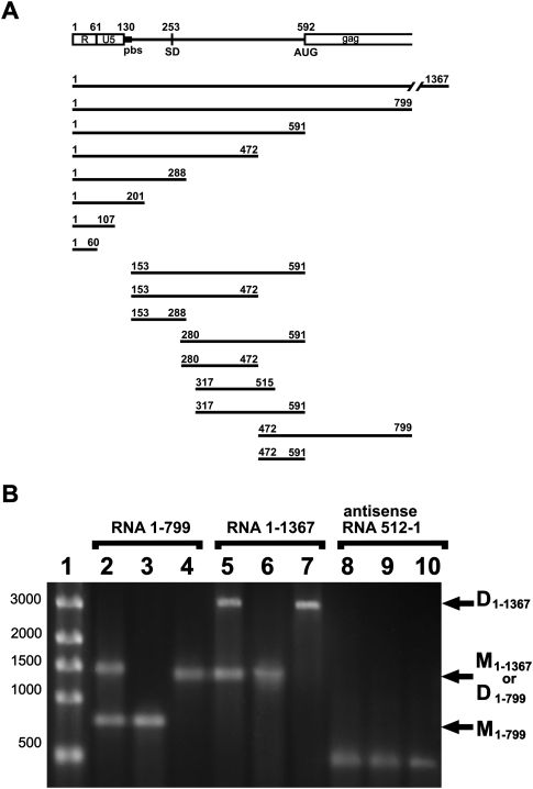
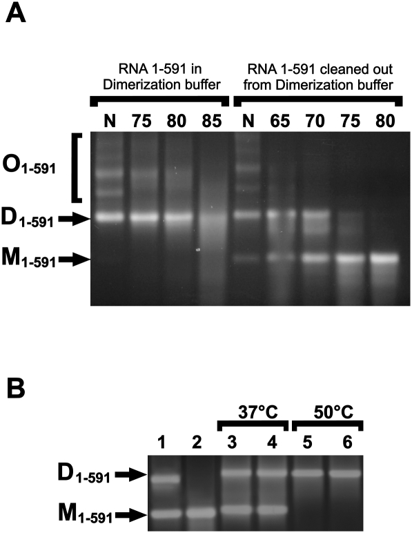
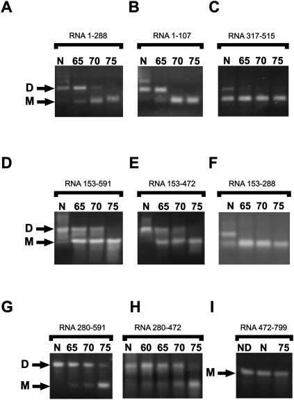
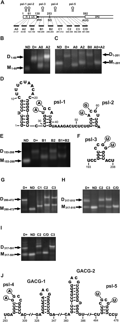
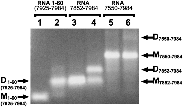
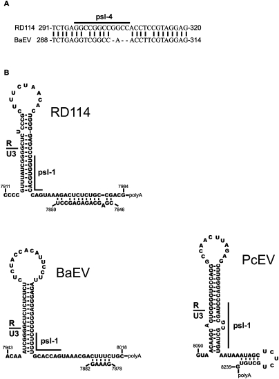
Retroviruses package their genome as RNA dimers linked together primarily by base-pairing between palindromic stem-loop (psl) sequences at the 5' end of genomic RNA. Retroviral RNA dimers usually melt in the range of 55 degrees C-70 degrees C. However, RNA dimers from virions of the feline endogenous gammaretrovirus RD114 were reported to melt only at 87 degrees C. We here report that the high thermal stability of RD114 RNA dimers generated from in vitro synthesized RNA is an effect of multiple dimerization sites located in the 5' region from the R region to sequences downstream from the splice donor (SD) site. By antisense oligonucleotide probing we were able to map at least five dimerization sites. Computational prediction revealed a possibility to form stems with autocomplementary loops for all of the mapped dimerization sites. Three of them were located upstream of the SD site. Mutant analysis supported a role of all five loop sequences in the formation and thermal stability of RNA dimers. Four of the five psls were also predicted in the RNA of two baboon endogenous retroviruses proposed to be ancestors of RD114. RNA fragments of the 5' R region or prolonged further downstream could be efficiently dimerized in vitro. However, this was not the case for the 3' R region linked to upstream U3 sequences, suggesting a specific mechanism of negative regulation of dimerization at the 3' end of the genome, possibly explained by a long double-stranded RNA region at the U3-R border. Altogether, these data point to determinants of the high thermostability of the dimer linkage structure of the RD114 genome and reveal differences from other retroviruses.


Figures

FIGURE 1.
FIGURE 1.
(A) Scheme of the first 799 nt of RD114 genomic RNA and representation of the different RNA fragments synthesized for dimerization analysis. (R) Repeat sequence; (U5) unique sequence at the 5′ end of the RNA genome; (pbs) primer-binding site; (SD) splice-donor site, mapped in this study, and (AUG) translation initiator codon of gag. (B) Representative 1% agarose gel electrophoresis of RD114 RNA fragments. (Lane 1) ssRNA ladder; (lanes 2–4) RD114 1–799 nt RNA fragment; (lanes 5–7) RD114 1–1367 nt RNA fragment; (lanes 8–10) RD114 antisense 512-1 nt RNA fragment. (Lanes 2,5,8) RNA was loaded on the gel just after in vitro transcription. (Lanes 3,6,9) RNA after in vitro transcription was cleaned on affinity columns and melted at 90°C for 2 min in water. (Lanes 4,7,10) RNA after melting was dimerized at 50°C for 30 min in the presence of dimerization buffer (see Materials and Methods). D, dimers; M monomers.
FIGURE 2.
FIGURE 2.
Formation of dimers and oligomers of 1–599 RD114 RNA determination of their thermal stability. One percent agarose gel electrophoresis of 1–599 RD114 RNA fragment. (A) RNA was cleaned out from in vitro transcription mix, denatured, and incubated with dimerization buffer as described. After dimerization 10 times diluted aliquots of RNA was incubated for 10 min at indicated temperatures or was cleaned out from dimerization buffer before heating in water. (B) RNA was cleaned out from in vitro transcription mix (lane 1), denatured (lane 2), and then incubated with dimerization buffer (lanes 3,5) or transcription buffer (lanes 4,6). RNA probes were incubated for 1 h at 37°C (lanes 3,4) or 20 min at 50°C (lanes 5 and 6). N, no heating; D, dimers; M monomers; O, oligomers.
FIGURE 3.
FIGURE 3.
Dimerization and determination of thermal stability of dimers of RD114 genomic RNA fragments. RNA was cleaned from in vitro transcription mix, denatured, and incubated with dimerization buffer as described. After dimerization, RNA was cleaned from dimerization buffer using affinity columns and aliquots incubated for 10 min in water at the indicated temperatures. (A) 1–288 RNA fragment; (B) 1–107 RNA fragment; (C) 317–515 RNA fragment; (D) 153–591 RNA fragment; (E) 153–472 RNA fragment; (F) 153–288 RNA fragment; (G) 280–591 RNA fragment; (H) 280–472 RNA fragment; (I) 472–799 RNA fragment. N, no heating; D, dimers; M monomers; ND, this probe was not incubated with dimerization buffer.
FIGURE 4.
FIGURE 4.
Antisense oligonucleotide mapping of dimerization sites on the RD114 leader RNA fragments. After synthesis and cleaning, RNA was denaturized at 90°C for 2 min and then incubated at 50°C for 30 min with dimerization buffer in the presence or absence of antisense DNA oligonucleotides as indicated. Probes were used for electrophoresis as described above. (A) Scheme of the first 799 nt of RD114 genomic RNA and representation of the antisense DNA oligonucleotides used in this study. Localization of the mapped palindromic stem–loops (psl) involved in dimerization indicated. (B) Antisense inhibition of dimerization of 1–60 RNA fragment (R-region); (C) antisense inhibition of dimerization of 1–201 RNA fragment; (D) predicted secondary structure of 1–65 nt RD114 genomic RNA, containing psl-1 and psl-2. The circles indicate nucleotide shifts in the created mutants; (E) antisense inhibition of dimerization of 153–288 RNA fragment; (F) predicted secondary structure of psl-3 (196–205 nt); (G) antisense inhibition of dimerization of 280–472 RNA fragment; (H) antisense inhibition of dimerization of 317–515 RNA fragment; (I) antisense inhibition of dimerization of 317–591 RNA fragment; (J) predicted secondary structure of psl-4 (296–207 nt), psl-5 (466–478 nt), and two stem–loops with GACG-loop motif (330–345 and 363–384). ND, this fragment was not incubated with dimerization buffer; D+ dimerized RNA with no antisense oligonucleotides; D, dimers; M, monomers.
FIGURE 5.
FIGURE 5.
Dimerization and determination of thermal dimer stability of RD114 genomic RNA fragments containing mutations in the psl sites. RNA was cleaned from in vitro transcription mix, denatured, and incubated with dimerization buffer as described. After dimerization, RNA was cleaned from dimerization buffer using affinity columns and aliquots incubated for 10 min in water at the indicated temperatures. (A) 1–288 RNA fragment with mutations in the psl-1, psl-2, and psl-3 sites; (B) 1–591 RNA fragment with mutations in the psl-4 and psl-5 sites; (C) 1–591 RNA fragment with mutations in the psl-1, psl-2, and psl-3 sites; (D) 1–591 RNA fragment with mutations in the psl-4 and psl-5 sites; (E) 1–591 RNA fragment with mutations in the psl-1, psl-2, psl-3, psl-4, and psl-5 sites. N, no heating; D, dimers; M, monomers.
FIGURE 6.
FIGURE 6.
Influence of upstream sequences on the 3′ R RNA fragment dimerization. After synthesis and cleaning RNA was denaturized at 90°C for 2 min (lanes 1,3,5) and incubated at 50°C for 30 min with dimerization buffer (lanes 2,4,6). Probes were used for electrophoresis immediately after induced dimerization. (Lanes 1,2) RNA fragment corresponding R region of RD114 genome, bases from 1 to 60 or from 7925 to 7984; (lanes 3,4) RNA fragment corresponding to RD114 genome, bases from 7852 to 7984; (lanes 5,6) RNA fragment corresponding to RD114 genome, bases from 7550 to 7984. D, dimers; M, monomers.
FIGURE 7.
FIGURE 7.
(A) Short stretch of homology between RD114 and BaEV containing the psl-4 sequence. (B) Predicted secondary structures at 3′ ends of the genomes of the three studied viruses. Note: The PcEV sequence used for the 3′ end of genomic RNA was reconstructed using in part a sequence from 5′ LTR of the available virus clone (accession number AF142988) due to the presence of a deletion and point mutations at the 3′LTR (Mang et al. 1999).

Similar articles

Cited by

References

    1. Aagaard L, Rasmussen SV, Mikkelsen JG, Pedersen FS. Efficient replication of full-length murine leukemia viruses modified at the dimer initiation site regions. Virology. 2004;318:360–370. - PubMed
    1. Andersen ES, Contera SA, Knudsen B, Damgaard CK, Besenbacher F, Kjems J. Role of the trans-activation response element in dimerization of HIV-1 RNA. J Biol Chem. 2004;279:22243–22249. - PubMed
    1. Badorrek CS, Gherge CM, Weeks KM. Structure of an RNA switch that enforces stringent retroviral genomic RNA dimerization. Proc Natl Acad Sci. 2006;103:13640–13645. - PMC - PubMed
    1. Basyuk E, Boulon S, Skou Pedersen F, Bertrand E, Vestergaard Rasmussen S. The packaging signal of MLV is an integrated module that mediates intracellular transport of genomic RNAs. J Mol Biol. 2005;354:330–339. - PubMed
    1. Bender W, Davidson N. Mapping of poly(A) sequences in the electron microscope reveals unusual structure of type C oncornavirus RNA molecules. Cell. 1976;7:595–607. - PubMed

Publication types

LinkOut - more resources