

Direct DNA amplification from crude clinical samples using a PCR enhancer cocktail and novel mutants of Taq
- PMID: 20075207
- PMCID: PMC2871721
- DOI: 10.2353/jmoldx.2010.090070
Direct DNA amplification from crude clinical samples using a PCR enhancer cocktail and novel mutants of Taq
Abstract
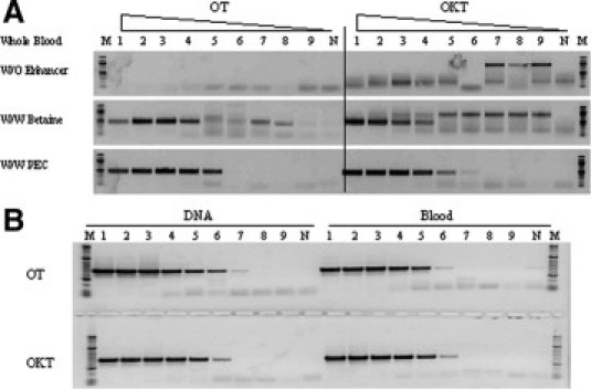
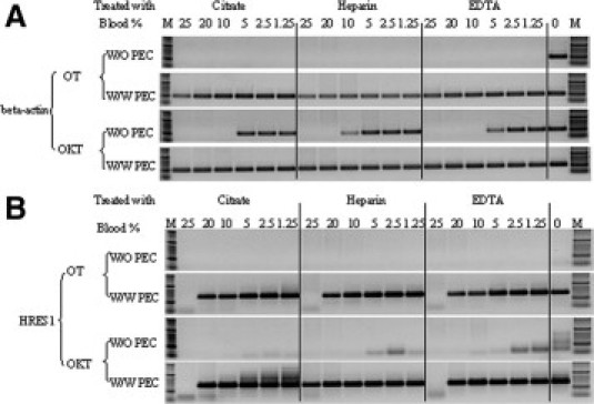
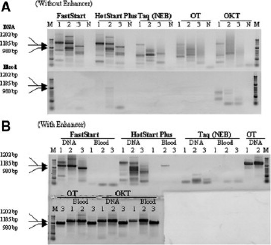
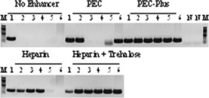
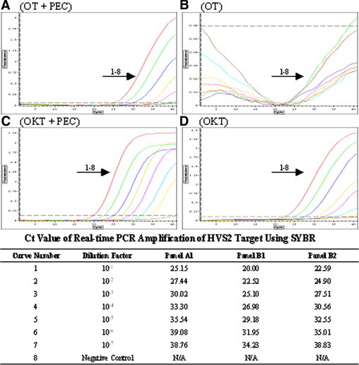
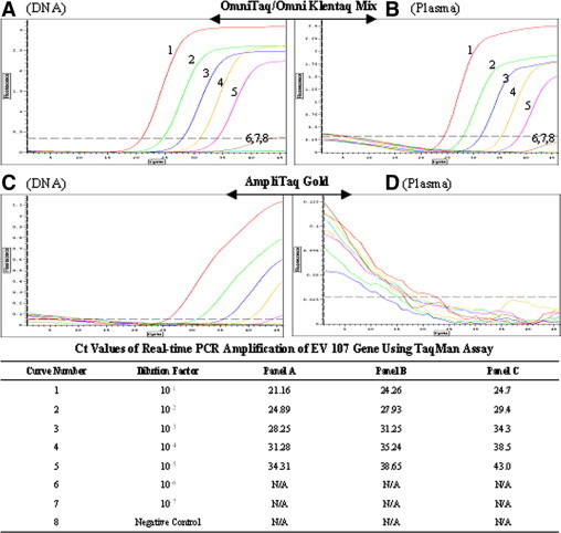
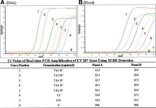
PCR-based clinical and forensic tests often have low sensitivity or even false-negative results caused by potent PCR inhibitors found in blood and soil. It is widely accepted that purification of target DNA before PCR is necessary for successful amplification. In an attempt to overcome PCR inhibition, enhance PCR amplification, and simplify the PCR protocol, we demonstrate improved PCR-enhancing cocktails containing nonionic detergent, l-carnitine, d-(+)-trehalose, and heparin. These cocktails, in combination with two inhibitor-resistant Taq mutants, OmniTaq and Omni Klentaq, enabled efficient amplification of exogenous, endogenous, and high-GC content DNA targets directly from crude samples containing human plasma, serum, and whole blood without DNA purification. In the presence of these enhancer cocktails, the mutant enzymes were able to tolerate at least 25% plasma, serum, or whole blood and as high as 80% GC content templates in PCR reactions. These enhancer cocktails also improved the performance of the novel Taq mutants in real-time PCR amplification using crude samples, both in SYBR Green fluorescence detection and TaqMan assays. The novel enhancer mixes also facilitated DNA amplification from crude samples with various commercial Taq DNA polymerases.
Figures
Similar articles
-
1,2-propanediol-trehalose mixture as a potent quantitative real-time PCR enhancer.BMC Biotechnol. 2011 Apr 18;11:41. doi: 10.1186/1472-6750-11-41. BMC Biotechnol. 2011. PMID: 21501492 Free PMC article.
-
Mutants of Taq DNA polymerase resistant to PCR inhibitors allow DNA amplification from whole blood and crude soil samples.Nucleic Acids Res. 2009 Apr;37(5):e40. doi: 10.1093/nar/gkn1055. Epub 2009 Feb 10. Nucleic Acids Res. 2009. PMID: 19208643 Free PMC article.
-
A strong strand displacement activity of thermostable DNA polymerase markedly improves the results of DNA amplification.Biotechniques. 2014 Aug 1;57(2):81-7. doi: 10.2144/000114198. eCollection 2014 Aug. Biotechniques. 2014. PMID: 25109293
-
[Quantitative PCR in the diagnosis of Leishmania].Parassitologia. 2004 Jun;46(1-2):163-7. Parassitologia. 2004. PMID: 15305709 Review. Italian.
-
PCR and DNA sequencing.Biotechniques. 1989 Jul-Aug;7(7):700-8. Biotechniques. 1989. PMID: 2698653 Review.
Cited by
-
Real-time PCR based detection of the lactase non-persistence associated genetic variant LCT-13910C>T directly from whole blood.Mol Biol Rep. 2019 Apr;46(2):2379-2385. doi: 10.1007/s11033-019-04696-9. Epub 2019 Feb 21. Mol Biol Rep. 2019. PMID: 30790118
-
Optimization of a Quantitative PCR Methodology for Detection of Aspergillus spp. and Rhizopus arrhizus.Mol Diagn Ther. 2022 Sep;26(5):511-525. doi: 10.1007/s40291-022-00595-1. Epub 2022 Jun 16. Mol Diagn Ther. 2022. PMID: 35710958 Free PMC article.
-
1,2-propanediol-trehalose mixture as a potent quantitative real-time PCR enhancer.BMC Biotechnol. 2011 Apr 18;11:41. doi: 10.1186/1472-6750-11-41. BMC Biotechnol. 2011. PMID: 21501492 Free PMC article.
-
Unlocking the potential of ancient fish DNA in the genomic era.Evol Appl. 2019 May 28;12(8):1513-1522. doi: 10.1111/eva.12811. eCollection 2019 Sep. Evol Appl. 2019. PMID: 31462911 Free PMC article.
-
Forensic animal DNA analysis using economical two-step direct PCR.Forensic Sci Med Pathol. 2014 Mar;10(1):29-38. doi: 10.1007/s12024-013-9521-8. Epub 2014 Jan 17. Forensic Sci Med Pathol. 2014. PMID: 24435950
References
-
- Cursons RT, Jeyerajah E, Sleigh JW. The use of polymerase chain reaction to detect septicemia in critically ill patients. Crit Care Med. 1999;27:937–940. - PubMed
-
- Rautenberg P, Lubbert C, Weers W, Boetel E, Schweichler J, Zhou L, Costard-Jackle A, Kraemer-Hansen H, Harder TC. Evaluation of the AmpliSensor PCR and the SHARP signal detection system for the early prediction of symptomatic CMV infection in solid transplant recipients. J Clin Virol. 1999;13:81–94. - PubMed
-
- Robertson JM, Walsh-Weller J. An introduction to PCR primer design and optimization of amplification reactions. Methods Mol Biol. 1998;98:121–154. - PubMed
-
- Bussani C, Cioni R, Mattei A, Fambrini M, Marchionni M, Scarselli G. Prenatal diagnosis of common aneuploidies in transcervical samples using quantitative fluorescent-PCR analysis. Mol Diagn Ther. 2007;11:117–121. - PubMed
Publication types
MeSH terms
Substances
Grants and funding
LinkOut - more resources
Full Text Sources
Other Literature Sources
Miscellaneous
