Review
. 2010 Apr;108(4):995-1002.
doi: 10.1152/japplphysiol.00712.2009. Epub 2010 Jan 14.

Central CO2 chemoreception and integrated neural mechanisms of cardiovascular and respiratory control

Affiliations
Review

Central CO2 chemoreception and integrated neural mechanisms of cardiovascular and respiratory control

Patrice G Guyenet et al. J Appl Physiol (1985). 2010 Apr.

Abstract

In this review, we examine why blood pressure (BP) and sympathetic nerve activity (SNA) increase during a rise in central nervous system (CNS) P(CO(2)) (central chemoreceptor stimulation). CNS acidification modifies SNA by two classes of mechanisms. The first one depends on the activation of the central respiratory controller (CRG) and causes the much-emphasized respiratory modulation of the SNA. The CRG probably modulates SNA at several brain stem or spinal locations, but the most important site of interaction seems to be the caudal ventrolateral medulla (CVLM), where unidentified components of the CRG periodically gate the baroreflex. CNS P(CO(2)) also influences sympathetic tone in a CRG-independent manner, and we propose that this process operates differently according to the level of CNS P(CO(2)). In normocapnia and indeed even below the ventilatory recruitment threshold, CNS P(CO(2)) exerts a tonic concentration-dependent excitatory effect on SNA that is plausibly mediated by specialized brain stem chemoreceptors such as the retrotrapezoid nucleus. Abnormally high levels of P(CO(2)) cause an aversive interoceptive awareness in awake individuals and trigger arousal from sleep. These alerting responses presumably activate wake-promoting and/or stress-related pathways such as the orexinergic, noradrenergic, and serotonergic neurons. These neuronal groups, which may also be directly activated by brain acidification, have brainwide projections that contribute to the CO(2)-induced rise in breathing and SNA by facilitating neuronal activity at innumerable CNS locations. In the case of SNA, these sites include the nucleus of the solitary tract, the ventrolateral medulla, and the preganglionic neurons.


Figures
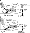
Fig. 1.
Fig. 1.
Cardiorespiratory coupling. A: arrowheads 1–4 indicate 4 lower brain stem or spinal locations where components of the central respiratory pattern generator may influence the sympathetic outflow to the heart and blood vessels. The sites are ranked according to the strength of the available supporting evidence from most plausible (1) to less documented (4). Site 1 is the main inhibitory relay of the baroreflex located in the caudal ventrolateral medulla (CVLM), a region that is coextensive with the pre-Bötzinger and rostral ventral respiratory group subdivisions of the ventral respiratory column. Site 2, the rostral ventrolateral medulla (RVLM), contains the main source of direct excitatory drive to the sympathetic vasomotor preganglionic neurons. The respiratory modulation of these neurons derives largely from the respiratory gating of their CVLM input but direct inputs from the central respiratory pattern generator (CRG) or from the retrotrapezoid nucleus (RTN) may also contribute to this modulation. Site 3 is the nucleus of the solitary tract (NTS). The NTS receives inputs from arterial baroreceptors and from the CRG. Site 4 is the spinal cord. Excitatory inputs from collaterals of bulbospinal inspiratory neurons were thought to contribute to the inspiratory modulation of SNA in cats either directly or via spinal interneurons. The required anatomic pathway has not been fully documented, and the theory is inconsistent with the phase-spanning nature of the respiration modulation of SNA present in rodents. Each arrow (1–4) describes one or a set of respiratory modulated inputs. The most plausible anatomic origins of these inputs are the ventral respiratory column (VRC), the dorsolateral pons (dlpons), and the RTN, which are highlighted in gray. B: possible role of the RTN in the central sympathetic chemoreflex. The RTN is a chemosensitive group of glutamatergic neurons that has a phase-spanning respiratory modulation (in rats) when the respiratory network is active and a tonic CO2-dependent activity below the apneic threshold or when the CRG is silenced pharmacologically. The RTN drives the CRG and probably integrates much of the chemical drive to breathe. The RTN also innervates the region that contains the vasomotor neurons (RVLM) and their CVLM antecedent neurons. The RTN could therefore contribute a respiratory-independent drive to the sympathetic outflow when CO2 is below the respiratory recruitment threshold and a respiratory-modulated drive to the sympathetic outflow when Pco2 exceeds this threshold. This theory remains to be thoroughly tested. NE, norepinephrine; SPGNs, preganglionic sympathetic neurons; medulla obl, medulla oblongata “7”, facial motor nucleus.

Similar articles

Cited by

References

    1. Abbott SBG, Stornetta RL, Fortuna MG, Depuy SD, West GH, Harris TE, Guyenet PG. Photostimulation of retrotrapezoid nucleus Phox2b-expressing neurons in vivo produces long-lasting activation of breathing in rats. J Neurosci 29: 5806–5819, 2009 - PMC - PubMed
    1. Ainslie PN, Duffin J. Integration of cerebrovascular CO2 reactivity and chemoreflex control of breathing: mechanisms of regulation, measurement, and interpretation. Am J Physiol Regul Integr Comp Physiol 296: R1473–R1495, 2009 - PubMed
    1. Alheid GF, McCrimmon DR. The chemical neuroanatomy of breathing. Respir Physiol Neurobiol 164: 3–11, 2008 - PMC - PubMed
    1. Antunes VR, Brailoiu GC, Kwok EH, Scruggs P, Dun NJ. Orexins/hypocretins excite rat sympathetic preganglionic neurons in vivo and in vitro. Am J Physiol Regul Integr Comp Physiol 281: R1801–R1807, 2001 - PubMed
    1. Aston-Jones G, Chen S, Zhu Y, Oshinsky ML. A neural circuit for circadian regulation of arousal. Nat Neurosci 4: 732–738, 2001 - PubMed

Publication types

MeSH terms