

Differential expression of tissue repair genes in the pathogenesis of chronic obstructive pulmonary disease
- PMID: 20075389
- PMCID: PMC2894408
- DOI: 10.1164/rccm.200812-1902OC
Differential expression of tissue repair genes in the pathogenesis of chronic obstructive pulmonary disease
Abstract
Rationale: The airflow limitation that defines severity of chronic obstructive pulmonary disease (COPD) is caused by a combination of small airway obstruction and emphysematous lung destruction.
Objectives: To examine the hypothesis that small airway obstructive and emphysematous destructive lesions are produced by differential expression of genes associated with tissue repair.
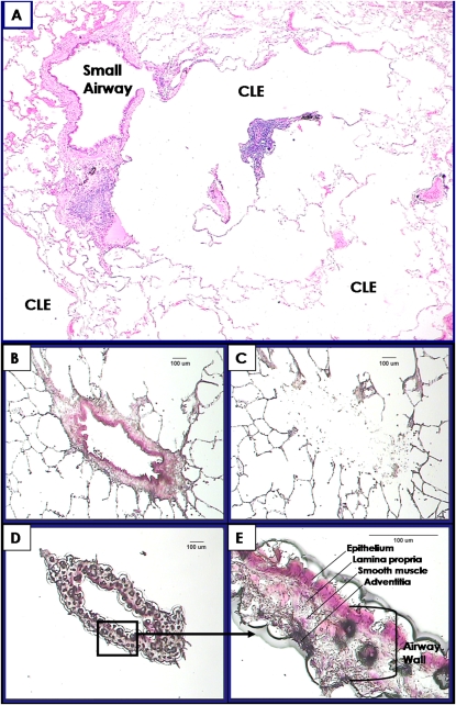
Methods: The expression of 54 genes associated with repair of repetitively damaged tissue was measured in 136 paired samples of small bronchioles and surrounding lung tissue separated by laser capture microdissection. These samples were collected from 63 patients at different levels of disease severity who required surgery for either lung cancer or lung transplantation for very severe COPD. Gene expression was measured by quantitative polymerase chain reaction in these paired samples and compared with the FEV(1) by linear regression analysis.
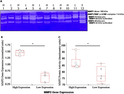
Measurements and main results: After corrections for false discovery rates, only 2 of 10 genes (serpin peptidase inhibitor/plasminogen activator inhibitor member 2 and matrix metalloproteinase [MMP] 10) increased, whereas 8 (MMP2, integrin-alpha1, vascular endothelial growth factor, a disintegrin and metallopeptidase domain 33, scatter factor/hepatocyte growth factor, tissue inhibitor of matrix metalloproteinase-2, fibronectin, and collagen 3alpha1) decreased in small airways in association with FEV(1). In contrast, 8/12 genes (early growth response factor 1, MMP1, MMP9, MMP10, plasminogen activator urokinase, plasminogen activator urokinase receptor, tumor necrosis factor, and IL13) increased and 4/12 (MMP2, tissue inhibitor of matrix metalloproteinase-1, collagen 1alpha1, and transforming growth factor-beta3) decreased in the surrounding lung tissue in association with progression of COPD.
Conclusions: The progression of COPD is associated with the differential expression of a cluster of genes that favor the degradation of the tissue surrounding the small conducting airways.
Figures
Similar articles
-
What drives the peripheral lung-remodeling process in chronic obstructive pulmonary disease?Proc Am Thorac Soc. 2009 Dec;6(8):668-72. doi: 10.1513/pats.200907-079DP. Proc Am Thorac Soc. 2009. PMID: 20008873 Free PMC article. Review.
-
Elevated circulating PAI-1 levels are related to lung function decline, systemic inflammation, and small airway obstruction in chronic obstructive pulmonary disease.Int J Chron Obstruct Pulmon Dis. 2016 Sep 26;11:2369-2376. doi: 10.2147/COPD.S107409. eCollection 2016. Int J Chron Obstruct Pulmon Dis. 2016. PMID: 27713627 Free PMC article.
-
[The role of matrix metalloproteinases in extracellular matrix remodelling in chronic obstructive pulmonary disease rat models].Zhonghua Nei Ke Za Zhi. 2002 Jun;41(6):393-8. Zhonghua Nei Ke Za Zhi. 2002. PMID: 12137602 Chinese.
-
Evaluation of small sample cDNA amplification for microdissected airway expression profiling in COPD.COPD. 2007 Jun;4(2):91-105. doi: 10.1080/15412550701246427. COPD. 2007. PMID: 17530502
-
Lung structure and function in COPD.Int J Tuberc Lung Dis. 2008 May;12(5):467-79. Int J Tuberc Lung Dis. 2008. PMID: 18419881 Review.
Cited by
-
A gene expression signature of emphysema-related lung destruction and its reversal by the tripeptide GHK.Genome Med. 2012 Aug 31;4(8):67. doi: 10.1186/gm367. eCollection 2012. Genome Med. 2012. PMID: 22937864 Free PMC article.
-
Matrix metalloproteinases in emphysema.Matrix Biol. 2018 Nov;73:34-51. doi: 10.1016/j.matbio.2018.01.018. Epub 2018 Mar 23. Matrix Biol. 2018. PMID: 29406250 Free PMC article. Review.
-
Gene coexpression networks reveal novel molecular endotypes in alpha-1 antitrypsin deficiency.Thorax. 2021 Feb;76(2):134-143. doi: 10.1136/thoraxjnl-2019-214301. Epub 2020 Dec 10. Thorax. 2021. PMID: 33303696 Free PMC article.
-
Modeling Extracellular Matrix-Cell Interactions in Lung Repair and Chronic Disease.Cells. 2021 Aug 20;10(8):2145. doi: 10.3390/cells10082145. Cells. 2021. PMID: 34440917 Free PMC article.
-
TGF-β Signaling Pathways in Different Compartments of the Lower Airways of Patients With Stable COPD.Chest. 2018 Apr;153(4):851-862. doi: 10.1016/j.chest.2017.12.017. Epub 2017 Dec 28. Chest. 2018. PMID: 29289685 Free PMC article.
References
-
- Rabe KF, Hurd S, Anzueto A, Barnes PJ, Buist SA, Calverley P, Fukuchi Y, Jenkins C, Rodriguez-Roisin R, van Weel C, et al. Global strategy for the diagnosis, management, and prevention of chronic obstructive pulmonary disease: GOLD executive summary. Am J Respir Crit Care Med 2007;176:532–555. - PubMed
-
- McLean KH. The macroscopic anatomy of pulmonary emphysema. Australas Ann Med 1956;5:73–88. - PubMed
-
- Hogg JC, Macklem PT, Thurlbeck WM. Site and nature of airway obstruction in chronic obstructive lung disease. N Engl J Med 1968;278:1355–1360. - PubMed
-
- Van Brabandt H, Cauberghs M, Verbeken E, Moerman P, Lauweryns JM, Van de Woestijne KP. Partitioning of pulmonary impedance in excised human and canine lungs. J Appl Physiol 1983;55:1733–1742. - PubMed
Publication types
MeSH terms
Grants and funding
LinkOut - more resources
Full Text Sources
Other Literature Sources
Medical
Miscellaneous
