. 2010 May;82(5):1008-17.
doi: 10.1095/biolreprod.109.082057. Epub 2010 Jan 14.

P-body loss is concomitant with formation of a messenger RNA storage domain in mouse oocytes

Affiliations

P-body loss is concomitant with formation of a messenger RNA storage domain in mouse oocytes

Matyas Flemr et al. Biol Reprod. 2010 May.

Abstract

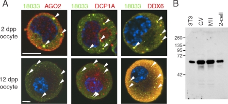
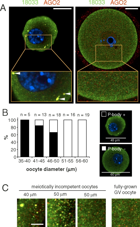
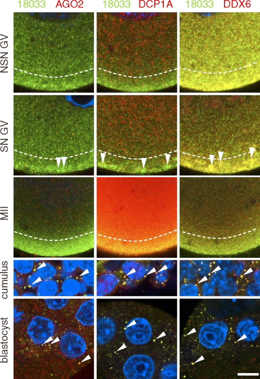
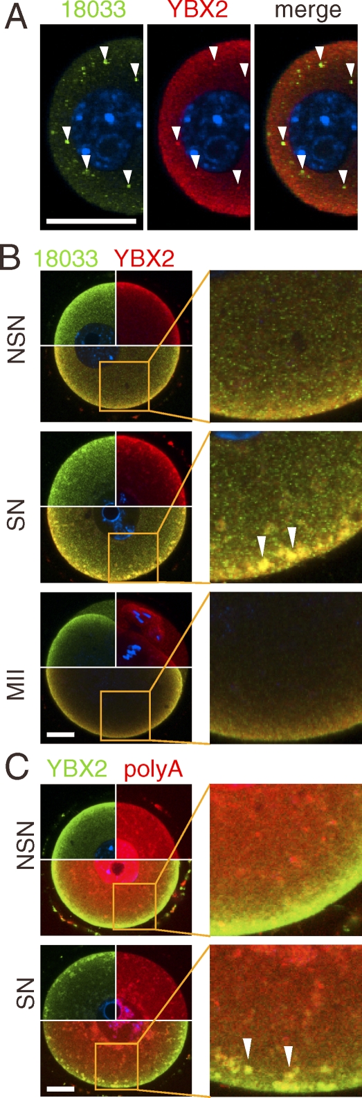
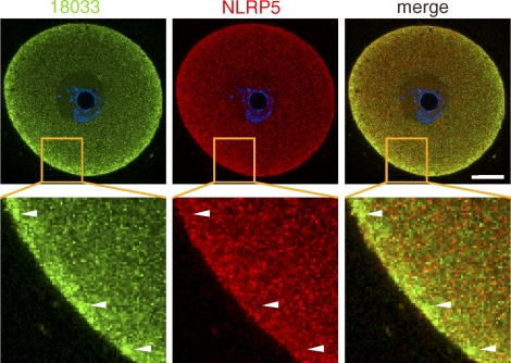
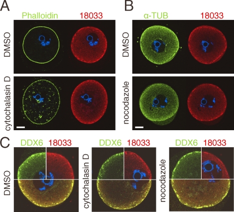
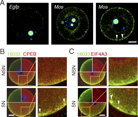
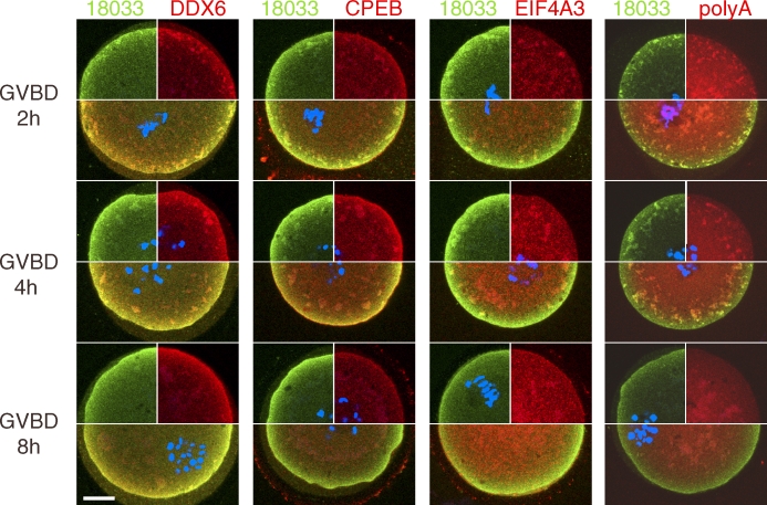
In mammalian somatic cells, several pathways that converge on deadenylation, decapping, and 5'-3' degradation are found in cytoplasmic foci known as P-bodies. Because controlled mRNA stability is essential for oocyte-to-zygote transition, we examined the dynamics of P-body components in mouse oocytes. We report that oocyte growth is accompanied by loss of P-bodies and a subcortical accumulation of several RNA-binding proteins, including DDX6, CPEB, YBX2 (MSY2), and the exon junction complex. These proteins form transient RNA-containing aggregates in fully grown oocytes with a surrounded nucleolus chromatin configuration. These aggregates disperse during oocyte maturation, consistent with recruitment of maternal mRNAs that occurs during this time. In contrast, levels of DCP1A are low during oocyte growth, and DCP1A does not colocalize with DDX6 in the subcortical aggregates. The amount of DCP1A markedly increases during meiosis, which correlates with the first wave of destabilization of maternal mRNAs. We propose that the cortex of growing oocytes serves as an mRNA storage compartment, which contains a novel type of RNA granule related to P-bodies.


Figures

FIG. 1.
FIG. 1.
P-bodies are present in small incompetent oocytes. A) Confocal images of mouse meiotically incompetent oocytes from 2 dpp and 12 dpp females after staining with 18033, AGO2, DCP1A, and DDX6 antibodies. Staining with 18033 is green colored, other proteins are shown in red, and DNA staining (DAPI) is shown in blue. Colocalization yields yellow color. The arrowheads depict P-bodies. For each sample, at least 10 oocytes were examined, and representative images are shown. Bar = 10 μm. B) DDX6 antibody specificity analyzed by Western blot analysis. DDX6 antibody used in this work detects a single band corresponding to the size of DDX6 (RCK/p54).
FIG. 2.
FIG. 2.
P-bodies disappear as oocytes increase in size. A) Representative confocal images showing colocalization of 18033 and AGO2 in small, meiotically incompetent (12 dpp; left panel) and fully grown GV SN oocytes (right panel). Staining with 18033 is green colored, AGO2 is shown in red, and DNA staining (DAPI) is shown in blue. Colocalization of 18033 and AGO2 yields yellow color. Horizontal arrowheads depict P-body-like structures. AGO2 signal was enhanced in the fully grown GV oocyte to visualize the lack of AGO and 18033 colocalization. It should be noted that the observed intensity of the subcortical staining depends on setting the dynamic range of the image acquisition. Bars = 20 μm. B) Quantification of P-body presence/absence in incompetent oocytes (12 dpp). Oocyte diameter was estimated by image analysis of the central confocal sections. The numbers of incompetent oocytes within each group are indicated above the columns. The representative images show what was classified as a P-body-containing oocyte (shown in the solid bars in the histogram) and an oocyte not containing P-bodies (shown as the open bars in the histogram). These images were acquired in one experiment with the same confocal microscope settings. C) A detail of cytoplasmic staining from incompetent and fully grown oocytes showing the loss of 18033 and AGO2 colocalization in larger oocytes. Staining with 18033 is green colored, and staining with AGO2 is shown in red. Colocalization yields yellow color. Bar = 5 μm.
FIG. 3.
FIG. 3.
Expression of P-body components during oocyte development. Confocal images of mouse GV NSN and SN oocytes, MII eggs, cumulus cells, and blastocysts after staining with 18033, AGO2, DCP1A, and DDX6 antibodies. A representative region of a whole specimen is shown. Staining with 18033 is green colored, other proteins are shown in red, and DNA staining (DAPI) is shown in blue. Colocalization yields yellow color. Diagonal arrowheads depict P-bodies in blastocysts and somatic cells. Vertical arrowheads depict subcortical aggregates found in SN GV oocytes. Dashed lines border the subcortical domain with enhanced staining of 18033 and DDX6. Note low colocalization of DDX6 and 18033 outside of the subcortical domain. Staining with the same primary antibodies were performed independently in both the United States and Czech laboratories with similar results; a representative staining is shown. GV and MII samples in each column come from the same staining experiment and were scanned with identical confocal microscope settings. SN, surrounded nucleolus; NSN, nonsurrounded nucleolus; GV, fully grown germinal-vesicle oocyte; MII, oocyte in meiosis II. Bar = 10 μm. For each sample, at least 10 oocytes, eggs, blastocysts, or cumulus cells were examined; representative images are shown.
FIG. 4.
FIG. 4.
Poly(A) RNA and RNA-binding protein YBX2, but not the SCMC complex, form subcortical RNP aggregates with P-body components. A) YBX2 (MSY2) is also present in P-bodies. Confocal images showing colocalization of 18033 and YBX2 in small, meiotically incompetent oocytes (12 dpp). The arrowheads point to subcortical P-bodies. The blue signal is DNA staining (DAPI). B) Confocal images showing colocalization of 18033 and YBX2 in fully grown GV NSN and SN oocytes and MII eggs. C) Confocal images of colocalization of YBX2 and poly(A) RNA, visualized by TAMRA-oligo(dT)18, in fully grown GV NSN and SN oocytes. Contrast of poly(A) samples was linearly enhanced for better presentation of colocalization in SCRD and SCAs. The identities of green and red channels are indicated above each panel, colocalization yields yellow color. The blue signal is DNA staining (DAPI). The area in the merged image outlined by solid lines is magnified in the adjacent image. The arrowheads point to subcortical RNP aggregates. For each sample, at least 10 oocytes were examined, and representative images are shown. SN, surrounded nucleolus GV oocyte; NSN, nonsurrounded nucleolus GV oocyte. Bars = 20 μm.
FIG. 5.
FIG. 5.
Confocal images showing immunolocalization of 18033 and NLRP5 in GV SN oocytes. Identities of green and red channels are indicated above each panel, and colocalization yields yellow color. The blue signal is DNA staining (DAPI). The area in the merged image outlined by solid lines is magnified in the adjacent image. The arrowheads point to subcortical RNP aggregates. Ten oocytes were examined, and representative images are shown. SN, surrounded nucleolus GV oocyte; NSN, nonsurrounded nucleolus GV oocyte. Bar = 20 μm.
FIG. 6.
FIG. 6.
SCRD and SCAs are independent on actin and tubulin cytoskeleton. A) Immunolocalization of 18033 and actin (Phaloidin-A488) in oocytes treated with cytochalasin D. B) Immunolocalization of 18033 and α-tubulin in oocytes treated with nocodazole. Staining of DNA (DAPI) is shown in blue. C) SCAs remain unchanged upon cytochalasin D and nocodazole treatments. Confocal images showing colocalization of 18033 and DDX6 in oocytes treated with cytochalasin D and nocodazole. Staining of DNA (DAPI) is shown in blue. Dimethyl sulfoxide (DMSO)-treated oocytes serve as a control. Bars = 20 μm.
FIG. 7.
FIG. 7.
Subcortical RNP domain holds dormant maternal mRNAs. A) In situ detection of Mos mRNA in the SCRD. The arrowheads depict SCA-like structures in SN oocytes, which are similar to the SCAs shown in Figures 2 and 3. Bright nucleolar signal is caused by nonspecific binding of hybridization probes. The left image shows an oocyte stained with a probe complementary to the Egfp-coding sequence, which served as a control for nonspecific background. For each sample, at least five oocytes were examined, and representative images are shown. B) Confocal images showing colocalization of 18033 and CPEB in fully grown GV SN and NSN oocytes. C) Confocal images showing colocalization of 18033 and EIF4A3 in fully grown GV SN and NSN oocytes. Areas in the merged images outlined by solid lines in B and C are magnified in the adjacent images. DNA staining (DAPI) is shown in blue. For each sample, at least 10 oocytes were examined, and representative images are shown. SN, surrounded nucleolus GV oocyte; NSN, nonsurrounded nucleolus GV oocyte; MII, oocyte in meiosis II. Bars = 20 μm.
FIG. 8.
FIG. 8.
Dynamics of SCRD during GVBD. Confocal images of oocytes undergoing GVBD were examined by immunofluorescent staining with 18033, DDX6, CPEB, and EIF4A3 antibodies and TAMRA-oligo(dT)18 at 2, 4, and 8 h after release from the first meiotic block. Individual green- and red-colored stainings are shown in upper quadrants, DNA staining (DAPI) is shown in blue. The lower half of each oocyte shows a merged image where colocalization of the red and green signal yields yellow color. For each sample, at least five oocytes were examined, and representative images are shown. Bar = 20 μm.

Comment in

Similar articles

Cited by

References

    1. Brower PT, Gizang E, Boreen SM, Schultz RM.Biochemical studies of mammalian oogenesis: synthesis and stability of various classes of RNA during growth of the mouse oocyte in vitro. Dev Biol 1981; 86: 373–383. - PubMed
    1. Richter JD.CPEB: a life in translation. Trends Biochem Sci 2007; 32: 279–285. - PubMed
    1. Anderson P, Kedersha N.RNA granules. J Cell Biol 2006; 172: 803–808. - PMC - PubMed
    1. Eulalio A, Behm-Ansmant I, Izaurralde E.P bodies: at the crossroads of post-transcriptional pathways. Nat Rev Mol Cell Biol 2007; 8: 9–22. - PubMed
    1. Parker R, Sheth U.P bodies and the control of mRNA translation and degradation. Mol Cell 2007; 25: 635–646. - PubMed

Publication types

MeSH terms