Review
. 2010;86(1):11-39.
doi: 10.2183/pjab.86.11.

Unique features of animal mitochondrial translation systems. The non-universal genetic code, unusual features of the translational apparatus and their relevance to human mitochondrial diseases

Affiliations
Review

Unique features of animal mitochondrial translation systems. The non-universal genetic code, unusual features of the translational apparatus and their relevance to human mitochondrial diseases

Kimitsuna Watanabe. Proc Jpn Acad Ser B Phys Biol Sci. 2010.

Abstract

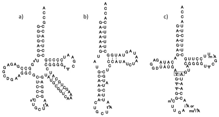
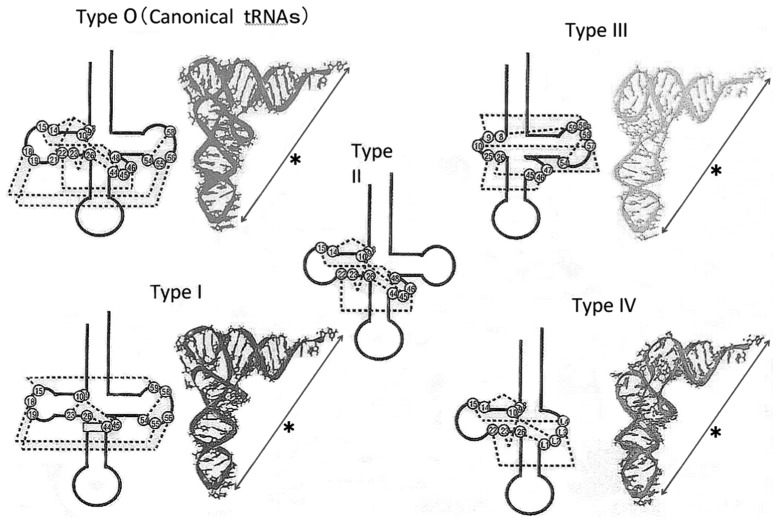
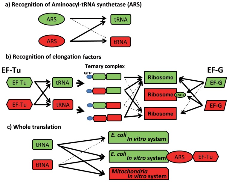
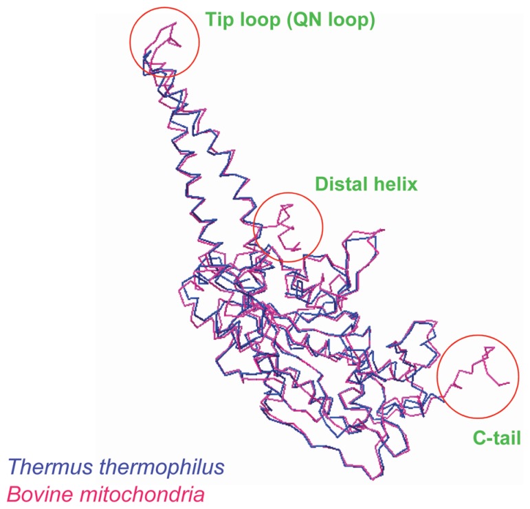
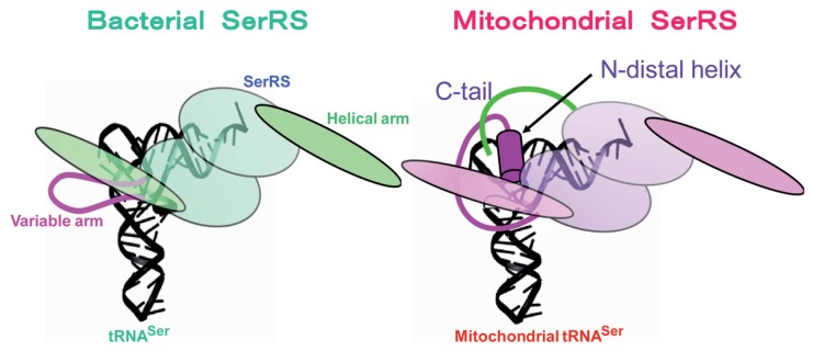
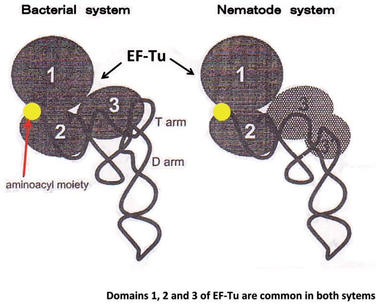
In animal mitochondria, several codons are non-universal and their meanings differ depending on the species. In addition, the tRNA structures that decipher codons are sometimes unusually truncated. These features seem to be related to the shortening of mitochondrial (mt) genomes, which occurred during the evolution of mitochondria. These organelles probably originated from the endosymbiosis of an aerobic eubacterium into an ancestral eukaryote. It is plausible that these events brought about the various characteristic features of animal mt translation systems, such as genetic code variations, unusually truncated tRNA and rRNA structures, unilateral tRNA recognition mechanisms by aminoacyl-tRNA synthetases, elongation factors and ribosomes, and compensation for RNA deficits by enlarged proteins. In this article, we discuss molecular mechanisms for these phenomena. Finally, we describe human mt diseases that are caused by modification defects in mt tRNAs.


Figures

Fig. 1
Fig. 1
Clover-leaf structures of tRNASerGCU of E. coli (a) and bovine mitochondria (b), and tRNASerUGA of bovine mitochondria (c), in which an additional base-pair in the anticodon stem is boxed.
Fig. 2
Fig. 2
Secondary (black) and tertiary structures (colored) of canonical tRNAs (Type 0) and animal mitochondrial tRNAs (Type I~Type IV). All tRNAs share the constraint that the distance (shown by*) and the mutual orientation between the anticodon and the CCA terminus are constant. Dotted lines in the secondary structure show internal base interactions.
Fig. 3
Fig. 3
Unilateral recognition relationships of tRNA with ARS (a), EF-Tu and EF-G (b), and ribosomes in whole translation reactions (c) in E. coli (green) and bovine mt (red) translation systems. Bold arrows and dotted arrows show functional and nonfunctional reactions, respectively.
Fig. 4
Fig. 4
Superposition of the Cα chain of bovine mt SerRS (red) onto the known structure of T. thermophilus SerRS (blue). The N-terminal helical region (Distal helix), the top region of the two long α helical arms (Tip loop) and the C-terminal region (C-tail) are additional regions in bovine mitochondria (circled) not present in T. thermophilus SerRS.
Fig. 5
Fig. 5
Model of tRNA recognition by SerRS in bacterial (left) and mitochondrial systems (right). The truncated parts of mt tRNA are compensated for by the Distal helix (red rod) for both tRNASerGCU and tRNASerUGA and the C tail (green line) for tRNASerUGA of mt SerRS.
Fig. 6
Fig. 6
Schematic representation of tRNA complexed with EF-Tu in bacterial (left) and nematode mt systems (right). An extended C-terminal region (57 amino acids) of nematode mt EF-Tu, Domain 3′, compensates for the absence of the T arm in nematode mt tRNA.
Fig. 7
Fig. 7
Secondary structures for large ribosomal RNAs of bacteria and mammalian mitochondria, shown with interacting ribosomal proteins. The secondary structure of human mt 16S RNA (red line) was superimposed on the secondary structure of E. coli 23S rRNA (black line), which is shown according to the format of Ban et al. The 5′ region of mt 16S rRNA (about 160 bases) could not be aligned with domain I of bacterial 23S rRNA. Ovals that represent large ribosomal proteins are mapped onto the secondary structure of rRNA, with interactions indicated by gray arrows. Solid arrows show interaction maps that were identified by the crystal structure of the 50S subunit. Broken arrows indicate interaction maps obtained from biochemical studies. The oval size of each protein indicates its molecular weight relative to that of its E. coli counterpart. The difference in molecular weight between the mt ribosomal protein and its E. coli counterpart is indicated by color: red, more than 15 kDa bigger than the E. coli counterpart; orange, 10~15 kDa; yellow, 5~10 kDa; green, less than 5 kDa. The colored large shaded regions represent the six domains from which the ribosome was constructed.
Fig. 8
Fig. 8
Three-dimensional models for large mt ribosomal RNA (gray) from mammalian (middle) and C. elegans (right) mitochondria, based on the crystal structure of a bacterial 50S subunit (left). The outline shows an edge line of the crystal structure of the 50S subunit from the crown view. Some functional rRNA domains are colored: red, P loop; blue, A loop; green, S/R loop; light blue, L2 binding helix (H66). The topological orientation of the ribosomal protein is based on the model for the mammalian mt ribosome.
Fig. 9
Fig. 9
a) Schematic of “chaplet” column chromatography. The DNA columns, in which a 3′-biotinylated DNA probe complementary to each mt tRNA is immobilized on streptavidin sepharose, are tandemly connected. Crude tRNA fractions from bovine liver or human placenta are circulated through this chaplet column to entrap each mt tRNA. b) Polyacrylamide gel electrophoretic pattern of purified mt tRNAs obtained by chaplet column chromatography. Each tRNA is isolated with almost 100% purity.
Fig. 10
Fig. 10
Chemical structures of τm5U (left) and τm5s2U (right).
Fig. 11
Fig. 11
Gene organization of the human mt genome (center), in which MELAS (A3243G and U3271C) and MERRF (A8344G) point mutations are indicated in the tRNALeu(UUR) (left) and tRNALys genes (right), respectively.
Fig. 12
Fig. 12
Wobble modification defect in mutant tRNAs, tRNALeu(UUR) (left) and tRNALys (right) from the mt diseases MELAS3243 and 3271, and MERRF8344. These point mutations act as negative determinants for taurine-containing wobble uridines in the tRNAs.
Fig. 13
Fig. 13
Translational activities of wild-type tRNALeu(UUR) (WT), operated tRNALeu(UUR) (OP), A3243G mutant tRNALeu(UUR) (3243), and U3271C mutant tRNALeu(UUR) (3271), in an in vitro mt translation assay using test mRNAs containing the UUA (left), UUG (center) or UUC (right) (negative control) codons. The radioactivity of the [3H]Leu-tRNA input in the reaction mixture was defined arbitrarily as 100%. The averages of three independent experiments with SD values are shown.
Fig. 14
Fig. 14
Usage of UUR Leu codons in 13 proteins encoded in human mtDNA. The numbers of UUA/UUG codons are shown for each gene.

Similar articles

Cited by

References

    1. Barrell B. G., Bankier A. T., Drouin J. (1979) A different genetic code in human mitochondria. Nature 282, 189–194 - PubMed
    1. Anderson S., Bankier A. T., Barrell B. G., de Bruijn M. H. L., Coulson A. R., Drouin J., et al. (1981) Sequence and organization of the human mitochondrial genome. Nature 290, 457–465 - PubMed
    1. de Bruijn M. H. L., Schreier P. H., Eepron I. C., Barrrell B. G., Chen E. Y., Armstrong P. W., et al. (1980) A mammalian mitochondrial serine transfer RNA lacking the “dihydrouridine” loop and stem. Nucleic Acids Res. 8, 5213–5222 - PMC - PubMed
    1. Arcari P., Brownlee G. G. (1980) The nucleotide sequence of a small (3S) seryl-tRNA (anticodon GCU) from beef heart mitochondria. Nucleic Acids Res. 8, 5207–5212 - PMC - PubMed
    1. Gesteland R. F., Ceck T. R., Atkins J. F. (eds.) (1999) The RNA World Second Edition – The nature of modern RNA suggests a prebiotic RNA world. Cold Spring Harbor Laboratory Press., New York, pp. 1–709

Publication types