. 2010 Feb 17;29(4):830-42.
doi: 10.1038/emboj.2009.395. Epub 2010 Jan 14.

Coordinated regulation of p53 apoptotic targets BAX and PUMA by SMAR1 through an identical MAR element

Affiliations

Coordinated regulation of p53 apoptotic targets BAX and PUMA by SMAR1 through an identical MAR element

Surajit Sinha et al. EMBO J. .

Abstract

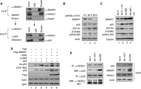
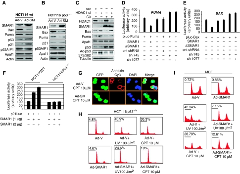
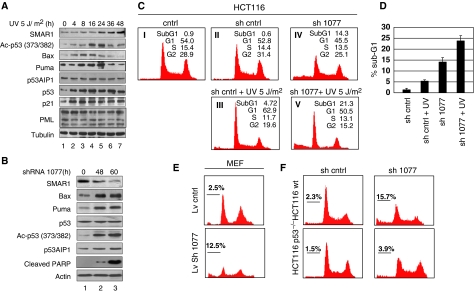
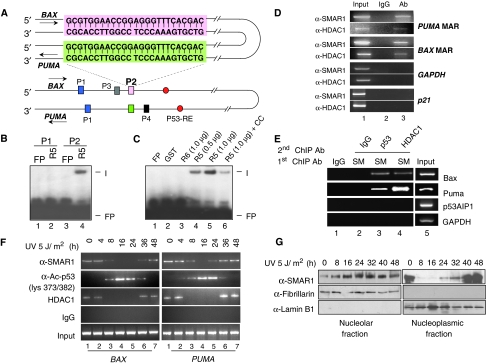
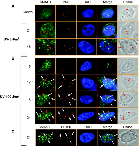
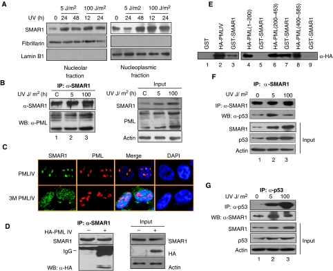
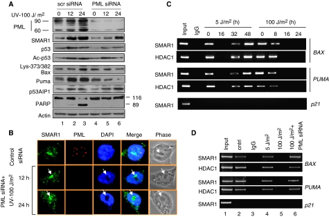
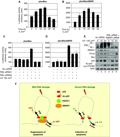
How tumour suppressor p53 bifurcates cell cycle arrest and apoptosis and executes these distinct pathways is not clearly understood. We show that BAX and PUMA promoters harbour an identical MAR element and are transcriptional targets of SMAR1. On mild DNA damage, SMAR1 selectively represses BAX and PUMA through binding to the MAR independently of inducing p53 deacetylation through HDAC1. This generates an anti-apoptotic response leading to cell cycle arrest. Importantly, knockdown of SMAR1 induces apoptosis, which is abrogated in the absence of p53. Conversely, apoptotic DNA damage results in increased size and number of promyelocytic leukaemia (PML) nuclear bodies with consequent sequestration of SMAR1. This facilitates p53 acetylation and restricts SMAR1 binding to BAX and PUMA MAR leading to apoptosis. Thus, our study establishes MAR as a damage responsive cis element and SMAR1-PML crosstalk as a switch that modulates the decision between cell cycle arrest and apoptosis in response to DNA damage.


Conflict of interest statement

The authors declare that they have no conflict of interest.

Figures

Figure 1
Figure 1
SMAR1 interacts with and regulates p53 acetylation endogenously. (A) Double co-immunoprecipitation assay to check the in vivo association of SMAR1–HDAC1 complex with p53. One milligram of cell lysate from HCT116 p53+/+ cells was immunoprecipitated sequentially with SMAR1 and HDAC1 antibodies. The eluted fraction was probed with p53 antibody. (B) HCT116 p53+/+ cells were transfected with SMAR1 shRNA (sh1077). Western blotting shows endogenous acetylation status of p53 at lys 373/382 and total p53 level 30 and 60 h after transfection. (C) HCT116 p53+/+ cells transduced with GFP expressing control and SMAR1 adenoviruses and treated with UV (100 J/m2) for 24 h. The levels of p53 and p53 acetylation status in comparison with SMAR1 expression are shown. (D) In vitro deacetylation assay of p53 by SMAR1. HCT116 p53−/− cells were transfected with p53, p300 expression plasmids in different combinations and treated with TSA (200 nM, 16 h) as given in the figure. GFP expression plasmid was transfected to monitor transfection efficiency. (E) Reversible co-immunoprecipitation assay in HCT116 p53+/+ cells showing differential association of p53 with HDAC1 in SMAR1 knockdown and overexpressed cells (left panel). Input controls are shown in right panel.
Figure 2
Figure 2
SMAR1 regulates the expression of key apoptotic molecules Bax and Puma and inhibits apoptosis. (A) HCT116 p53+/+ and (B) HCT116 p53−/− cells were transduced with GFP expressing control adenovirus (Ad-V) and SMAR1 Adenovirus (Ad-SM). Forty eight hours post-transduction, the levels of p53, p21, Bax, Puma, Apaf1 and p53AIP1 were determined. (C) HCT116 p53+/+ cells were transfected with HDAC1 siRNA (Santacruz) and levels of Bax, Puma and p53 acetylation were determined in the presence and absence of SMAR1 (C3). (D, E) Luciferase activity of full-length promoters of BAX and PUMA on SMAR1 overexpression and knockdown using two different shRNAs (sh745 and sh1077) in HCT116 p53−/− cells. A deletion mutant of SMAR1 lacking the DNA binding and the protein interacting domain (ΔSMAR1) was used as a control. Bars indicate s.d. from three independent experiments. (F) p21 luciferase assay on SMAR1 overexpression in HCT116 p53+/+ and p53−/− cells. (G) Annexin staining of HCT116 p53+/+ cells treated with Camptothecin (10 μM, 12 h) after transduction of GFP expressing control and SMAR1 adenoviruses. Cell cycle analysis in (H) HCT116 p53+/+ cells and (I) MEFs transduced with GFP expressing control (Ad-V) and SMAR1 (Ad-SM) adenoviruses. Forty eight hours post-transduction, cells were treated with UV (100 J/m2, 24 h) and Campthothecin (10 μM, 12 h) and thereafter stained with propidium iodide.
Figure 3
Figure 3
SMAR1 induces an anti-apoptotic signal in response to low-dose DNA damage. (A) HCT116 p53+/+ cells were UV irradiated with 5 J/m2 UV and cells collected at different time points as indicated. Immunoblot analysis was carried out as shown. (B) Expression levels of Bax, Puma, p53, acetylated p53 and PARP cleavage on shRNA (sh1077)-mediated knockdown of SMAR1 in HCT116 p53+/+ cells. (C) Propidium iodide staining in control HCT116 p53+/+ cells (48 h), cells treated with UV (5 J/m2, 48 h) and SMAR1 knockdown UV-treated cells (5 J/m2, 48 h) showing percentage apoptosis represented by sub-G1 population. (D) Statistical representation of percentage sub-G1 population on low-dose UV treatment and SMAR1 knockdown from three independent experiments. Error bars represent standard deviation. (E) Propidium iodide staining in MEFs transduced with control and SMAR1 lentivirus (F) Knockdown of SMAR1 by transient transfection in HCT116 p53+/+ and HCT116 p53−/− cells showing differential sub-G1 population.
Figure 4
Figure 4
SMAR1 regulates BAX and PUMA through MAR. (A) Schematic representation of BAX and PUMA promoter aligned with complementary strands showing the locations of two identical sequences P1 (blue) and P2 (pink in BAX and green in PUMA) in proximity to p53 response element (red). The sequence of P2 is boxed. (B) EMSA showing specific binding of SMAR1 (R5) to probe P2 (lane 4), but not to probe P1. (C) Purified GST (lane 2), R6 (lane 3) and R5 in increasing doses (lanes 4 and 5) were incubated with probe P2. Formation of complex (I) was visualized by autoradiography. Binding specificity of the complex in the presence of 10-fold molar excess of cold competitor (cold probe P2) is shown in lane 6. Free probe is denoted as FP. (D) Chromatin from HCT116 p53+/+ cells was immunoprecipitated with SMAR1 and HDAC1 antibodies (lane 3). PCR amplification was performed on MAR regions of BAX and PUMA. GAPDH and p21 promoters were used as control. Parallel immunoprecipitation with control IgG antibody is shown in lane 2. Lane 1 denotes input control. (E) Sequential ChIP on BAX and PUMA promoter MARs using anti-SMAR1/p53 (lane 3) and anti-SMAR1/HDAC1 (lane 4) antibodies in nuclear matrix fraction of HCT116 cells. Lanes 1 and 5 shows IgG and input control, respectively. (F) ChIP showing occupancy of SMAR1 on BAX (left panel) and PUMA (right panel) promoter on low-dose UV irradiation. Cross-linked chromatins from UV-irradiated HCT116 p53+/+ cells were pulled with SMAR1, p53, acetylated p53 lys373/382 and HDAC1 antibodies and bound chromatin fragments were detected by PCR. (G) Western blotting of SMAR1 in nucleolar and nucleoplasmic fraction of UV-irradiated (5 J/m2) HCT116 p53+/+ cells at different time points.
Figure 5
Figure 5
Apoptotic DNA damage translocates SMAR1 into PML nuclear bodies. Immunofluorescence analysis showing SMAR1–PML co-localization in HCT116 p53+/+ cells at (A) low-dose 5 J/m2, (B) high-dose 100 J/m2 UV and (C) Co-localization of SMAR1 with Sp100 at high dose. Cells were stained with SMAR1 (green), PML (red) and Sp100 (red) antibodies and analysed by confocal microscopy. Nuclei were stained with DAPI (blue). Co-localization of SMAR1 and PML bodies are shown in white arrows. Red arrows indicate SMAR1 in nucleolus. Images shown are representative of >50 images (n>50) taken in different fields from two independent experiments.
Figure 6
Figure 6
SMAR1 interacts strongly with PML after apoptotic DNA damage. (A) Western blotting of SMAR1 in nucleolar and nucleoplasmic fractions of low and high dose UV-irradiated HCT116 p53+/+ cells at different time points. (B) Two hundred microgram of total cell extract from HCT116 p53+/+ control and UV-irradiated cells were immunoprecipitated (IP) with SMAR1 antibody and complexes detected by immunoblotting (IB) with antibody against PML (left panel). A total of 20% of total cell lysate was used for input (right panel). (C) Immunostaining showing SMAR1–PML co-localization in HCT116 p53+/+ cells after overexpression of PML-IV and PML-IV sumoylation mutant (3 M PML-IV) constructs. PML is stained with red (Cy3) and SMAR1 is stained with green (FITC). Nucleus is stained with blue (DAPI). (D) Co-immunoprecipitation of SMAR1 and PML in HA-PML-IV-transfected HCT116 p53+/+ cells as detected by HA antibody. (E) GST pull-down assays were performed using purified GST and GST-SMAR1 in HCT116 p53+/+ cells overexpressing full-length HA-PML-IV and truncated HA-PML-IV proteins as shown in the figure. Specific binding was detected using anti-HA. (F, G) HCT116 p53+/+ cell extracts (0.5 mg) from control and UV treated with 5 and 100 J/m2 were immunoprecipitated with SMAR1 and p53 antibodies and amount of p53 and SMAR1 in the immunoprecipitated complex were detected by antibodies against p53 and SMAR1, respectively.
Figure 7
Figure 7
Silencing of PML facilitates transrepression of BAX and PUMA by SMAR1. (A) Comparative expression levels of SMAR1, Ac-p53 and p53 in HCT116 p53+/+ cells transfected with scrambled siRNA (left half) versus PML siRNA (right panel) on UV (100 J/m2) treatment. (B) Confocal staining of SMAR1 and PML in HCT116 p53+/+ cells transfected with PML siRNA and treated with UV. Images are representative of >50 fields (n>50) from two independent experiments. (C) ChIP assay in HCT116 p53+/+ cells exposed to low dose (5 J/m2) and high dose (100 J/m2) UV irradiation. (D) ChIP showing SMAR1 occupancy on BAX and PUMA promoter after low dose (5 J/m2, 48 h) and at high dose (100 J/m2, 24 h) in PML knockdown HCT116 p53+/+ cells.
Figure 8
Figure 8
Repression of Bax and Puma is specific through the MAR by SMAR1. (A, B) Luciferase activity of the full-length BAX (plucBax) and MAR-deleted (plucBaxΔMAR) reporters in HCT116 p53+/+ cells treated with low dose 5 J/m2 UV. (C, D) Promoter read out of plucBax and plucBaxΔMAR reporters in HCT116 p53+/+ cells transfected with two different PML siRNAs and analysed 24 h after treatment with high dose UV (100 J/m2). (E) Expression analyses of Bax and Puma on double knockdown of PML and SMAR1 in HCT116 p53+/+ cells. (F) Model showing the MAR element (blue box) of BAX and PUMA promoter juxtaposed to each other causing the looping out of the intervening sequence. SMAR1 (orange) binds to this identical MAR element along with HDAC1 (green) and p53 (red) forming a repressor complex switching off transcription after mild DNA damage (suppression of apoptosis). After severe apoptotic DNA damage, SMAR1 is no longer bound to the MAR element facilitating p53 acetylation (p53-Ac) and transcription of BAX and PUMA (induction of apoptosis).

Similar articles

Cited by

References

    1. Andersen JS, Lyon CE, Fox AH, Leung AK, Lam YM, Steen H, Mann M, Lamond AI (2002) Directed proteomic analysis of the human nucleolus. Curr Biol 12: 1–11 - PubMed
    1. Aylon Y, Oren M (2007) Living with p53, dying of p53. Cell 130: 597–600 - PubMed
    1. Bernardi R, Pandolfi PP (2007) Structure, dynamics and functions of promyelocytic leukaemia nuclear bodies. Nat Rev Mol Cell Biol 8: 1006–1016 - PubMed
    1. Bernardi R, Scaglioni PP, Bergmann S, Horn HF, Vousden KH, Pandolfi PP (2004) PML regulates p53 stability by sequestering Mdm2 to the nucleolus. Nat Cell Biol 6: 665–672 - PubMed
    1. Brooks CL, Gu W (2006) p53 ubiquitination: Mdm2 and beyond. Mol Cell 21: 307–315 - PMC - PubMed

Publication types

MeSH terms