. 2010 Mar 3;29(5):956-68.
doi: 10.1038/emboj.2009.406. Epub 2010 Jan 14.

PIKE-A is required for prolactin-mediated STAT5a activation in mammary gland development

Affiliations

PIKE-A is required for prolactin-mediated STAT5a activation in mammary gland development

Chi-Bun Chan et al. EMBO J. .

Abstract

PI 3-kinase enhancer A (PIKE-A) is critical for the activation of Akt signalling, and has an essential function in promoting cancer cell survival. However, its physiological functions are poorly understood. Here, we show that PIKE-A directly associates with both signal transducer and activator of transcription 5a (STAT5a) and prolactin (PRL) receptor, which is essential for PRL-provoked STAT5a activation and the subsequent gene transcription. Depletion of PIKE-A in HC11 epithelial cells diminished PRL-induced STAT5 activation and cyclin D1 expression, resulting in profoundly impaired cell proliferation in vitro. To confirm the function of PIKE-A in PRL signalling in vivo, we generated PIKE knockout (PIKE-/-) mice. PIKE-/- mice displayed a severe lactation defect that was characterized by enhanced apoptosis and impaired proliferation of mammary epithelial cells. At parturition, STAT5 activation and cyclin D1 expression were substantially reduced in the mammary epithelium of PIKE-/- mice. The defective mammary gland development in PIKE-/- mice was rescued by overexpression of a mammary-specific cyclin D1 transgene. These data establish a critical function for PIKE-A in mediating PRL functions.


Conflict of interest statement

The authors declare that they have no conflict of interest.

Figures
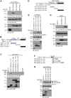
Figure 1
Figure 1
PIKE-A interacts with STAT5a and PRLR. (A) PIKE-A specifically binds STAT5a. HEK293 cells were co-transfected with various GFP-STAT plasmids and mGST-PIKE-A. The GFP proteins were immunoprecipitated and the bound PIKE-A proteins were detected using anti-GST-HRP antibody (top panel). The expression of mGST-PIKE-A (middle panel) and different GFP-STAT proteins (bottom panels) were verified. (B) Diagrammatic representation of GST-PIKE truncation constructs. (C) PIKE-A N-terminus associates with STAT5a. In vitro mapping of PIKE-A domains that associate with STAT5a. Purified GST-tagged PIKE-A proteins were incubated with lysates of HEK293 cells transfected with GFP-STAT5a. The N-terminal end (1–72 a.a.) of PIKE-A, but not the C-terminal domain associates with STAT5a (middle panel). The GST-fused PIKE-A fragments (as indicated with asterisk) used in the in vitro binding were detected using anti-GST-HRP antibody (bottom panel). (D) Diagrammatic representation of myc-STAT5a truncation constructs. (E) STAT5a C-terminal associates with PIKE-A. HEK293 cells were co-transfected with GFP-PIKE-A and various deletion mutants of myc-STAT5a as shown in (D). The expressed STAT5a mutants were immunoprecipitated and the bound GFP-PIKE-A was detected using anti-GFP antibody (second panel). The expression of GFP-PIKE-A (third panel) and myc-STAT5a mutants (fourth panel) were also examined. (F) Diagrammatic representation of GFP-PRLR truncation constructs. (G) PIKE-A binds C-terminal PRLR intracellular-domain. HEK293 cells were co-transfected with mGST-PIKE-A and various deletion mutants of GFP-PRLR intracellular domains. The expressed PRLR mutants were immunoprecipitated and the bound mGST-PIKE-A was detected using anti-GST-HRP antibody (top panel). The expression of mGST-PIKE-A (bottom panel) and GFP-PRLR mutants (middle panel, indicated with asterisk) were also examined. (H) PIKE-A does not interact with other STAT5a-associated cytokine receptor. HEK293 cells were transfected with mouse EPO receptor (EPOR) plasmid and various GFP constructs as indicated. After 24 h of serum starvation, the transfected cells were stimulated with 3 ng/ml EPO for 15 min. The GFP-tagged proteins were immunoprecipitated and the associated EPOR was examined by using anti-EPOR antibody (top panel). The expression of EPOR (middle panel) and GFP-tagged proteins (bottom panel) were also verified.
Figure 2
Figure 2
PRL stimulation provokes the formation of PRLR/PIKE-A/STAT5a complex. (A) PRL stimulation interferes with the PIKE-A and Stat5 interaction, but enhances the formation of PRLR/PIKE-A complex. HC11 cells infected with adenovirus-expressing wild-type PIKE-A were stimulated with 10 nM recombinant mouse PRL for various time intervals as indicated. PIKE-A was then immunoprecipitated by control IgG or anti-PIKE-A antibody and the bound STAT5 and PRLR were detected using anti-Stat5 (first panel) and anti-PRLR antibodies (second panel). The expression of STAT5 (fourth panel) and PIKE-A (fifth panel) were examined. Phosphorylation of total STAT5 was also verified to confirm the activation of HC11 by PRL (third panel). (B) Phosphorylation of PRLR is essential for PIKE-A/PRLR interaction. HC11 cells were infected with adenovirus-expressing wild-type PIKE-A for 48 h followed by serum starvation for 24 h. Before 10 mM PRL stimulation, the cells were pre-treated with tyrosine-kinase inhibitor genistein (1 μM) for 45 min. The expressed PIKE-A was immunoprecipitated and the associated PRLR was detected using an anti-PRLR antibody (first panel). Phosphorylation of PRLR was examined using anti-phospho-tyrosine antibody on immunoprecipitated PRLR (second panel). The expression of PRLR (third panel) and PIKE-A (fourth panel) was also verified. (C) PIKE-A/PRLR interaction is JAK2-kinase dependent. HC11 cells were infected with adenovirus-expressing wild-type PIKE-A for 48 h followed by serum starvation for 24 h. Before 10 mM PRL stimulation for 15 min, the cells were pre-treated with JAK2-kinase inhibitor AG490 (50 μM) for 1 h. PIKE-A was immunoprecipitated and the associated STAT5 and PRLR was detected using specific antibody (first and second panels). Phosphorylation of JAK2 and STAT5 were determined to examine the effect of JAK2 inhibition (fourth and sixth panels). The expressions of PRLR, JAK2, STAT5 and PIKE were also verified (third, fifth, seventh and eighth panels). (D) PRL-provoked STAT5 phosphorylation is abolished in PIKE-A-depleted HC11 cells. HC11 cells were infected with either control adenovirus or adenovirus-expressing shPIKE. Two days after infection, the cells were serum-starved for 24 h and stimulated with 10 nM recombinant mouse PRL for 15 min. PRLR was immunoprecipitated and the associated STAT5 were determined by anti-STAT5 antibody (first panel). Phosphorylation of STAT5 was determined using specific antibody against phosphorylated STAT5 (third panel). Total PRLR (second panel), STAT5 (fourth panel) and PIKE-A (seventh panel) expressions was examined. Phosphorylation of ERK (fifth panel) by PRL was not affected in PIKE-depleted HC11 cells. Total ERK and tubulin were also determined (sixth and eighth panels). (E) PRL triggers nuclear translocation of STAT5a, but not PIKE-A. GFP-STAT5a and myc-PIKE-A were co-transfected in HC11 cells, serum-starved for 24 h and stimulated with 10 nM recombinant mouse PRL for 45 min. Cellular localization of PIKE-A and STAT5a was examined by confocal microscopy. Exclusive cytoplasmic localization of myc-PIKE-A was detected in both control and PRL-stimulated HC11 cells, whereas GFP-STAT5a accumulated in the nucleus after PRL stimulation.
Figure 3
Figure 3
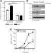
PIKE-A is essential for epithelial cell proliferation. (A) PIKE-A is required for PRL-stimulated cyclin D1 promoter activity. After transient transfection with the PRE3-luciferase vector, PIKE-A expression in HC11 cells was suppressed by infecting the cells with adenovirus-expressing sh-PIKE. After 48 h, the infected cells were then serum-starved for 24 h and treated with (solid bar) or without (open bar) 10 nM PRL in serum-free medium for another 24 h, and luciferase activity was then determined (**P<0.01, ***P<0.001, Student's t-test). (B) PIKE-A is critical for basal and PRL-induced cyclin D1 expression in HC11 cells. HC11 cells were infected with either control adenovirus or adenovirus-expressing sh-PIKE. Two days after infection, the cells were serum-starved for 24 h and stimulated with 10 nM recombinant mouse PRL for 24 h. Expression of cyclin D1 was examined by immunoblotting (top panel). PIKE-A expression in HC11 after shRNA infections was confirmed (middle panel). Tubulin level was also examined to show equal input of the proteins (bottom panel). (C) Diminished proliferation in PIKE-depleted mammary gland epithelial cells. HC11 cells were infected with control adenovirus or adenovirus-expressing sh-PIKE. Three days after infection, cell proliferations of the infected cells were examined by trypan blue exclusion assay (***P<0.001, two-way ANOVA).
Figure 4
Figure 4
Lobuloalveolar hypoplasia in PIKE knockout mice. (A) Impaired lobuloalveolar development in PIKE−/− mammary glands. Carmine alum-stained whole mount of wild-type and PIKE−/− mammary glands (fourth inguinal) collected from 14-week-old virgin, pregnant (7.5, 13.5 and 18.5 days post-coitus) and lactating mice (1 day postpartum). Representative results of three individual animals from each genotype were shown. (B) Histological analysis (H&E staining) of mammary glands is shown in (A). Scale bar represents 100 μm. (C) Structure of mammary alveolus of lactating wild-type and PIKE−/− mice. Scale bars represent 10 μm. Representative results from of three individual animals from each genotype were shown. (D) Alveolar lumen size of lactating mammary gland (1 day postpartum). The results are expressed as mean±s.e.m. of three mice (>30 alveoli in each animal) from each genotype. Scar bars represent 10 μm. (E) Alveoli density of pregnant and lactating mammary gland (1 day postpartum). The results are expressed as mean±s.e.m. of three mice (four individual fields in each sample) of each genotype. (F) Impaired protein expression in PIKE−/− mammary gland. Mammary tissues (fourth inguinal) from late gestation (18.5 dpc) and lactating (1 day postpartum) animals were collected and various milk proteins (β-casein and WAP), phosphorylation of signal transduction molecules (STAT5, Akt and ERK) and cyclin D1 levels were analysed by immunoblotting. Expression of β-tubulin was also performed to show equal loading. The results are representative blot of three individual animals from each genotype.
Figure 5
Figure 5
Defective proliferation and enhanced apoptosis in lactating mammary epithelial cells from PIKE−/− mice. (A) Ki67 staining of lactating mammary glands (1 day postpartum, fourth inguinal) from wild-type and PIKE−/− mice. Representative results of three individual animals from each genotype are shown. Scale bar represents 50 μm. (B) Quantitative measurement of cells showing positive Ki67 staining from different gestation stage (n=3, ***P<0.001, Student's t-test). (C) Apoptosis occurs in PIKE−/− mammary gland. Paraffin-embedded fourth inguinal mammary glands from wild-type or PIKE−/− mice at virgin, mid-pregnancy (13.5 dpc), late pregnancy (18.5 dpc) and lactation (1 day postpartum) were examined by TUNEL (green) and the total nuclei were stained with DAPI (blue). Apoptotic nuclei (as indicated by arrows) were found in PIKE−/− mammary gland, but not in the wild-type control. Representative results of three individual animals from each genotype at specific time point are shown. (D) Akt phosphorylation in lactating mammary epithelial cells (fourth inguinal, 1 day postpartum) was determined by immunohistochemical staining using phosphor-Ser473 antibody. Diminished Akt phosphorylation was found in PIKE−/− mice. Representative results of three individual animals from each genotype are shown. Scale bar represents 50 μm. (E) Apoptotic signalling in PIKE−/− mammary gland. Expression of various apoptotic signalling components was examined in late gestation (18.5 dpc) and lactating mammary gland (1 day postpartum) collected from PIKE−/− and wild-type mice. Expression of β-tubulin was also performed to show equal loading. Shown are representative blots from three individual animals from each genotype. (F) Overexpression of Bcl-2 in PIKE−/− mammary gland. PIKE−/− mice in mid-gestation (12.5 dpc) were administrated with control adenovirus (Ad-control) or adenovirus carrying Bcl-2 (Ad-Bcl-2) through intaductal injection. Mammary glands were collected at lactation (1 day postpartum) and proteins were extracted for western blot analysis as indicated.
Figure 6
Figure 6
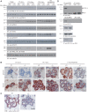
STAT5 phosphorylation is diminished in PIKE−/− mammary gland. (A) Wild-type (+/+) and PIKE (−/−) mammary gland (fourth inguinal) at different times of pregnancy (14-week-old virgin, 7.5 days, 13.5 days, 18.5 post-coitus and 1 day postpartum) were collected and homogenized. The phosphorylation of JAK2 (third panel) and STAT5 (fifth panel) were determined using specific antibodies as indicated. Phosphorylation of PRLR was determined by immunoprecipitation followed by phospho-tyrosine determination (first panel). The expression of PRLR (second panel), JAK2 (fourth panel), total STAT5 (sixth panel) and PIKE-A (seventh panel) were also examined. Tubulin expression of each sample was determined to show equal loading (eighth panel). (B) Phosphorylation of STAT5 was impaired in lactating PIKE−/− mammary gland epithelial cells. Phosphorylation of STAT5 was determined by immunohistochemical analysis in mammary gland (fourth inguinal) collected at mid-gestation (13.5 dpc), late gestation (18.5 dpc) and lactation day 1 (upper panel). Total STAT5 levels were also determined (lower panel). Representative results from three individual mice of each genotype were shown. Scale bar represents 50 μm. (C) Expression of cyclin D1 was impaired in PIKE−/− mammary gland epithelial cells. Cyclin D1 expression was determined by immunohistochemical analysis in lactating mammary gland (1 day postpartum). Positive cells showing nuclear cyclin D1 staining are indicated by arrows. Representative results from three individual mice of each genotype were shown. Scale bar represents 50 μm. (D) Association of endogenous STAT5 and PIKE-A in fourth inguinal mammary gland (1 day postpartum). Endogenous STAT5 was immunoprecipitated using specific antibody against total STAT5 and the bound PIKE-A was detected using anti-PIKE-A antibody. Association of STAT5 and PIKE-A was detected in wild type, but not PIKE−/− mammary gland (first panel). PRLR bound to STAT5 in wild type, but not PIKE−/− mice (third panel). PRLR in the cell lysates was immunoprecipitated using anti-PRLR antibody and the associated STAT5 was detected using anti-STAT5 antibody (second panel). The presence of STAT5 (third panel), PRLR (fourth panel) and PIKE-A (fifth panel) in the cell lysates was verified.
Figure 7
Figure 7
Rescue of PIKE−/− mammary gland defect by a cyclin D1 transgene. (A) Cell autonomous defect in PIKE−/− mammary gland. Whole-mount analysis of transplanted mammary glands were stained with carmine alum on lactation day 1. Directions of transplantation are indicated. (B) PCR screening of mice from heterozygous mating of PIKE+/−MMTV-cyclinD1+/− mice. (C) Western blot analysis of cyclin D1 expression in the mammary gland (fourth inguinal) of lactating mice as shown in (B). (D) Carmine alum-stained whole mount of cyclin D1 transgene bearing wild-type and PIKE−/− mammary glands (fourth inguinal) collected from lactating mice (1 day postpartum). Representative results from two individual animals of each genotype were shown. (E) Characterization of mammary gland from PIKE−/−MMTV-cyclin D1 mice. Proteins were extracted from lactating mammary gland (1 day postpartum) and used for western blot analysis as indicated. (F) Proposed function of PIKE-A in mammary PRL signalling cascade.

Similar articles

Cited by

References

    1. Ahn JY, Hu Y, Kroll TG, Allard P, Ye K (2004a) PIKE-A is amplified in human cancers and prevents apoptosis by up-regulating Akt. Proc Natl Acad Sci USA 101: 6993–6998 - PMC - PubMed
    1. Ahn JY, Rong R, Kroll TG, Van Meir EG, Snyder SH, Ye K (2004b) PIKE (phosphatidylinositol 3-kinase enhancer)-A GTPase stimulates Akt activity and mediates cellular invasion. J Biol Chem 279: 16441–16451 - PubMed
    1. Ahn JY, Rong R, Liu X, Ye K (2004c) PIKE/nuclear PI 3-kinase signaling mediates the antiapoptotic actions of NGF in the nucleus. EMBO J 23: 3995–4006 - PMC - PubMed
    1. Ben-Jonathan N, LaPensee CR, LaPensee EW (2008) What can we learn from rodents about prolactin in humans? Endocr Rev 29: 1–41 - PMC - PubMed
    1. Brisken C (2002) Hormonal control of alveolar development and its implications for breast carcinogenesis. J Mammary Gland Biol Neoplasia 7: 39–48 - PubMed

Publication types

MeSH terms