. 2010 Jan 14;463(7278):245-9.
doi: 10.1038/nature08669.

An allosteric mechanism of Rho-dependent transcription termination

Affiliations

An allosteric mechanism of Rho-dependent transcription termination

Vitaly Epshtein et al. Nature. .

Erratum in

  • Nature. 2010 Aug 19;466(7309):1006

Abstract

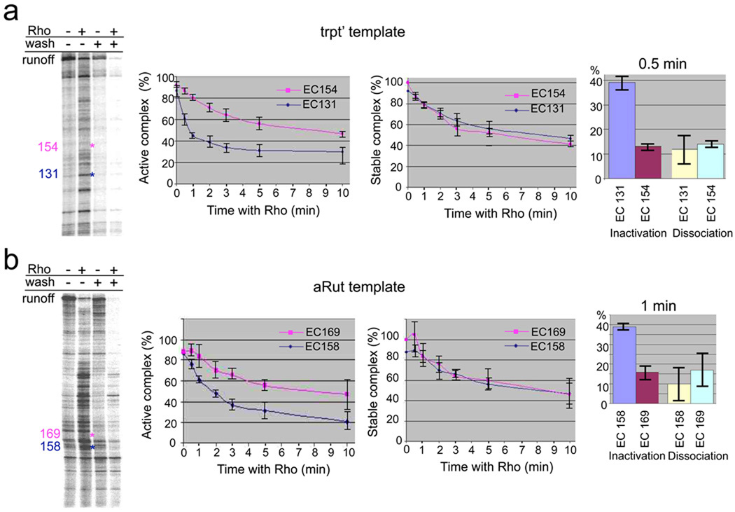
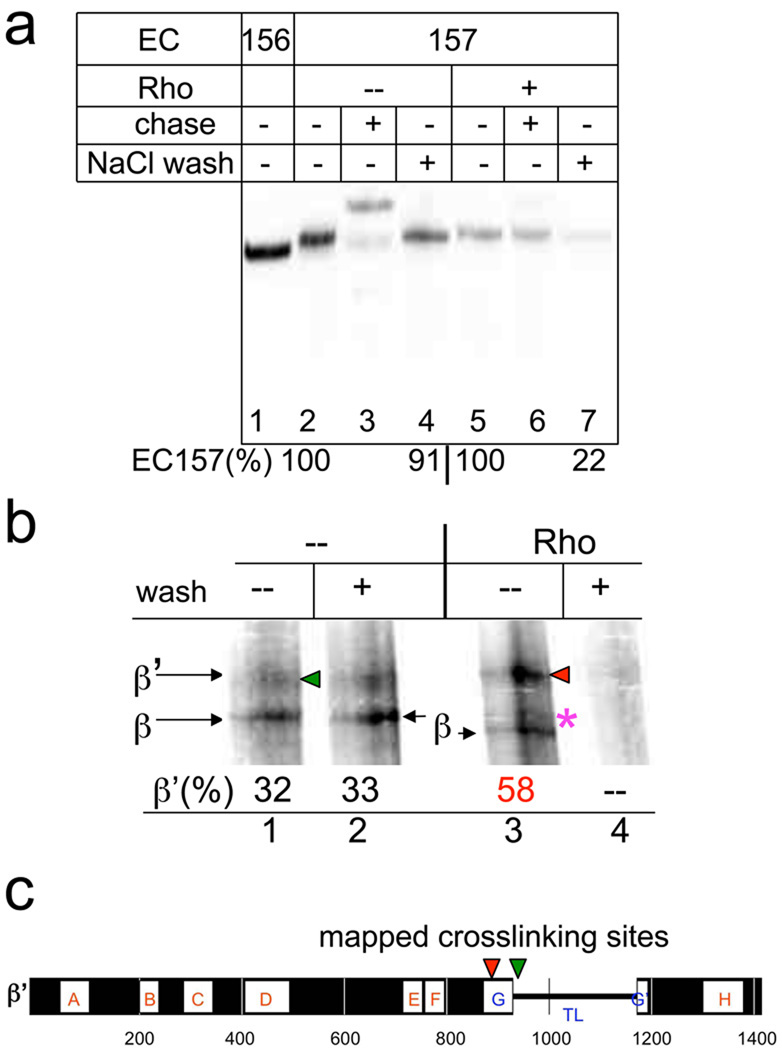
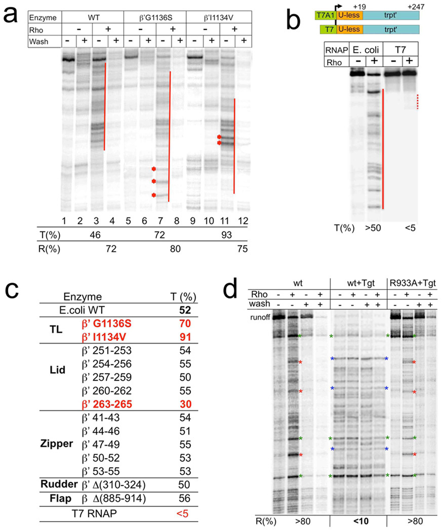
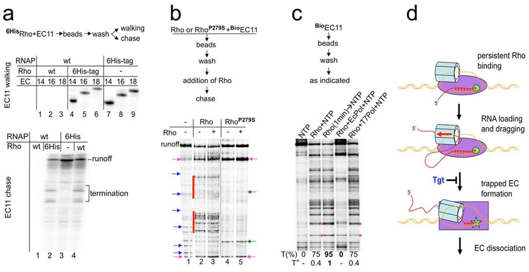
Rho is the essential RNA helicase that sets the borders between transcription units and adjusts transcriptional yield to translational needs in bacteria. Although Rho was the first termination factor to be discovered, the actual mechanism by which it reaches and disrupts the elongation complex (EC) is unknown. Here we show that the termination-committed Rho molecule associates with RNA polymerase (RNAP) throughout the transcription cycle; that is, it does not require the nascent transcript for initial binding. Moreover, the formation of the RNAP-Rho complex is crucial for termination. We show further that Rho-dependent termination is a two-step process that involves rapid EC inactivation (trap) and a relatively slow dissociation. Inactivation is the critical rate-limiting step that establishes the position of the termination site. The trap mechanism depends on the allosterically induced rearrangement of the RNAP catalytic centre by means of the evolutionarily conserved mobile trigger-loop domain, which is also required for EC dissociation. The key structural and functional similarities, which we found between Rho-dependent and intrinsic (Rho-independent) termination pathways, argue that the allosteric mechanism of termination is general and likely to be preserved for all cellular RNAPs throughout evolution.


Figures

Fig. 1
Fig. 1. Uncoupling EC inactivation from dissociation at Rho termination sites
a, Comparison of naturally terminating and non-terminating ECs formed on the trpt’ template. Left panel shows the distribution of Rho termination sites on the trpt’ template. The initial radiolabeled EC21 was immobilized on metal-chelating beads and chased in the presence (lane 2) or absence (lane 1) of Rho. The locations of EC131 (a representative terminating EC) and EC154 (a representative non-terminating EC) are indicated in blue and pink, respectively. “Wash” lanes demonstrate disappearance of released RNA from immobilized terminated complexes. The kinetics of Rho-mediated inactivation and dissociation of EC131 and EC154 were measured by walking the EC to the indicated position, incubating the corresponding ECs for the indicated time intervals with or without Rho, followed by either a chase (GTP, UTP, ATP) or a wash with transcription buffer (TB) (Supplementary Fig. 2). Data are shown as mean ±S.E. from four independent experiments. The Y-axis indicates either the proportion of EC that remained active (active complex %) or that is retained RNA (stable complex %); The X-axis indicates the time of incubation with Rho. The right panel shows the extent of EC inactivation and dissociation after 30 sec of incubation with Rho. A similar analysis was performed for several other ECs on this template (Supplementary Fig. 2). b, Comparison of the naturally terminating and non-terminating ECs obtained at the aRut template. The left panel shows the distribution of Rho termination sites at the aRut template. The initial radiolabeled EC43 was chased in the presence (lane 2) or absence (lane 1) of Rho. The locations of EC158 (a representative terminating EC) and EC169 (a representative non-terminating EC) are indicated in blue and pink, respectively. “Wash” lanes demonstrate disappearance of released RNA from immobilized terminated complexes. The kinetics of Rho-mediated inactivation and dissociation of EC158 and EC169 were measured by walking the EC to the indicated position, incubating the corresponding ECs for the indicated time intervals with or without Rho, followed by either a chase (GTP, UTP, CTP) or a wash with transcription buffer (TB) (Supplementary Fig. 3). Data are shown as mean ±S.E. from four independent experiments. The Y-axis indicates either the proportion of EC that remained active (active complex %) or that is retained RNA (stable complex %); The X-axis indicates the time of incubation with Rho. The right panel shows the extent of EC inactivation and dissociation after 1 min of incubation with Rho. A similar analysis was performed for several other ECs on this template (Supplementary Fig. 3). Taken together, the results indicate that rapid EC inactivation is the determining event at natural Rho termination sites.
Fig. 2
Fig. 2. EC inactivation (trap) by Rho is accompanied by a rearrangement of protein-RNA contacts in the catalytic center
a, EC157 carries the crosslinking probe (4-thioU) at the RNA 3’-terminus. EC157 was incubated with Rho for 3 min and either chased (with GTP, ATP, CTP) or washed with TB containing 0.7 M NaCl (NaCl wash). The panel shows that Rho traps and destabilizes EC157 (renders it salt sensitive). “%” indicates the relative amount of RNA retained in the immobilized EC157 after the beads were washed. b, RNA-protein crosslinking in the trapped (lanes 3, 4) and intact (lanes 1, 2) EC157. A 4% SDS gel showing β’ and β subunits derivatized by UV-induced RNA crosslinking. Numbers indicate the fraction of radiolabeled β’ in relation to the total amount of derivatized subunits. An asterisk indicates the change in β mobility. The control (lane 4) demonstrates that all crosslinking takes place in the terminating (trapped) EC, because all label was eliminated by washing. c, Summary of RNA crosslink mapping results (see Supplementary Fig. 5–7 for mapping details). The horizontal bar represents the E.coli β’ subunit. Lettered boxes show evolutionarily conserved regions. G helix-loop-helix (TL) domain is indicated. The locations of crosslinking sites from derivatized β’ probes obtained with and without Rho are indicated by red and green arrowheads, respectively.
Fig. 3
Fig. 3. Effect of RNAP mutations, heterologous (T7) RNAP, and tagetitoxin (Tgt) on Rho termination
a, A representative runoff assay. Wild type and TL mutant ECs were immobilized on beads and chased in the presence or absence of Rho. The termination efficiency (%T) was calculated as described in Methods. %R indicates the fraction of released RNA at the termination zones after washing the beads. Red lines show the termination zones. Asterisks show the termination sites, which are particularly strong in the case of “fast” G1136S and “slow” I1134V mutants as compared to wt. b, Rho is not effective against heterologous RNAP. The initial EC19 was prepared with either E.coli or T7 RNAP using identical DNA templates except for the corresponding promoters (schematically shown on top). The initial ECs were then chased in the presence of equal amount of Rho and dATP. NTP concentration was selected to ensure the slower elongation rate of T7 RNAP as compared to that of E. coli RNAP (Supplementary Fig. 9). Red solid and dotted lines indicate the robust and weak termination zones, respectively. c, The table summarizes the effects of E.coli RNAP mutations and T7 RNAP on Rho termination. %T represents the average values from at least two independent experiments. Mutations leading to profound changes in Rho termination are highlighted in red. d, Suppression of Rho termination by Tgt. Rho and Tgt were added as indicated to the initial EC immobilized on beads. After the completion of the chase reaction, the beads were washed (as indicated) to monitor the RNA release. %R indicates the amount of released (washed out) RNA due to Rho action. Red asterisks indicate representative major Rho termination sites, which were absent in Tgt probes. Blue asterisks indicate representative major EC stop sites caused by Tgt. These stalled complexes became resistant to Rho termination, i.e. remained mostly intact after incubation with Rho. Green asterisks indicate representative major pause/arrest sites present in all probes, and serve as reference points. Tgt also inhibited Rho-mediated dissociation of these ECs. WT represents the wild type RNAP. R933A represents the point mutation in the TL, which prevents Tgt binding to RNAP. This control demonstrates that all the negative effects of Tgt on Rho termination are due to alterations in the properties of the RNAP catalytic center.
Fig. 4
Fig. 4. Functional Rho binds RNAP throughout the transcription cycle
a, Rho binds to the EC at the start of transcription and terminates the complex at downstream sites. The flowchart describes the experimental design. The initial EC11-Rho6His and 6HisEC11-Rho were prepared on the aRut template, immobilized on Co2+-chelating sepharose beads, and then washed multiple times with TBGlu (100 mM KGlu, 10 mM MgGlu). Radioautograms show [32P] RNA isolated from the EC immobilized via 6HisRho or 6HisRNAP that was walked three steps in TB to the indicated positions (top panel; lanes 4–9) or chased with all four NTPs (bottom panel, lanes 2–4). Major termination sites are indicated. Without the 6His tag no RNA could be recovered on the beads after the washes (top panel, lanes 1–3; bottom panel, lane 1). b, The same Rho molecule that binds the EC at the start of transcription terminates transcription at downstream sites. The flowchart describes the experimental design. Wild type (Rho) and mutant Rho (RhoP279S), the ATPase activity of which was abolished, were used in the experiment. Biotinilated RNAP was used to prepare the startup BioEC11 on the trpt’ template that carried [32P] RNA. Rho or RhoP279S were added to BioEC11. Next, BioEC11-Rho and BioEC11-RhoP279S were immobilized on NeutrAvidin beads, washed multiple times with TBGlu (100 mM KGlu, 10 mM MgGlu), and then chased in the presence of excess Rho as indicated. The radioautogram shows [32P] RNA isolated from the chased reactions. Lane 1 shows the chased reaction without Rho. Blue arrows indicate natural pause/arrest sites. Major Rho termination signals are indicated by red stripes (lanes 2, 3). No increase in Rho termination was observed if excess Rho was added to the washed BioEC11-Rho (lane 3), indicating that the initially-bound Rho was responsible for the majority of the termination signals. Initially-bound RhoP279S did not cause any termination (lane 4), and it also prevented termination by subsequently-added wild type Rho (lanes 5). Note that RhoP279S dramatically altered the pause distribution profile: the majority of pauses disappeared, some new pauses appeared (indicated by green arrows) and some pauses became much stronger (indicated by pink arrows). A similar effect on elongation can be observed with wild type Rho, although they are masked by strong termination signals. c, Early binding of Rho to the EC is crucial for termination. The initial radiolabeled EC11 was chased by the addition of NTPs without Rho (lane 1), together with Rho (lanes 2, 4, and 5), or after a 1 min preincubated with Rho (lane 3). In lanes 4 and 5, Rho was premixed with a 2-fold molar excess of E.coli or T7 RNAP, respectively. Rifampicin was added to ensue a single round of transcription. The overall termination efficiency (%T) was calculated as described in Methods. Red asterisks indicate early Rho termination sites, which became particularly strong if Rho preincubation occurred (lane 3). T* indicates the relative amount of these terminated products. d, The molecular pathway to Rho termination. Schematic shows the newly identified critical steps in the Rho termination process. Rho binds to RNAP at the start of transcription. Once RNA becomes long enough, Rho has an opportunity to load onto it (forming the RNA loop) and to thread the transcript through its cavity using energy derived from ATP. Eventually a topological strain results in the inactivation of RNAP (trapped complex formation), which involves a conformational change of the TL domain at the catalytic center. The trapped EC is sensitive to salt, indicating that its hybrid is partially unwound and/or that the clamp is opened due to the motion of the TL. Unwinding of the hybrid results from the direct action of Rho-mediated RNA:DNA helicase activity and/or to the clash caused by Rho between the lid domain and the hybrid. The unstable, trapped complex eventually dissociates. Rho, however, remains bound to RNAP for the next cycle of transcription. The antibiotic tagetitoxin (Tgt) abolishes Rho termination by interfering with the formation of the trapped complex.

Similar articles

Cited by

References

    1. Adhya S, Gottesman M. Control of transcription termination. Annu. Rev. Biochem. 1978;47:967–996. - PubMed
    1. Richardson JP. Preventing the synthesis of unused transcripts by Rho factor. Cell. 1991;64:1047–1049. - PubMed
    1. Cardinale CJ, et al. Termination factor Rho and its cofactors NusA and NusG silence foreign DNA in E. coli. Science. 2008;320:935–938. - PMC - PubMed
    1. Roberts JW. Termination factor for RNA synthesis. Nature. 1969;224:1168–1174. - PubMed
    1. Geiselmann J, Yager TD, Gill SC, Calmettes P, von Hippel PH. Physical properties of the Escherichia coli transcription termination factor Rho. 1. Association states and geometry of the Rho hexamer. Biochemistry. 1992;31:111–121. - PubMed

Publication types

LinkOut - more resources