

Development of the human choriocapillaris
- PMID: 20075975
- PMCID: PMC4848024
- DOI: 10.1038/eye.2009.318
Development of the human choriocapillaris
Abstract
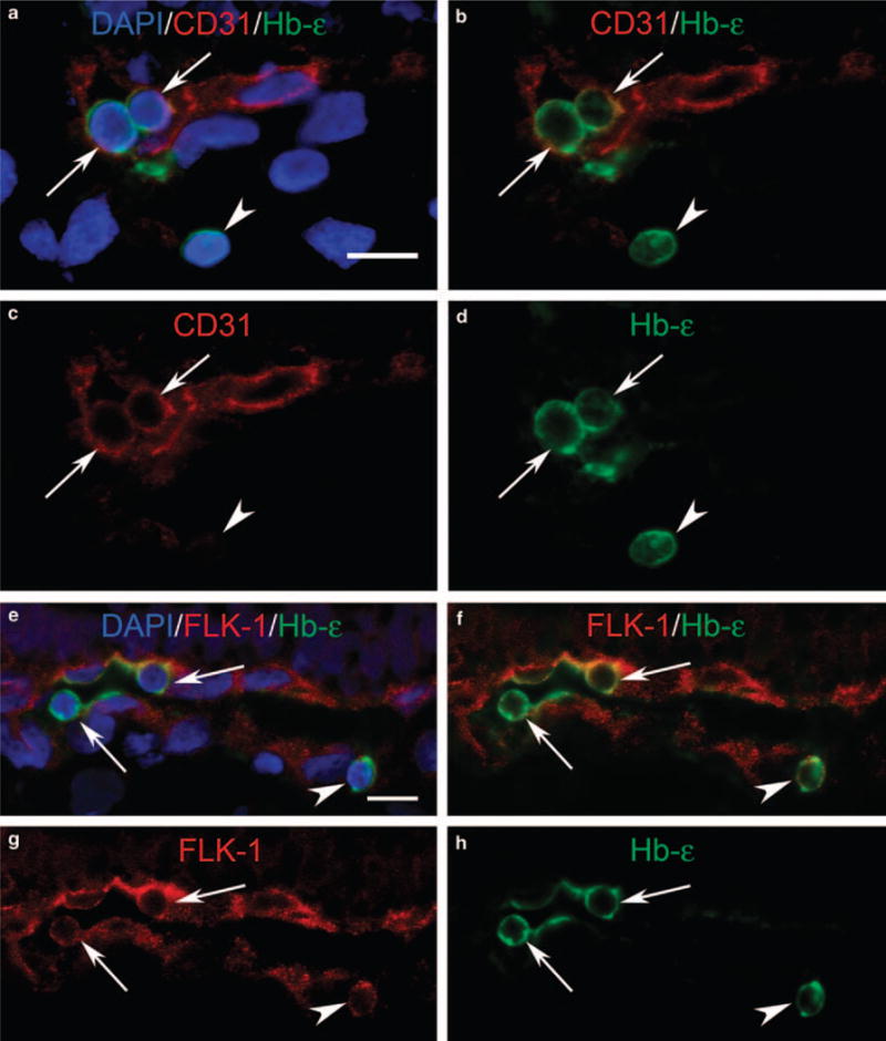
Vasculogenesis and/or angiogenesis are thought to be the major mechanisms for new vessel formation during development. A third mechanism, haemo-vasculogenesis, has been described in which blood vessel and blood cells (haematopoiesis (expression of CD34(+)) and erythropoiesis (presence of epsilon chain of haemoglobin or Hb-epsilon(+))) differentiate from a common precursor, the haemangioblast. This review describes the mechanism(s) for development of human choroidal vascular from 6 until 22 weeks gestation (WG). Endothelial cell or EC (CD31, CD34, CD39, VEGFR-2) and angioblast (CD39, VEGFR-2) markers were present in choriocapillaris (CC) by 7 WG through 22 WG. From 6 to 8 WG, many erythroblasts (nucleated Hb-epsilon(+) RBCs) were observed in the CC layer. Erythroblasts (Hb-epsilon(+)) were also positive for CD34, CD31, and/or VEGFR-2. Proliferation of vascular cells (Ki67+), suggesting angiogenesis, was not observed until 12 WG. TEM analysis demonstrated that CC was structurally immature even at 11 WG: no basement membrane, absence of pericytes, and poorly formed lumens that were filled with filopodia. Contiguous fenestrations and significant PV-1 (protein in diaphragms of fenestrations) were not observed until 21-22 WG. Smooth muscle actin was prominent at 20 WG and the maturation of pericytes was confirmed by TEM. Therefore, the embryonic CC appears to form initially by haemo-vasculogenesis (Hb-epsilon(+)/CD31(+) cells), whereas angiogenesis (CD34(+)/Ki67(+)) appears to be the mode of intermediate and large choroidal vessel development later in the foetus. Contiguous fenestrations, mature pericytes, and EC basal lamina occur late in development, around 22 WG, which coincides with photoreceptors developing inner segments.
Conflict of interest statement
The authors declare no conflict of interest.
Figures


Similar articles
-
The embryonic human choriocapillaris develops by hemo-vasculogenesis.Dev Dyn. 2007 Aug;236(8):2089-100. doi: 10.1002/dvdy.21231. Dev Dyn. 2007. PMID: 17654716 Free PMC article.
-
Maturation of the fetal human choriocapillaris.Invest Ophthalmol Vis Sci. 2009 Jul;50(7):3503-11. doi: 10.1167/iovs.08-2614. Epub 2009 Mar 5. Invest Ophthalmol Vis Sci. 2009. PMID: 19264887 Free PMC article.
-
Evidence of hematopoietic differentiation, vasculogenesis and angiogenesis in the formation of human choroidal blood vessels.Exp Eye Res. 2011 May;92(5):361-76. doi: 10.1016/j.exer.2011.02.009. Epub 2011 Feb 24. Exp Eye Res. 2011. PMID: 21354137
-
Development of the hyaloid, choroidal and retinal vasculatures in the fetal human eye.Prog Retin Eye Res. 2018 Jan;62:58-76. doi: 10.1016/j.preteyeres.2017.10.001. Epub 2017 Nov 2. Prog Retin Eye Res. 2018. PMID: 29081352 Free PMC article. Review.
-
Comparison of the Behavior of Perivascular Cells (Pericytes and CD34+ Stromal Cell/Telocytes) in Sprouting and Intussusceptive Angiogenesis.Int J Mol Sci. 2022 Aug 12;23(16):9010. doi: 10.3390/ijms23169010. Int J Mol Sci. 2022. PMID: 36012273 Free PMC article. Review.
Cited by
-
Engineering a blood-retinal barrier with human embryonic stem cell-derived retinal pigment epithelium: transcriptome and functional analysis.Stem Cells Transl Med. 2013 Jul;2(7):534-44. doi: 10.5966/sctm.2012-0134. Epub 2013 Jun 3. Stem Cells Transl Med. 2013. PMID: 23734062 Free PMC article.
-
Structural and molecular changes in the aging choroid: implications for age-related macular degeneration.Eye (Lond). 2017 Jan;31(1):10-25. doi: 10.1038/eye.2016.216. Epub 2016 Oct 7. Eye (Lond). 2017. PMID: 27716746 Free PMC article. Review.
-
Physiologic upper limits of pore size of different blood capillary types and another perspective on the dual pore theory of microvascular permeability.J Angiogenes Res. 2010 Aug 11;2:14. doi: 10.1186/2040-2384-2-14. J Angiogenes Res. 2010. PMID: 20701757 Free PMC article.
-
Retinal Vasculature in Development and Diseases.Annu Rev Vis Sci. 2018 Sep 15;4:101-122. doi: 10.1146/annurev-vision-091517-034018. Annu Rev Vis Sci. 2018. Retraction in: Annu Rev Vis Sci. 2020 Oct 15;0. doi: 10.1146/annurev-vs-04-091720-200001. PMID: 30222533 Free PMC article. Retracted. Review.
-
Caffeine Inhibits Choroidal Neovascularization Through Mitigation of Inflammatory and Angiogenesis Activities.Front Cell Dev Biol. 2021 Oct 14;9:737426. doi: 10.3389/fcell.2021.737426. eCollection 2021. Front Cell Dev Biol. 2021. PMID: 34722519 Free PMC article.
References
-
- Bhutto IA, Lutty GA. The vasculature of choroid. In: Schepro D, D’Amore PA, editors. Encyclopedia of Microvasculatures. Elsevier; San Diego: 2004.
-
- Daufenbach DR, Ruttum MS, Pulido JS, Keech RV. Chorioretinal colobomas in a pediatric population. Ophthalmology. 1998;105(8):1455–1458. - PubMed
-
- Lutty G, Grunwald J, Majji AB, Uyama M, Yoneya S. Changes in choriocapillaris and retinal pigment epithelium in age-related macular degeneration. Mol Vis. 1999;5:35. - PubMed
Publication types
MeSH terms
Substances
Grants and funding
LinkOut - more resources
Full Text Sources
Other Literature Sources
Research Materials
